19 de marzo de 2024

Leucocoprinus cretaceus - An edible mushroom?

Abstract

Leucocoprinus species are generally not counted amongst the edible mushrooms that can be safely foraged or are worthwhile to cultivate. Their superficial similarity to toxic Lepiota species and the known or presumed toxicity of L. birnbaumii and L. brebissonii may have resulted in suspicion of other members of this genus. However reviewing the literature on Leucocoprinus cretaceus will turn up many mentions of its edibility, lack of toxicity and even its pleasant flavour. A review of this literature is presented here with several erroneous entries in the taxonomic history highlighted and a number of synonyms suggested. The culmination of this research was the determination that L. cretaceus is indeed safe to consume and to this end it was cultured, cooked and consumed numerous times without ill effects. It is not only a very pleasant tasting mushroom with a satisfying, meaty consistency but it is also easily grown on a variety of substrates including cheap, commercially available potting soil and coir. Given that this species prefers warm climates it may represent a viable edible species to culture in regions where conventional fungiculture is limited by high temperatures. Additionally with Summer temperatures increasing globally and many conventional agricultural practices suffering as a result it may be worth considering for seasonal cultivation in temperate regions, for cultivation in greenhouses alongside edible plants or small scale cultivation at home as growth at room temperature can be adequate. In order to undertake cultivation it is necessary to understand the abundant sclerotia produced by this species and as of yet their edibility is not known and careful effort has been made to avoid consuming them.


1. Introduction

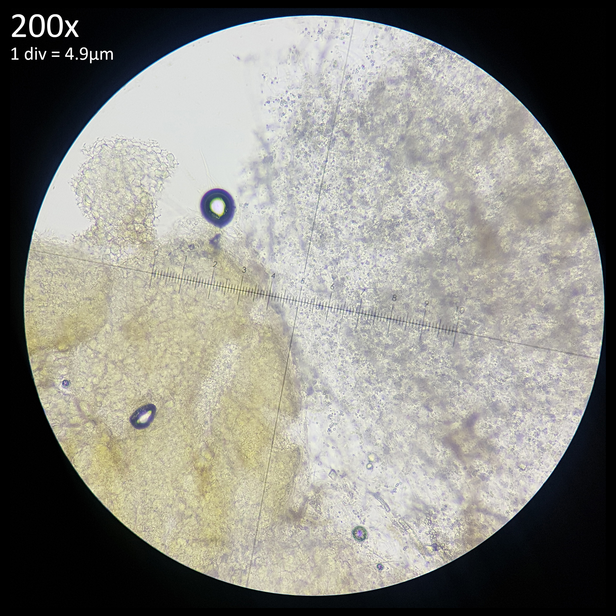
The taxonomic history of Leucocoprinus cretaceus is filled with mentions of edibility and descriptions of it smelling and tasting very pleasant. This species however has been surrounded in confusion ever since it was first described and has regularly been conflated with L. cepistipes, L. birnbaumii and others. As such these accounts are unreliable especially as they are in contrast with more recent and reliable descriptions that note an unpleasant smell and taste. In 'Flora Agaricina Neerlandica' Vellinga notes that descriptions of the smell are variable with it having been recorded as weak, Lepiotoid, similar to that of Inocybe bongardii or strong, unpleasant and nauseating even in young specimens. A herbaceous smell similar to geraniums (Pelargonium) is noted when crushed and this comparison has also been made by other authors with some Inocybe species. The taste is likewise described as strong, unpleasant and astringent though not bitter. A spore size of 8.0-12 x 5.5-7.5 μm is given or 8.7-10.5 x 5.9-6.8 μm on average.[1]

One explanation for the variation in the smell noted by different authors may be due to the confusion over identifying this species since even some modern papers and books still conflate it with L. cepistipes. It may also be worth considering the variation in how different people perceive smells and tastes since there are many species with descriptive smells noted by some authors whilst others note nothing discernible or simply say it is indistinct. Even the strong, pleasant aniseed smell of Agaricus arvensis or unpleasant phenol, inky smell of the cut flesh of Agaricus xanthodermus are not noted by some observers rendering these methods of identification unreliable.

However this experiment in culturing L. cretaceus has revealed that the mushroom simply does have a very variable smell depending on the maturity of the specimens and which part is tested. It does indeed range from insignificant, pleasant, floral and simply unpleasant or even nauseating depending on the age and condition of the specimen. The result is a mushroom that varies substantially between smelling appetising and highly off putting.

Whether or not Leucocoprinus cretaceus is edible seemed worth exploring out of more than simple curiosity - many cultivated mushrooms are species which would fruit naturally in Autumn, preferring cooler environments which may not be easily created in some locations. Whereas L. cretaceus is noted to thrive in hot and humid environments with it often appearing in greenhouses and indoor plant pots during the summer. With climate change rendering the Summer months increasingly hot and hostile to conventional agricultural practices it seems important to try and assess which tropical species can be cultivated for consumption. Leucocoprinus species already do well in the environments we have created, routinely appear in plant pots and are quite versatile saprotrophs with observations on soil, leaf litter, wood and in compost. As such they seem ideal for cultivation.

In order to explore the edibility of L. cretaceus it is necessary to first address some possible synonyms which suggest edibility before going into the further taxonomy and historic literature.


2. Possible synonyms

Genbank sequences for Leucocoprinus cretaceus are very consistent with many sequences with a 99-100% match and no outliers. Whereas what is seen with Leucocoprinus birnbaumii is a much wider range with many at only 90-95% suggesting other species submitted under the same name. Other Leucocoprinus species also have more of a range and none seem to have so many consistent samples as L. cretaceus.

It therefore seems likely that the three species listed here, all of which are regarded as current species in both Species Fungorum and Mycobank, should be considered synonyms of L. cretaceus rather than being something that is merely similar. The species described by Beeli, Heinemann and Bouriquet make no mention of any of the synonyms of L. cretaceus so do not appear to have considered this species when describing their own. The species described by Smith and Weber is compared to a number of species that are considered synonyms though there appears to be no significant distinction between their species and L. cretaceus. Subsequently the information they present regarding the taste, smell and potential edibility seem like they may be applicable to L. cretaceus.


Leucocoprinus elaeidis

Lepiota elaeidis (or elaidis) was described in 1927 by Maurice Beeli from DR Congo where they were found growing in groups at the base of palms. The description and illustration of an all white mushroom suggests that it may be synonymous with Leucocoprinus cretaceus. The spore size given is a match and the cap and stem are described as white and covered in flakes or scales with a yellow discolouration noted on the stem when touched or damaged. It also notes yellowing of the gills and sometimes a yellow or brown tinge to the cap. This correlates with many observations of L. cretaceus that show yellow on the stem if the white covering is removed or some yellow-brown discolouration towards the centre of the cap. This can routinely be observed during cultivation of L. cretaceus with the yellow colouration developing in old specimens but being absent in younger ones. Beeli says the locals called the mushroom elela and so it was evidently known to them but no information on edibility is mentioned. The smell and taste are described as pleasant however. Beeli gives a spore size of 9-10 (12) x 6-8 μm.[2] The species was illustrated by M Goossens-Fontana in Paul Heinemann's Flore Iconographique des Champignons du Congo[3] and Heinemann would later go on to reclassify this species as Leucocoprinus elaeidis, providing a spore size of 8.5-11 x 5.3-7.4 μm.


Leucocoprinus nanianae

Leucocoprinus nanianae was described from Madagascar by Gilbert Bouriquet in 1946 but this species seems poorly known as the text it is found in appears to be quite rare. However it likewise sounds like a synonym of Leucocoprinus cretaceus with a similar description of a white and powdery cap and stem and a spore size that again is a match. The cap is noted as having some very light brown fibrils but the illustration appears to show yellowing at the centre of the cap and possibly elsewhere around the gills and stem, though this is not noted in the text. The taste and smell are described as pleasant and some of the natives said the mushroom was edible so Bouriquet fed 400 grams of the cooked mushroom to a dog, which apparently did not suffer any ill effects. A spore size of 8.5-12.25 x 5.5-8.5 μm is given.[4] In 'Nomenclatural Overview of Lepiotaceous Fungi (Agaricaceae)' Vellinga notes that it looks like L. cretaceus.[5]


Leucocoprinus breviramus

Leucocoprinus breviramus was described by Helen Vandervort Smith and Nancy Smith Weber in 1982 from specimens collected in Brazoria county, Texas. The species is described very similarly to Leucocoprinus cretaceus noting the white colouration, floccose scales and a slightly yellowish brown tinge in the centre of the cap. Yellow staining is noted when the stem is handled with a floccose coating below the annulus and a smooth surface above. The specimens studied were collected by Ervin Hillhouse in 1971 and he regularly ate them describing the raw taste as mild and the cooked taste as very good and like that of young button mushrooms. Even with large servings no symptoms were noted. A spore size of 7.5-9.0 (10) x 5.5-6.5 μm is given.[6]

The reasons for Smith and Weber describing this as a new species rather than recognising it as L. cretaceus appear to once again come down to the confusion in the early taxonomy of Leucocoprinus species and the erroneous illustration provided by Bulliard. Smith and Weber note that their species does not resemble Bulliard's illustration of Agaricus cretaceus but assume that the illustration is the type for the species. They say that their species is closely related to Lepiota cretata Locquin giving a citation of Haller, 1950.

Haller refers to Lepiota cretata Locquin 1949 citing letters sent to him by Marcel Locquin.[7] However Locquin had already described the species as Leucocoprinus cretaceus in 1945 so the reason for the difference in name is unclear.[8] Haller says it is similar to Bulliard's illustration but fluffier and whiter noting that Bulliard's somewhat grey illustration does not correspond to his description but that good illustrations are found in the work of Mattirolo and Cooke. He notes L. cepaestipes as being a collective species with the white, brown and yellow forms. The photo that is included is easily recognisable as L. cretaceus even in black and white.[7]

Smith and Weber also cite the 'excellent description and discussion on L. cretata' that is presented by Marcel Josserand. Much of the history and taxonomic confusion is discussed here but Josserand remarks that the species is so distinctive that it unclear how it ever got confused. Josserand rejects Bulliard's illustration as representing the species and agrees with Locquin and Haller on the identity of Lepiota cretata. A discrepancy is noted in the description of the taste with Haller noting it as bitter but Josserand describing it as sour but not bitter, though noting it is not a serious discrepancy.[9]

However Smith and Weber reject both Bulliard's illustration and Lepiota cretata for reasons that are not entirely clear. They do however note that Leucocoprinus cretatus Locq. ex Lanzoni appears to be a European species which differs by virtue of being more robust, fruiting in large clusters, having firm, hard flesh when young and a bitter taste. They also note a larger spore size. This may be worth exploring further however I have yet to manage to locate this text and these differences do not seem significant. In culture it can be observed that sometimes singular fruiting bodies of L. cretaceus will mature and other times vast clusters together. The flesh of the stipe is very firm when fresh but becomes soft within a few days and when conditions are colder the mushrooms appear more prone to growing short and thick or aborting entirely rather than displaying the more slender appearance. Temperature and humidity may also be factors in whether a single large mushroom grows or many small caespitose fruiting bodies form. An appreciable difference in spore size can be noted if comparing a spore print to a gill section as it appears that many larger spores in some Leucocoprinus species may remain attached to the gills rather than falling when spore printing them.

Smith and Weber do not compare their species to Leucocoprinus cretaceus (Bull.) Locq. (1945) and the text in which it was described is not listed in the references so it is unclear if they were aware of this name amidst all the other competing and confusing names. In the nomenclatural overview, Vellinga notes L. breviramus is 'probably synonym of Leucocoprinus cretaceus'[5] Additionally in Nancy Smith Weber and Alexander H. Smith's 1985 book 'A Field Guide to Southern Mushrooms' a photograph of Leucocoprinus breviramus is provided and clearly shows L. cretaceus.[10]

It therefore seems probable that the species which Ervin Hillhouse consumed, often in large servings, was Leucocoprinus cretaceus. This seems to provide good supporting evidence for the species being edible but similar accounts are found in the taxonomic history of this species.


3. Taxonomic history

Like many of the most common Leucocoprinus species the taxonomic history of L. cretaceus is confusing and full of misidentifications and erroneous information and so requires significant time to unpack and evaluate. The species presented here are ones that are currently listed as synonyms in Species Fungorum and/or Mycobank and which include mentions of edibility. Whilst it was the mention of edibility in some of these species which provided the impetus to explore this subject some of these synonyms are clearly erroneous when the texts are further investigated.


Agaricus cretaceus

In 1788 Jean Baptiste François Pierre Bulliard described Agaricus cretaceus from greenhouses in France and said the mushrooms were very pleasant to smell and taste. However the illustration provided more closely resembles Leucocoprinus cepistipes so it is not clear which mushroom he was tasting.[11] This illustration would go on to create much confusion with Agaricus cepaestipes often being used interchangeably with this species, however as that species itself was often illustrated as yellow like L. birnbaumii the confusion was only exacerbated and all three species were often considered to be the same.

In 'Flora Agaricina Neerlandica', Vellinga notes that Bulliard's description clearly notes a cottony, chalk white mushroom which does not confer with the illustration resulting in some authors arguing that this illustration represents Leucoagaricus leucothites. Subsequently some authors have used Leucoagaricus cretaceus as a name for Leucoagaricus leucothites.[1]

It is possible that this confusion may have resulted in synonyms being listed for Leucocoprinus cretaceus that would make more sense as synonyms of Leucoagaricus leucothites (now known as Leucocoprinus leucothites.)


Pluteus cretaceus

In 1836 Elias Magnus Fries described Pluteus cretaceus and said they were edible and tasted better than other mushrooms. A common name of Krithvita champignonen was given which translates as 'chalk white mushroom'. Species Fungorum and Mycobank list this as a synonym of Leucocoprinus cretaceus however the description that Fries gives makes this doubtful.

The description itself provides little more detail beyond it being a completely white, edible mushroom with wide gills which retain their white gill colour until the mushroom starts to degrade. The stem is described as hollow, with a ring and no volva. It was found growing around Lund, Sweden in late Autumn.[12]

It seems unlikely that the tropical L. cretaceus would find late Autumn in Sweden to be a satisfactory environment in which to fruit but Fries does not specify any further details on the habitat, like if they were in greenhouses or compost. The more important information may be the gills retaining their colour until they start to degrade. This species is listed alongside Pluteus bombycinus and Pluteus campestris and the section on the Plutei is introduced by saying these species have reddish or brown spores but do not deliquesce into black liquid like the 'completely useless' Coprinoids. It would appear then that Fries was grouping Volvariella bombycina and Agaricus campestris (for which he notes several similar species exist that are not important to separate) together with Pluteus cretaceus because of the whitish caps and pinkish gills. When he notes that the gills retain their white colour until they start to degrade he may be describing a species with white gills that discolour pinkish with age.[12]

This suggests that Fries was actually describing Leucocoprinus leucothites, an all white mushroom which develops a pink colour to the gills with age. This species is often regarded as being edible with some sources cautioning a gastrointestinal reaction in some people or not recommending them due to the potential for novice foragers to confuse them with dangerous Amanita species. It does not explain the reddish brown spores noted but perhaps Fries simply did not spore print it and was only grouping it together with these species based on the similar appearance and gill colour. In any case it seems a far more likely candidate to find in Sweden in Autumn than L. cretaceus. Subsequently Fries' description of this as an edible and tasty mushroom must be disregarded for this research.


Psalliota cretacea & Pratella cretacea

In 1871 Paul Kummer described Psalliota cretacea as growing in fields, meadows and gardens in Germany in Autumn. He stated that the mushroom was tasty and suggested the common name Kreideweißer champignon which translates as 'chalk white mushroom'. He does not state whether he is reclassifying Fries' Pluteus cretaceus though his description is also more likely to be Leucocoprinus leucothites as many of the species he is describing as Psalliota are now considered Agaricus species. His key to the Psalliota genus notes gills that start pale or grey before blackening, turning faint olive green, reddish brown, purplish or brown.[13]

In 1878 Claude Casimir Gillet reclassified Fries' Psalliota cretacea as Pratella cretacea agreeing that the flavour was pleasant and said they were an edible mushroom. Gillet's description for the Pratella genus says they have brown or purple-black spores and most of the species he describes and illustrates within this genus have since been reclassified as Agaricus.[14]

Therefore Kummer and Gillet's description of the species as edible should also be disregarded here. It isn't clear if these species ended up listed as synonyms due to a mistaken reclassification or because of the former classification of Leucoagaricus leucothites as Leucoagaricus cretaceus. Regardless they do not appear to be descriptions of Leucocoprinus cretaceus.


4. Further descriptions

1835 Carlo Vittadini describes Agaricus (Lepiota) cretaceus with a description that seems accurate and correct for this species. It is described as growing in groups on rich substrates comprised of decomposing animal and vegetable matter, so presumably was observed on compost. Vittadini does not specify that they were found in a greenhouse however with the temperatures in Italy it wouldn't be surprising to find them growing outside. He says the mushrooms are edible and similar in taste and smell to Pelliccione, the common name given for Agaricus procerus, now known as Macrolepiota procera.[15]

E affatto innocente, e raccolto giovinetto può essere, al pari del Pelliccione, per gli usi della tavola destinato.

'He is completely innocent and collected as a young man, like Pelliccione, he can be destined for the uses of the table.' (Translated from Italian via Google).

However a strange footnote is included:

(t) Donnèe aux animaux, à la dose d'un seul , elle excite deux heurss après un vomissement considérable j l'animai se piami, ne veul rien prendre , tombe dans l'assoupissement et meurt. Paul. 2, pag. 360.

This is taken from Jean-Jacques Paulet's 1793 text Traité des champignons.[16] (Translated from French via Google).

XXIV. The Palette with darts or three-carts (pi. CLXIII, fig. 3). This species, which I do not find described, is a mushroom which rises to the height of five or six inches, white, but with [gills] which take on a green eye. The surface of the [cap] is white & covered with triangular, equal, pyramidal points; these points of a dirty white, adhere strongly by their base to your skin which covers the [cap]. This [cap] is regularly circular. The [gills] are usually covered with a dust similar to fine flour, and with a fine veil which ends up hanging only on the stem, and acts as a collar. The stem is white, cylindrical, of an equal diameter, full of a soft substance furnished by a bulb which pivots a little in the earth, and is exhausted, it becomes hollow like the stem.

This plant, which one finds in autumn in the park of Saint Maur, is of a soft substance, & has a very pleasant odor of ordinary mushroom; however, it is very evil. Given to animals, in a single dose, it excites considerable vomiting two hours after; the animal complains, does not want to take anything, falls into drowsiness & dies.

Paulet's text is filled with mentions of him feeding various mushrooms to animals to see if they would die... but it is not clear which animals were the unfortunate subjects of his questionable tests. Searching the ~500 page text for 'animaux' turns up 197 results, 'chien' turns up 33 and a quick look at some of these shows he was indeed deliberately feeding mushrooms to dogs to see if they would die. Other searches for common test animals like rats, mice and rabbits don't turn up results and there were only a couple mentions for cats. So it could be that the animal which died from eating this mushroom was a dog.

The green discolouration mentioned in the gills is interesting. This immediately suggests Chlorophyllum molybdites, which the size and scaly cap would support however this is generally regarded as a North American species. A study in 2022 noted that Chlorophyllum molybdites was widespread on the coast of Sicily in Italy[17] and an online post from the University of Veterinary Medicine Budapest cites identifications of the species in Scotland, Australia and Cyprus with the first poisoning cases being in Sicily and Spain in 2014.[18] though citations are not given to explore further.

It doesn't seem impossible that the species could have found its way to Europe in the late 1700s and that may explain the author being unable to find a description of it however the base isn't especially bulbous as noted in this description and combined with the pyramidal scales it starts to sound more like an Amanita species.

Whilst C. molybdites is regarded as highly toxic with serious gastrointestinal distress, it generally is not considered as fatal with no such incidents recorded in adults. However there are a few isolated cases of small children dying after consuming these mushrooms and many sources mention that they may be deadly to dogs and horses. This information is circulated on several sites but the original source of it is unclear so it cannot be evaluated for veracity.

Another possibility is Leucocoprinus cepistipes, which is sometimes observed to develop greenish gills with age or upon drying and Leucocoprinus cepistipes var. rorulentus specifically does note this trait. The bulbous base would match, the height of the stem given is within the range described and this species was documented from Europe. There is no information on toxicity with this variant but it seems doubtful that it would be fatal if consumed however.

Paulet gives no scientific name for the mushroom he is describing and instead provides only a common name of La Palette à dards ou à trois-carts and a reference for an illustration on plate CLXIII, fig. 3. The other mushroom described on this page is given the common name La petite Rape and is illustrated in fig. 1 & 2.[16] These plates do not appear in this text however, at least not in the scanned versions available online. Other species in this text correlate with the illustrations in Iconographie des champignons de Paulet[19] but the common name given does not for this mushroom. Plate CLXIII figures 1 & 2 have the common name Oronge à pointes de rape and the scientific name Hypophyllum radula. Figure 3 has the common name Oronge à pointes de trois quart and the scientific name Hypophyllum tricuspidatum. The citation for each points back to page 359 in Traité des champignons so does confer with the previous text.

To further confuse the matter page 89 of Iconographie des champignons de Paulet gives a description for these illustrations and gives the name Agaricus echinocephalus[20] (now known as Amanita echinocephala). This species is known from Europe and descriptions do note a slight greenish tinge to the gills so this would seem to be a good contender for the species described by Paulet.

Neither Hypophyllum radula or H. tricuspidatum are listed in Species Fungorum with any synonyms and Mycobank lacks a full citation for either but lists H. radula as having the current name Agaricus pauletii (no page citation) with the later classified Lepiota pauletii listed as a synonym. The citation for Lepiota pauletii however does not seem to point to anything by this name but does include an illustration of Lepiota echinocephala.

Further time has not been spent exploring this because it seems that regardless of which species poisoned the dog (or rather which species Paulet poisoned the dog with), it does not match the description of Leucocoprinus cretaceus and so Paulet's description of toxicity can be disregarded. Vittadini's information however on edibility is useful nonetheless as his own description matches that of L. cretaceus.


In 1918 Oreste Mattirolo described Lepiota cretacea as a reclassification of Bulliard's Agaricus cretaceus. He did not provide a description however and instead referred to that provided by Vittadini which he compared to 'a photograph made with words'. Mattirolo says the similarity to Pelliccione, Lepiota procera as described by Vittadini is remarkable. Mattirolo says he collected them for food but it is not clear which species he is referring to and whether the 'remarkable' similarity he is noting is in reference to the taste or appearance.

Translated from Italian via Google:

In order to have an idea of the type of our mushroom, it seems to me very opportune to mention the observation made by Vittadini, that the Cretaceous Lepiota resembles the common Pelliccione, that is, the Lepiota procera Quel. Having collected in the soil deriving from the decomposition of the leaves in the Royal Botanical Garden of Turin, numerous specimens, which I also used as food, I was able to convince myself that, especially in the more developed individuals, this similarity is remarkable.

It sounds like Mattirolo was eating Lepiota cretacea but it is unclear. The only description he provides personally are measurements of the spores at 8-10 x 4-6 μm which is within the correct sort of range for Leucocoprinus cretaceus.[21]

Fig. 1 - Mattirolo's illustration of Lepiota cretacea (1918) Fig. 2 - Hard's photograph of Lepiota cepaestipes (1908)

Mattirolo's illustration (fig. 1) is also a match for Leucocoprinus cretaceus and could not really represent anything similar like L. cepistipes as has been confused by other authors. Mattirolo also provided a list of illustrations which he said best compared with Lepiota cretacea. Fortunately these are all long since out of copyright and so they can be included here to easily compare side by side.

Figure 2 - Miron Elisha Hard's photograph of Lepiota cepaestipes [22] in his 1908 text 'The mushroom, edible and otherwise' is described by Mattirolo as 'very significant'. With the scales visible on the stipe it does seem to resemble L. cretaceus more than L. cepistipes. Hard lists it as an edible species however he also describes there being yellow, yellowish and white forms so it appears that he was unclear on the identity of these species.
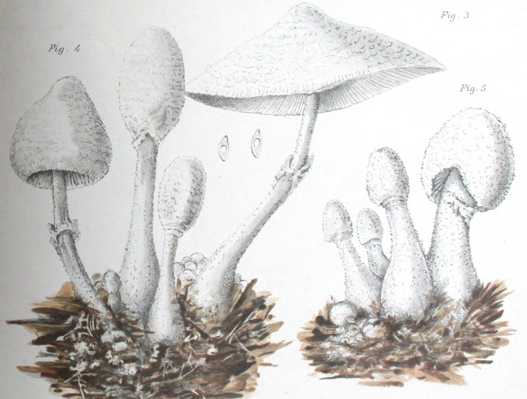
Fig. 3 - Bulliard (1788) Fig. 4 - Sowerby (1797) Fig. 5 - Hornemann (1823) Fig. 6 - Greville (1828)

Figure 3 - Pierre Bulliard's illustration of Agaricus cretaceus [11] was described as corresponding well to the truth but being too exaggerated with its reddish tint and lacking the cottony character of the mushroom. This illustration appears to more closely resemble L. cepistipes rather than L. cretaceus and so Mattirolo's critique of the image suggests he was not collecting L. cepistipes mushrooms.

Figure 4 - James Sowerby's illustration [23] was not seen by Mattirolo personally but cited only based on the mention by other authors.

Figure 5 - Jens Wilken Hornemann's illustration of Agaricus cepaestipes from Floræ Danicæ Iconum [24] was described as 'leaving much to be desired'. Mattirolo notes that Elias Magnus Fries based his forma pumila upon this illustration. Mattirolo provides a citation of 'Flora danica (vol. X, fasc. XXVIII a XXX; tav. 1721, anno 1800)' for this illustration however tab 1721 is Stereocaulon incrustatum so this appears to be a citation error. Agaricus cepaestipes is found on tab 1798 of vol. 10, fasc. XXX but this was published in 1823 not 1800. This illustration is presumably the one that he meant to cite however as he said that it leaves much to be desired and indeed it does lacking in both detail and colour.

Figure 6 - Robert Kaye Greville's illustration of Agaricus cepaestipes [25] was not seen personally by Mattirolo but was examined by Pier Andrea Saccardo who said it corresponded well to the fresh specimens sent to him by Mattirolo.
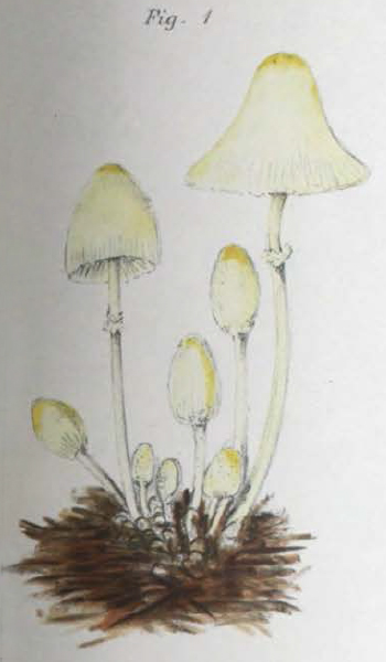
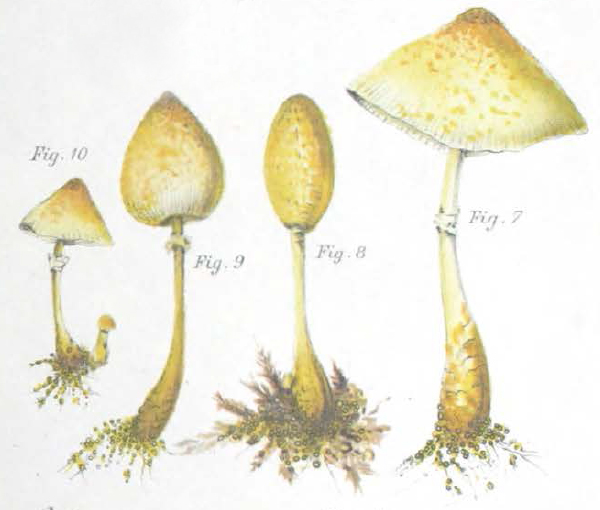
Fig. 7 - Barla (1889) Fig. 8 - Patouillard (1889) Fig. 9 - Gillet (1874-1898) Fig. 10 - Cooke (1881-1891) Fig. 11 - Cooke (1881-1891)

Figure 7 - Jean-Baptiste Barla's illustrations of Lepiota cepaestipes [26] found in figures 7-11 amongst several other species also regarded as Lepiota at the time do not appear to contain much detail to distinguish them from other white mushrooms. Mattirolo cites figures 5 and 6 of this compilation, which represent Lepiota rorulenta (now known as Leucocoprinus cepistipes var. rorulentus) which are said to correspond well to the species type.

Figure 8 - Narcisse Patouillard's illustration of Leucocoprinus cepaestipes [27] from 1889 is described by Mattirolo as 'recommendable, on the whole'. This illustration, with its distinctly scaly cap and stipe clearly does represent Leucocoprinus cretaceus rather than L. cepistipes.

Figure 9 - Claude-Casimir Gillet's illustration of Lepiota cepaestipes [28] is described by Mattirolo as being one of the best depictions. This illustration does resemble L. cretaceus so may support that being the species Mattirolo was collecting.

Figure 10 - Mordecai Cubitt Cooke's illustration of Agaricus (Lepiota) Cepaestipes var. cretaceus [29] is only described as 'excellent'. The base of the stipe in this illustration strongly resembles that of L. cretaceus. Cooke also illustrated Agaricus (Lepiota) Cepaestipes in this text (fig. 11) with white and yellow forms owing to the confusion between the different species. However Mattirolo does not cite this illustration and since it appears in the first volume of 'Illustrations of British Fungi' 1881-1883 as opposed to the volume VIII supplement published 1889-1891 it is not clear if he also saw and compared this one.

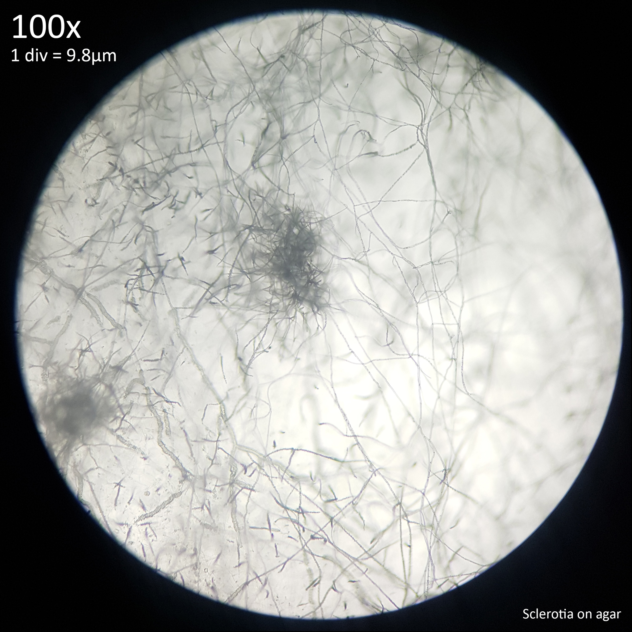
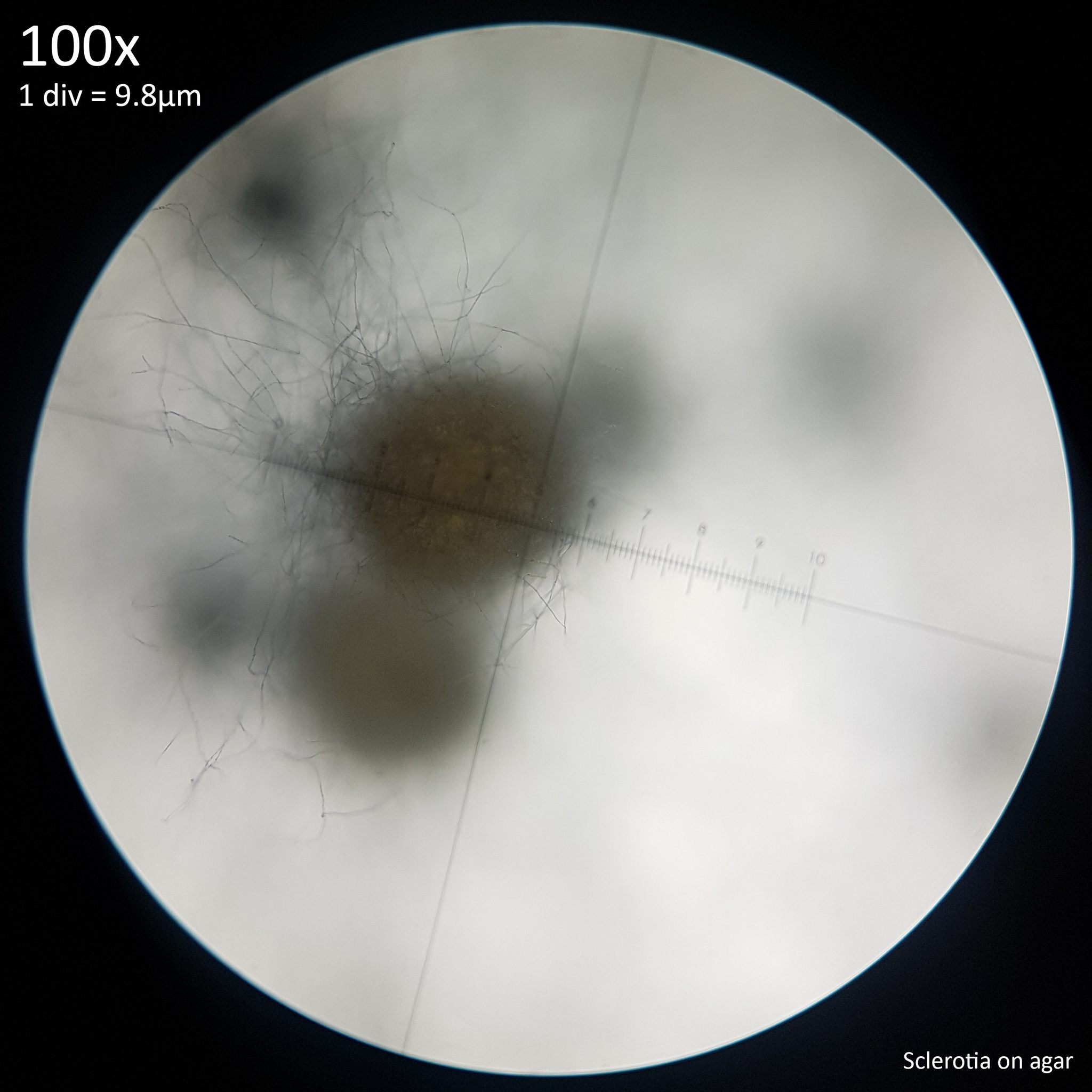
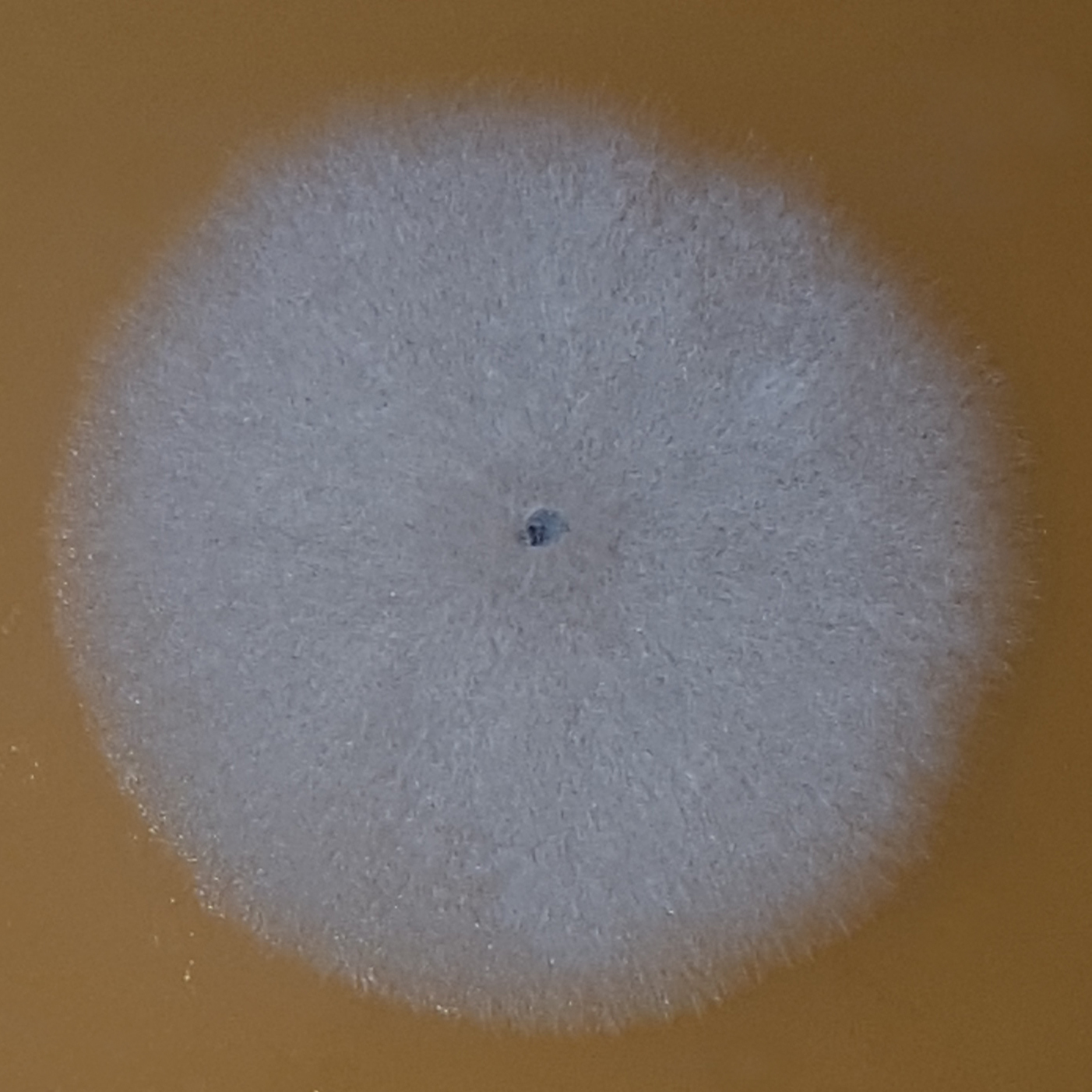
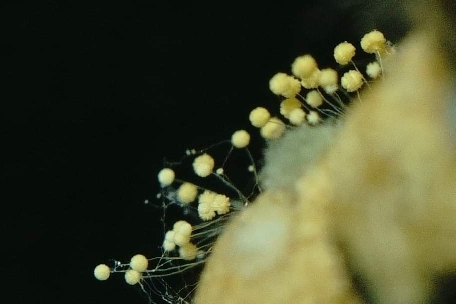
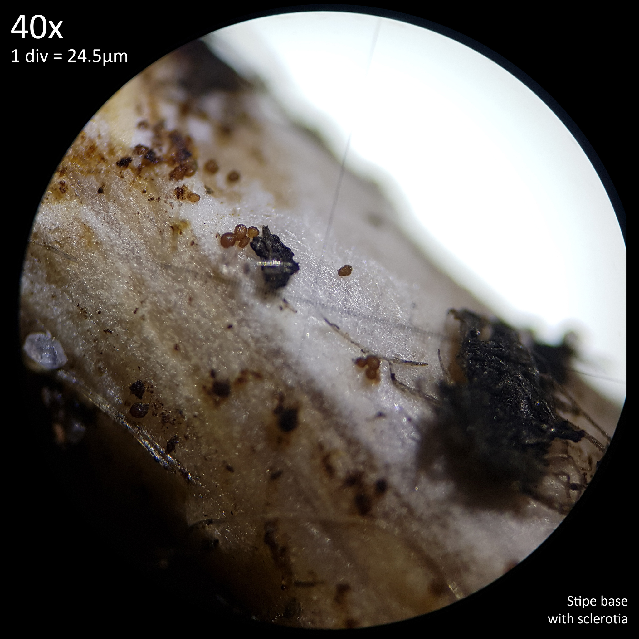
The text does not make it perfectly clear if Mattirolo actually did eat L. cretaceus but his list of illustrations do point to that being the species he was observing. Mattirolo was never able to successfully cultivate any of the Leucocoprinus species he was studying due to contamination issues and so his observations were made only on specimens that grew in the greenhouses, where the mushrooms had been introduced with tropical plants. Interestingly he did not observe sclerotia in this species and stated that it had none however when it is cultivated the sclerotia are abundantly noticeable and seem to always appear before the mushrooms.


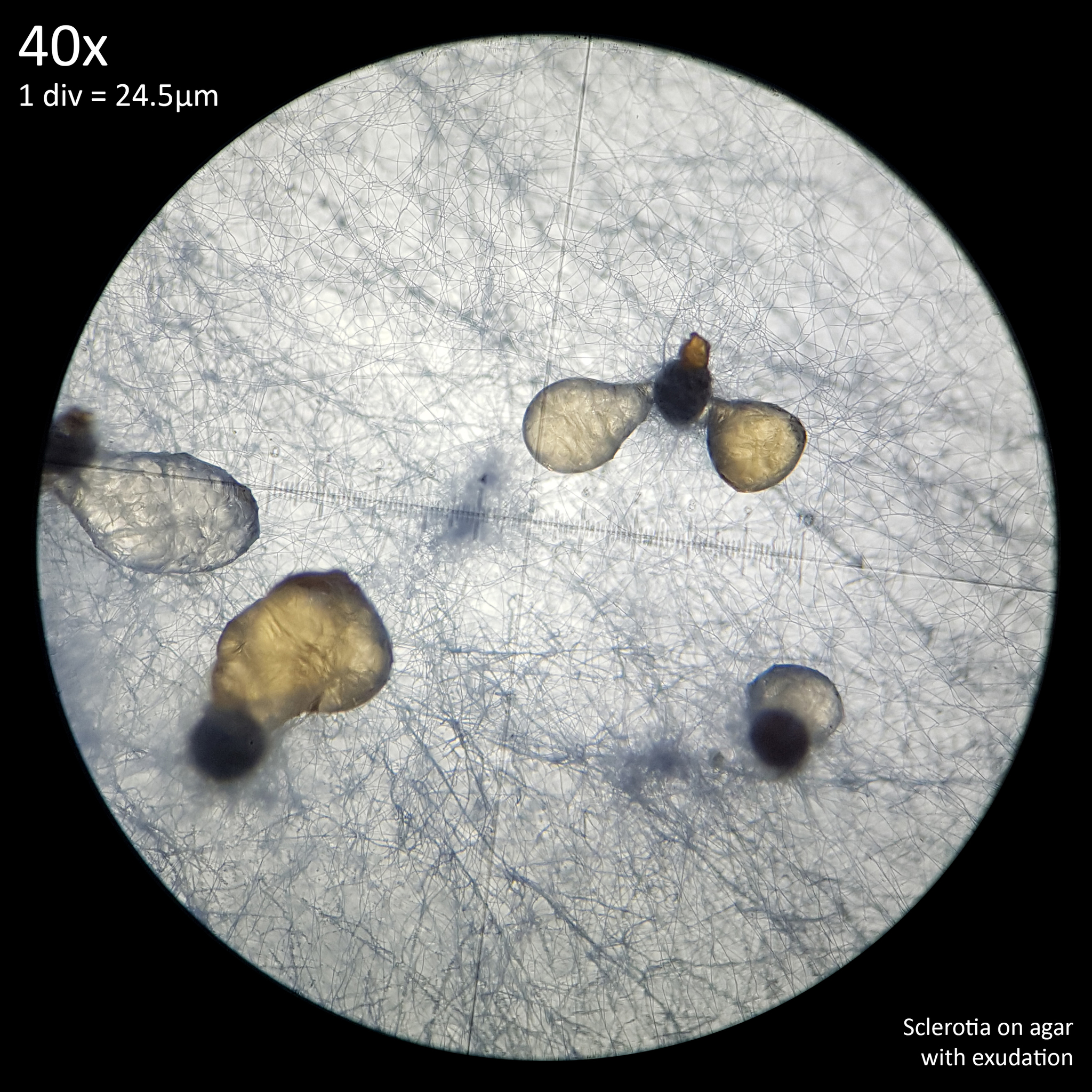
5. Cultivating Leucocoprinus cretaceus

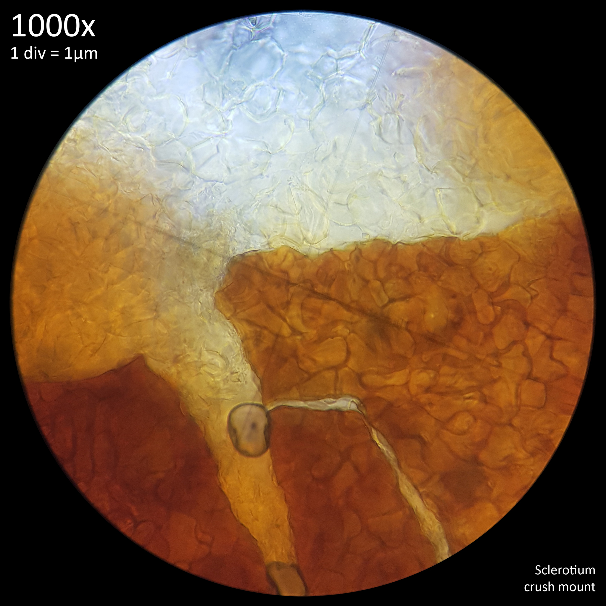
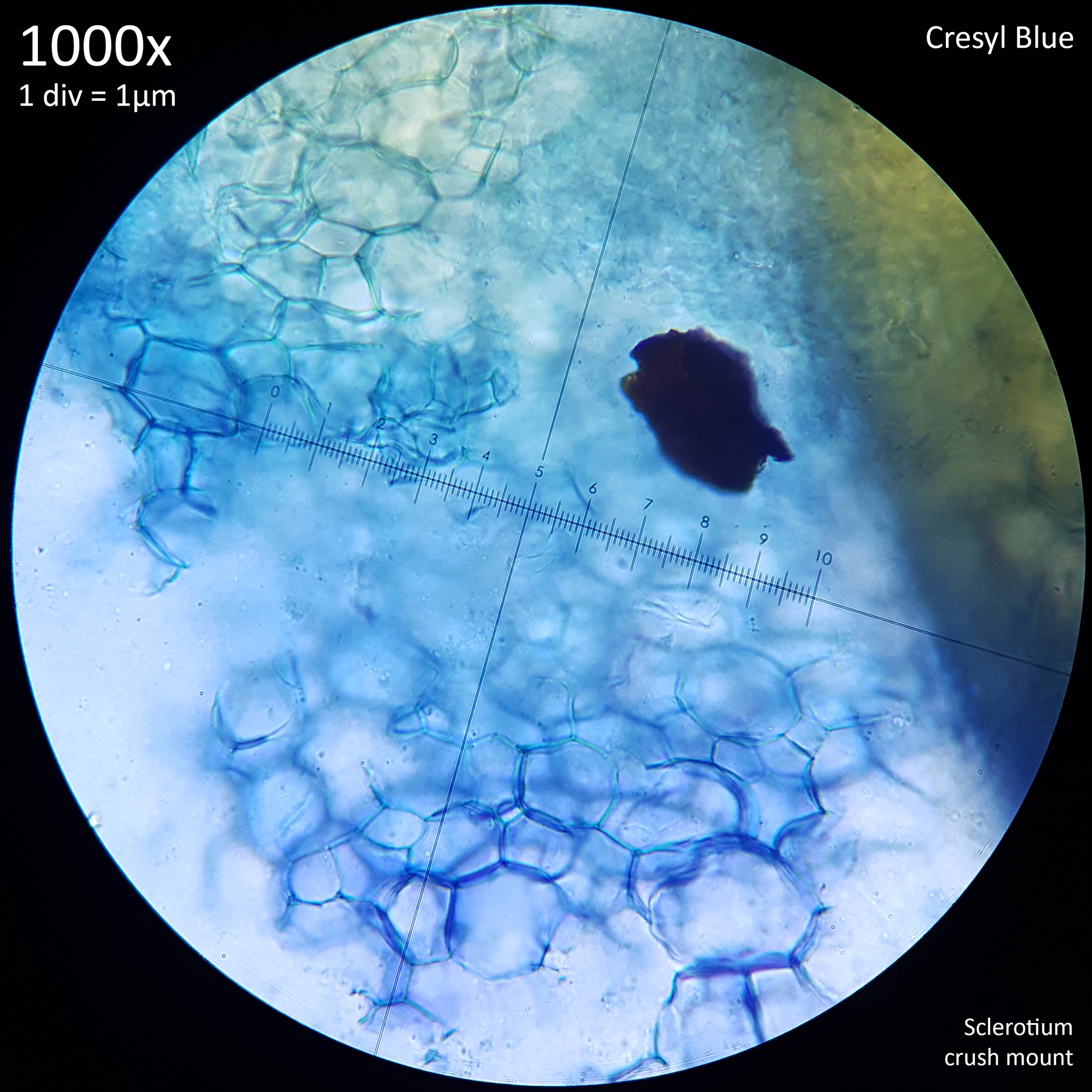
The bulk of the information on culturing L. cretaceus for this experiment is presented in 'A preliminary investigation of sclerotia in Leucocoprinus cretaceus' as it was the growth of the sclerotia that was first noticed. The first mushrooms which were observed began growing only after sclerotia were harvested for examination though subsequent observations have since been made of numerous specimens grown on various substrates. In sealed jars at room temperature or with heating elements the mushrooms appear to grow readily and easily on a variety of substrates and have a rapid rate of growth such that regularly harvesting them becomes necessary.

The duration of time between immature primordia developing and the mushrooms maturing to the point at which the cap opens appears quite variable and dependant on environmental conditions with heat and humidity accelerating the process. When conditions are less than optimal the primordia appear to develop more slowly and sometimes seem to stall in their growth to grow more or abort later. Once the cap opens however the mushrooms only last for around two days before deteriorating so the window of time in which to harvest them is relatively short. Likewise the stems of the mushrooms, if kept in the fridge will not last more than a couple days before withering and darkening so dehydration is necessary for storage. The caps and stems dry very readily however and can be preserved even if just left to dry at room temperature without the use of a dehydrator.

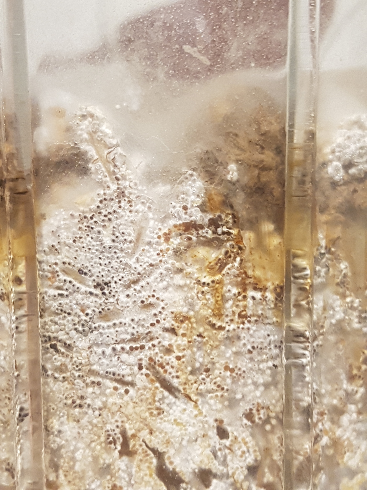
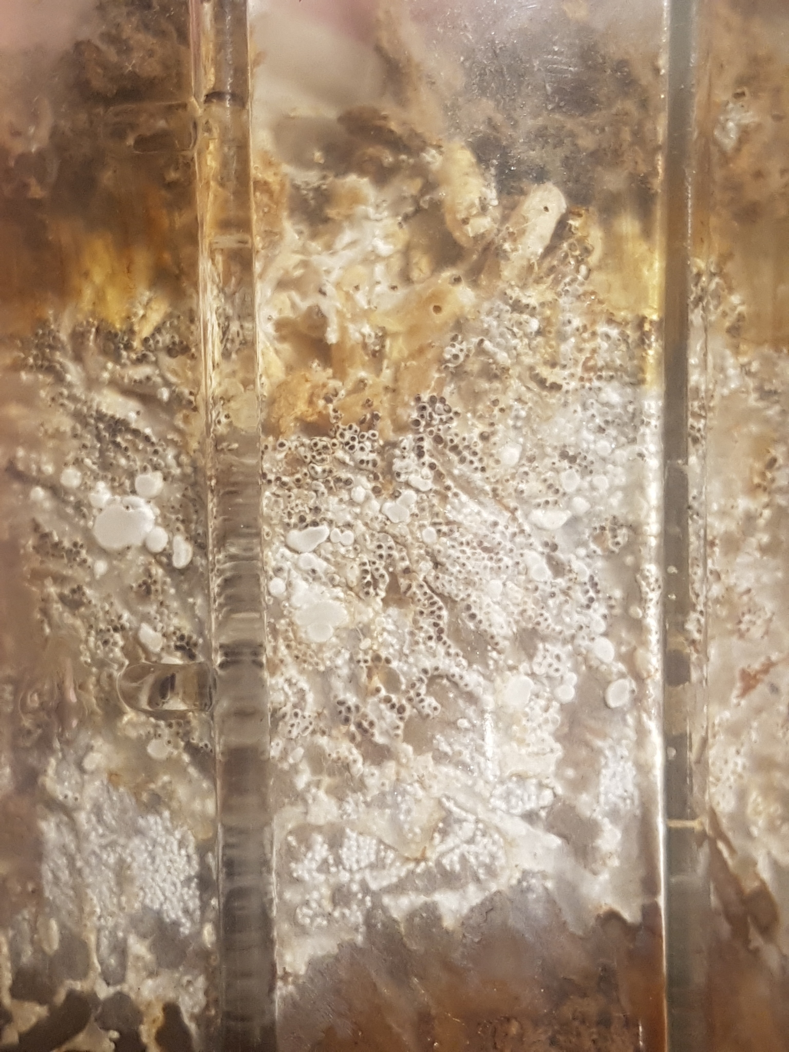
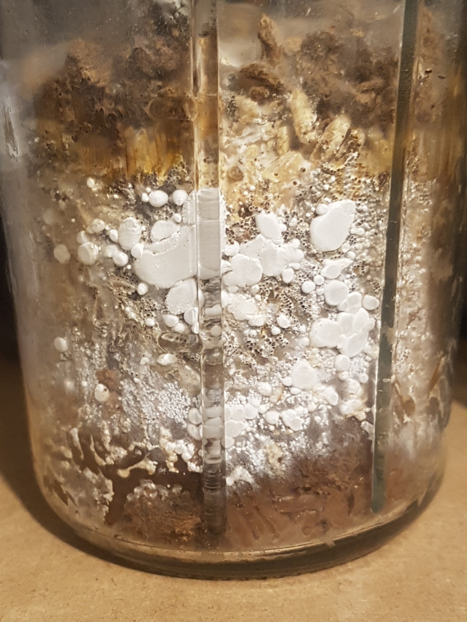
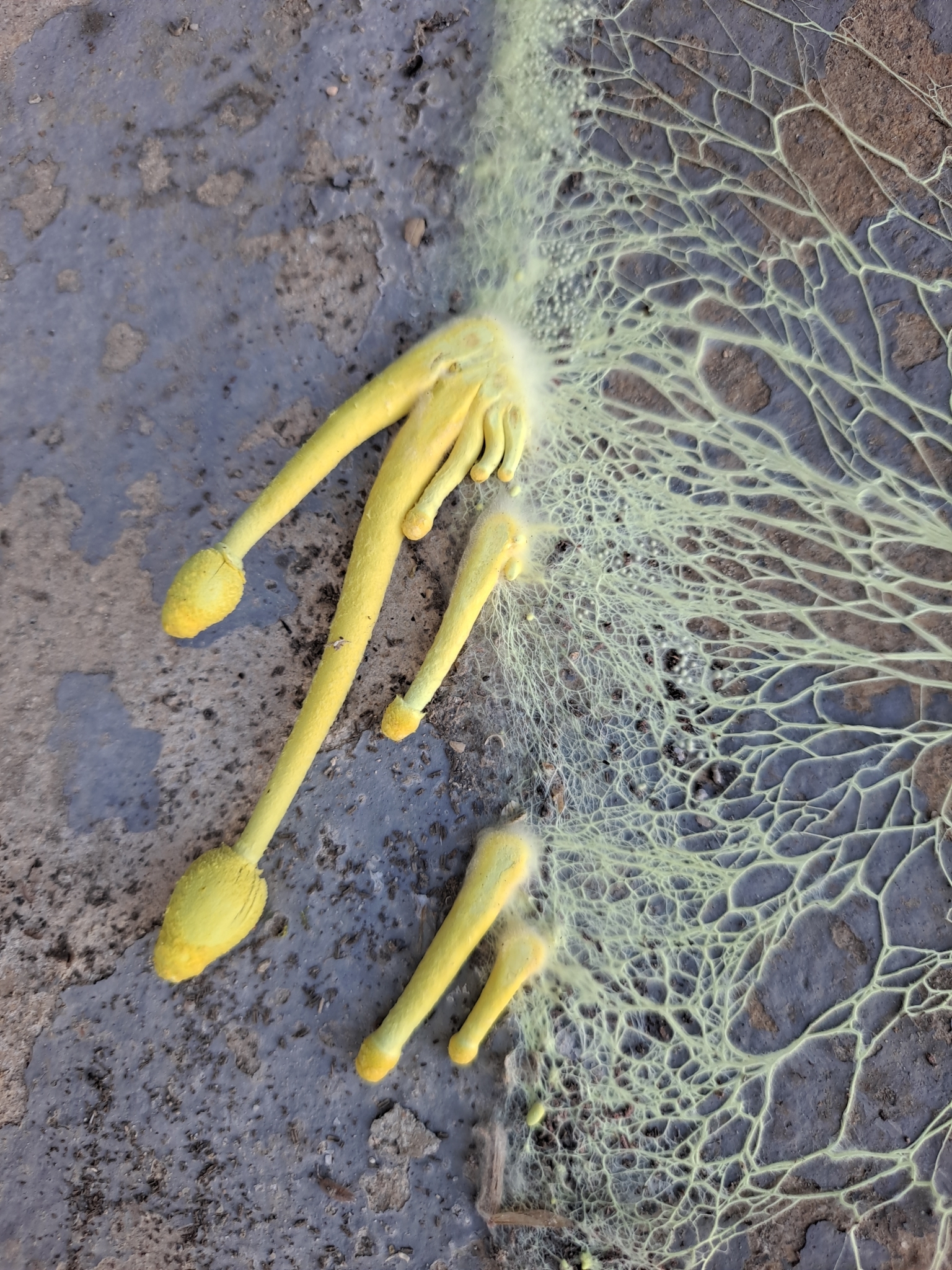
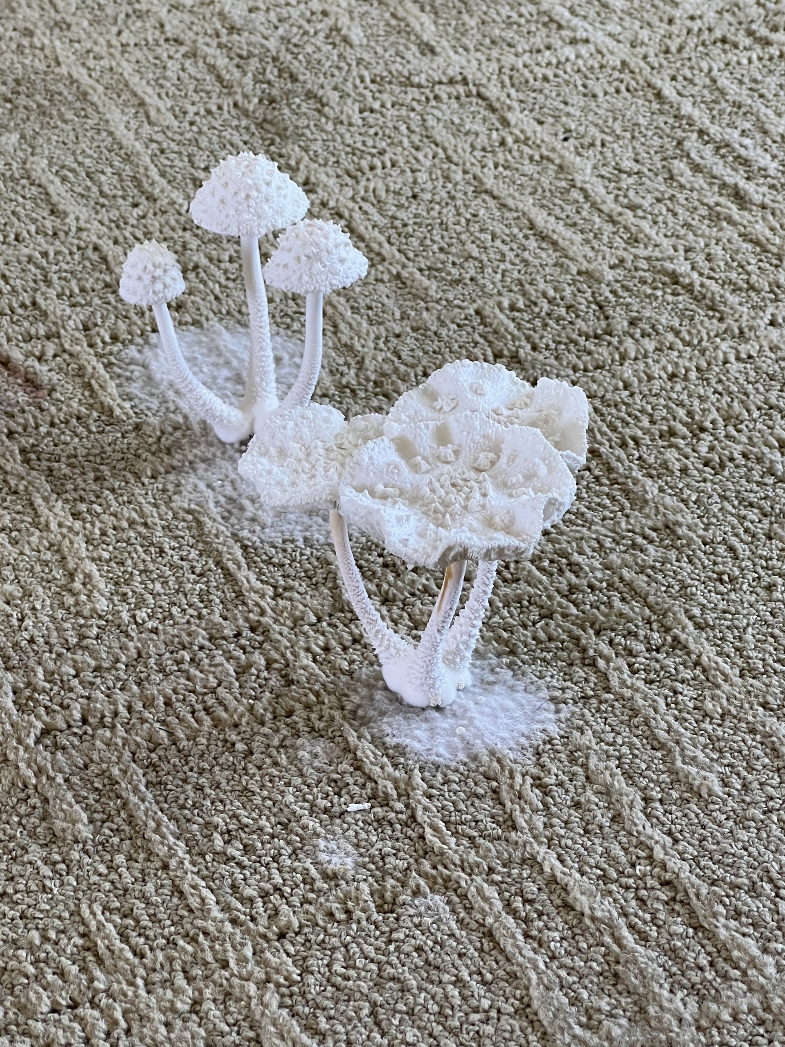
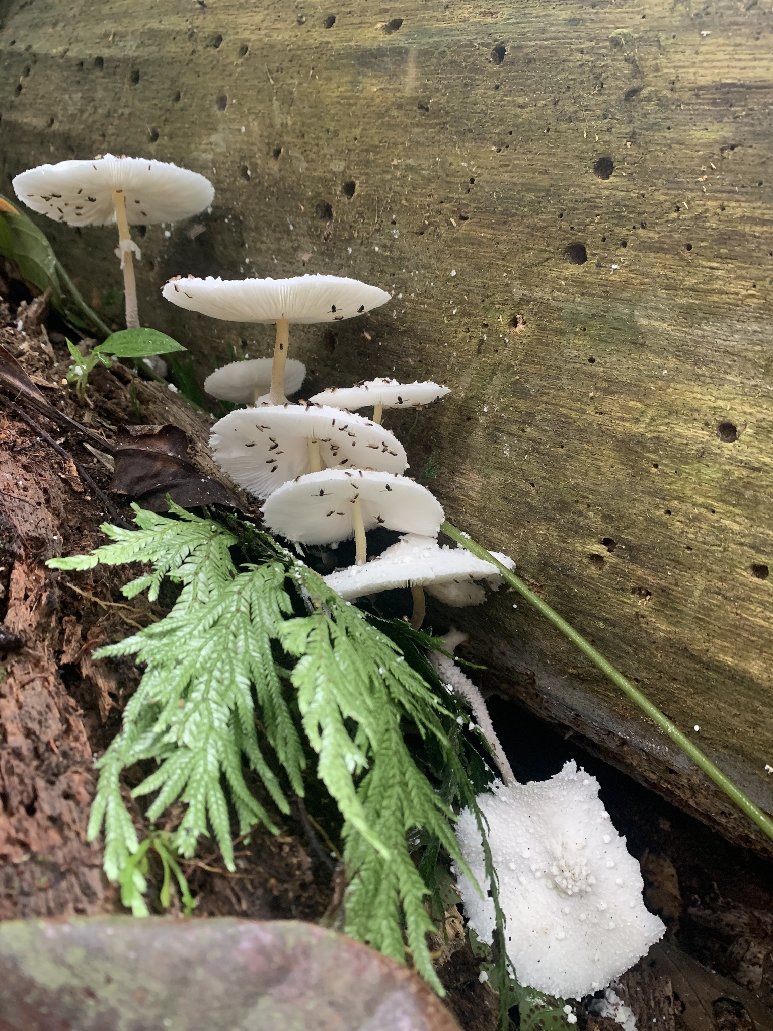
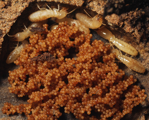
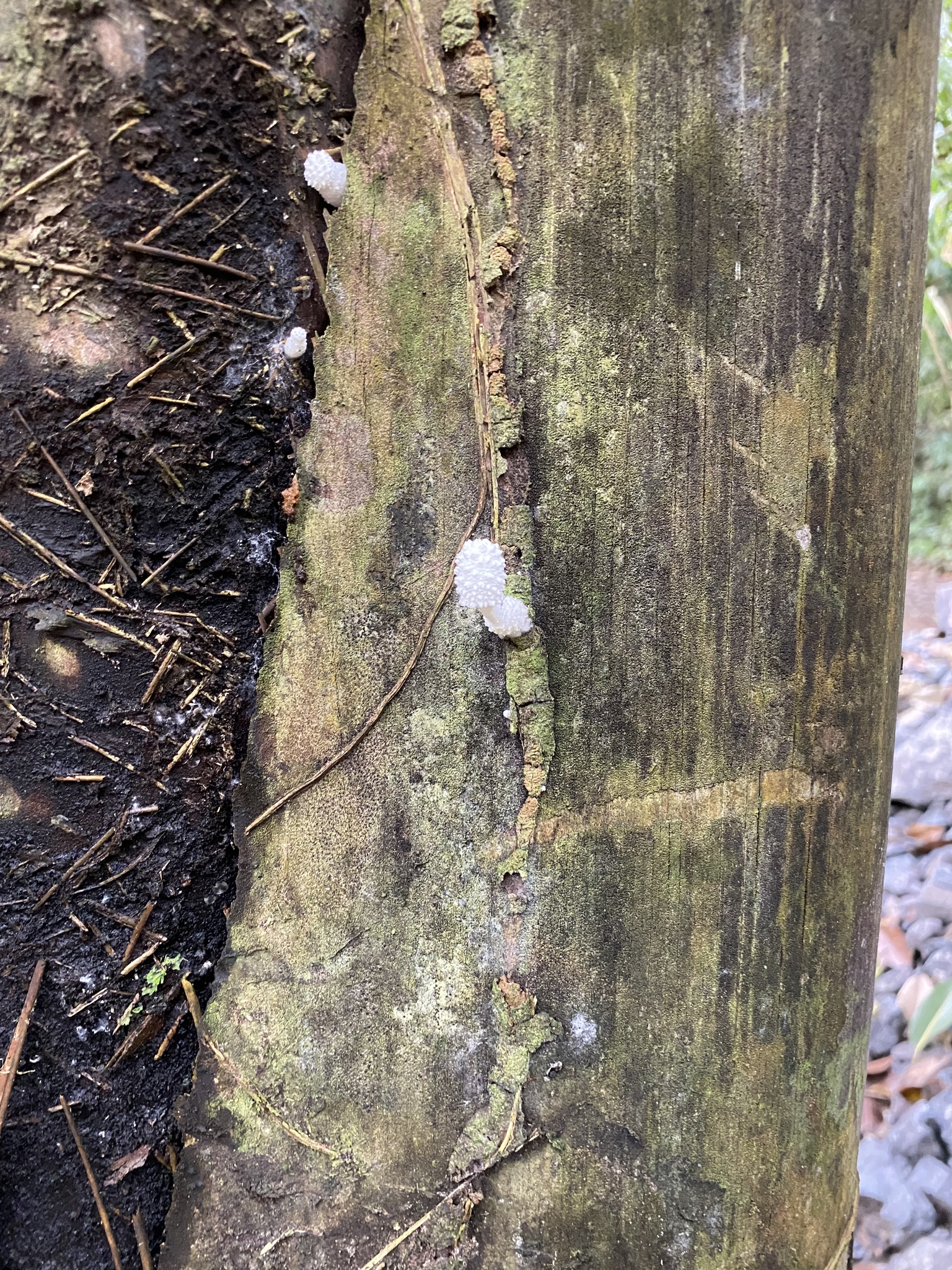
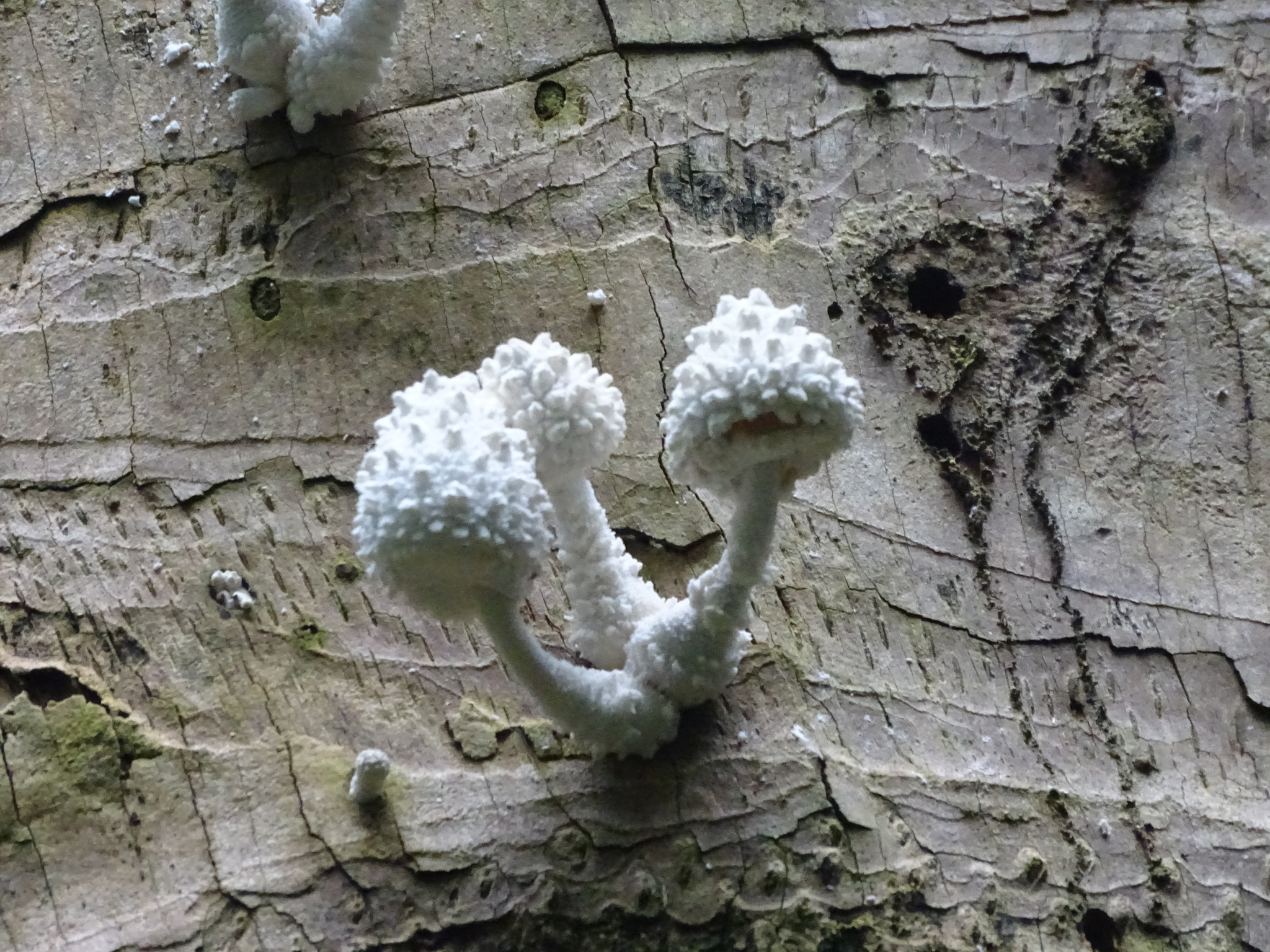
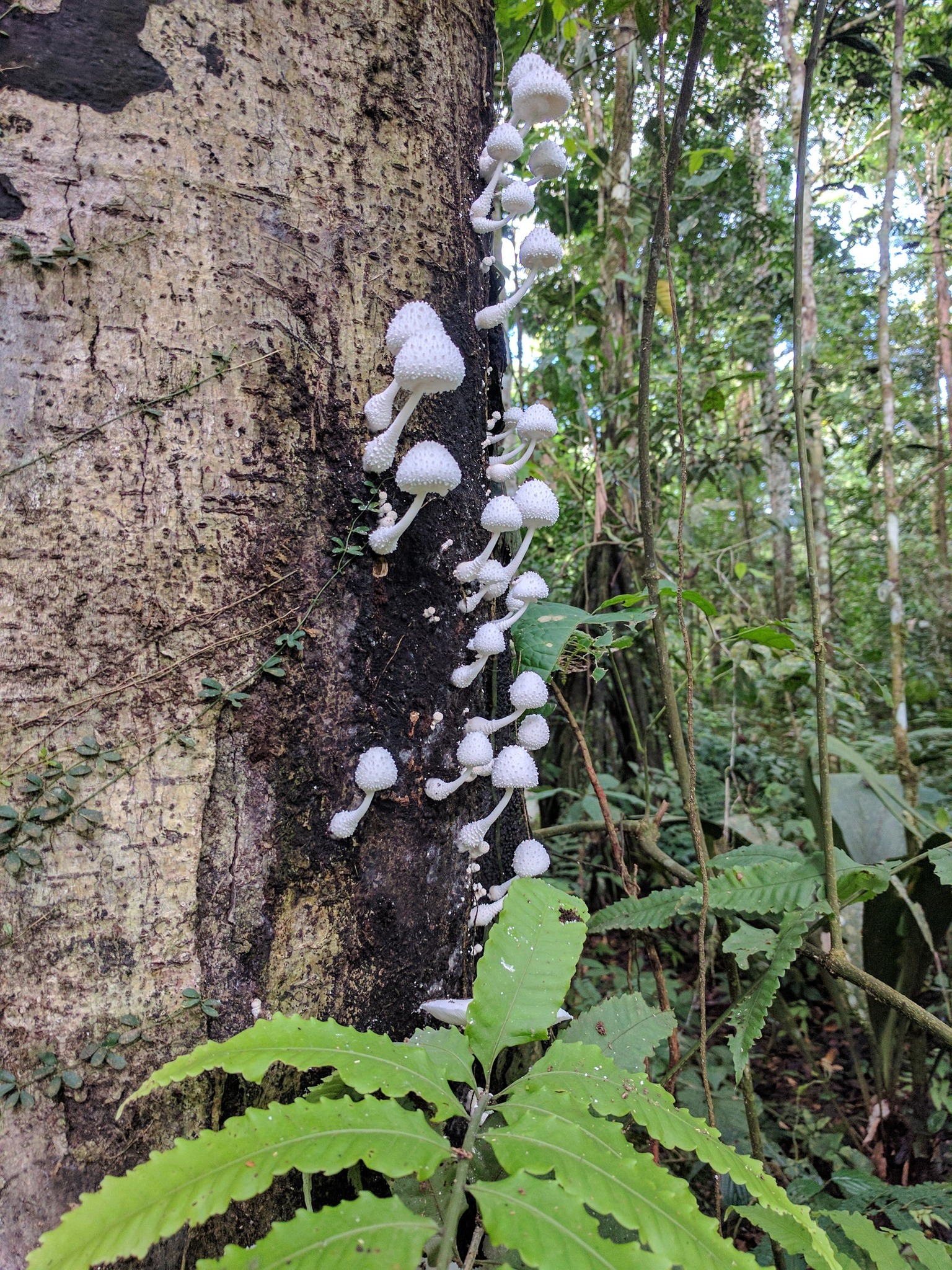
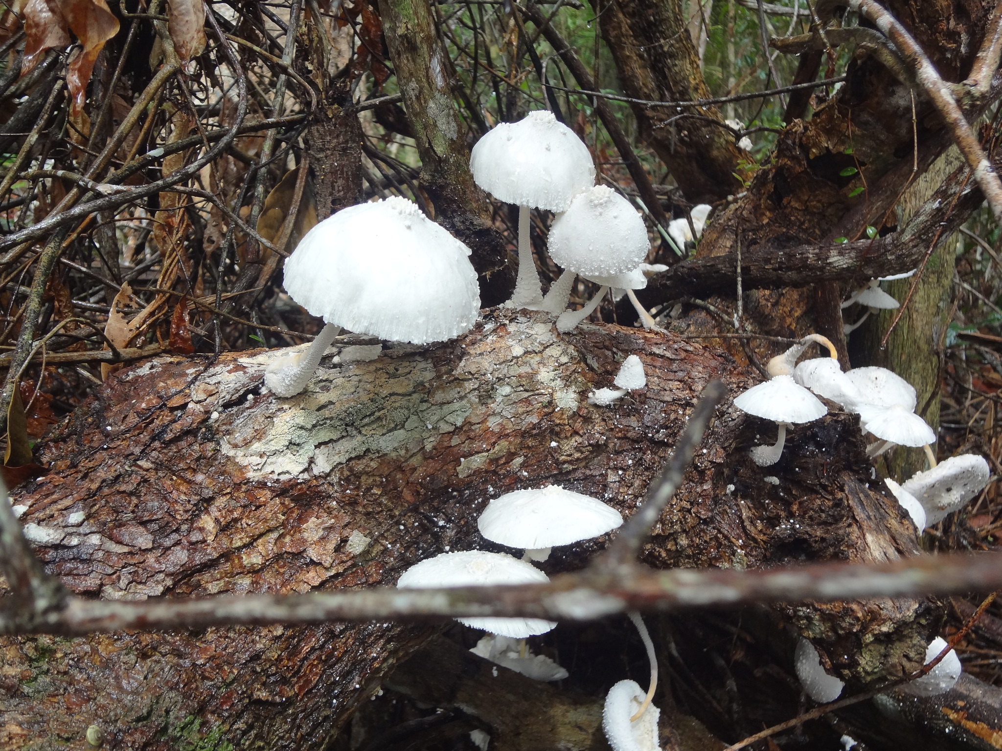
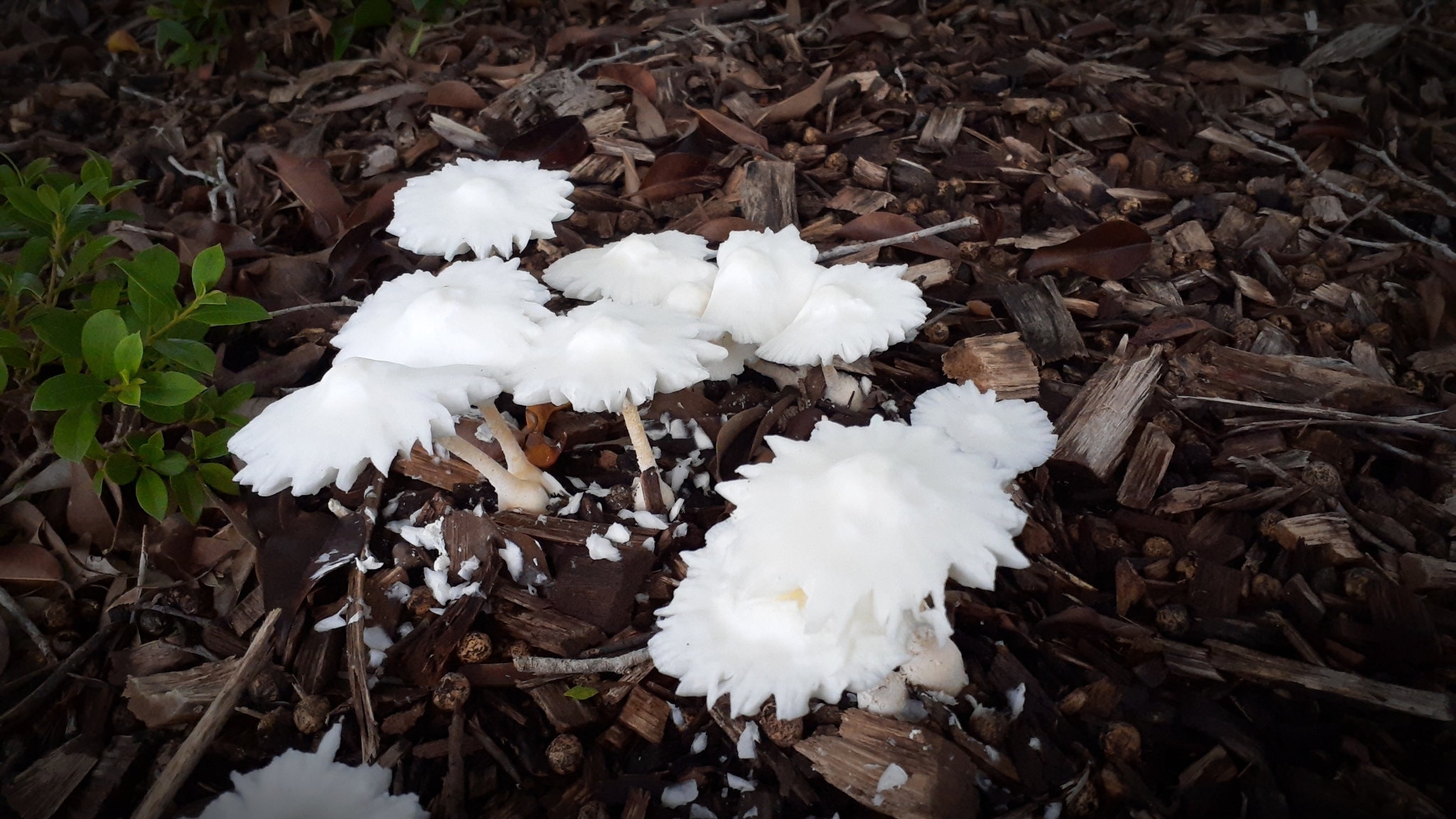
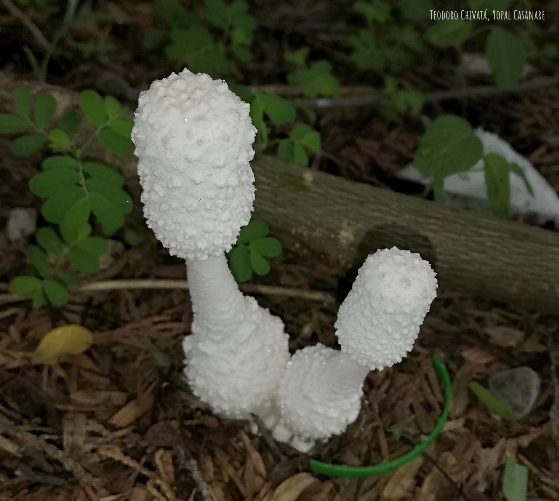
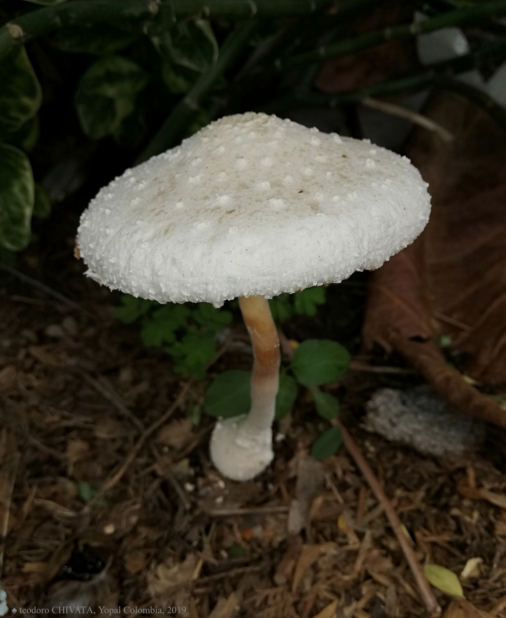
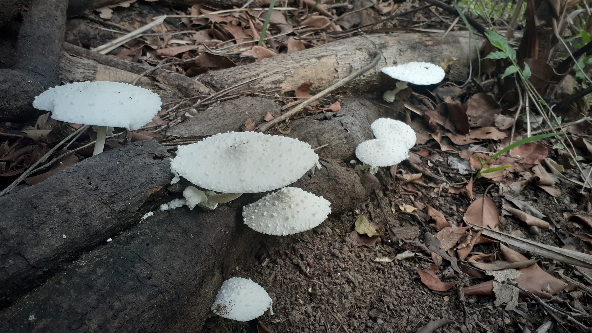
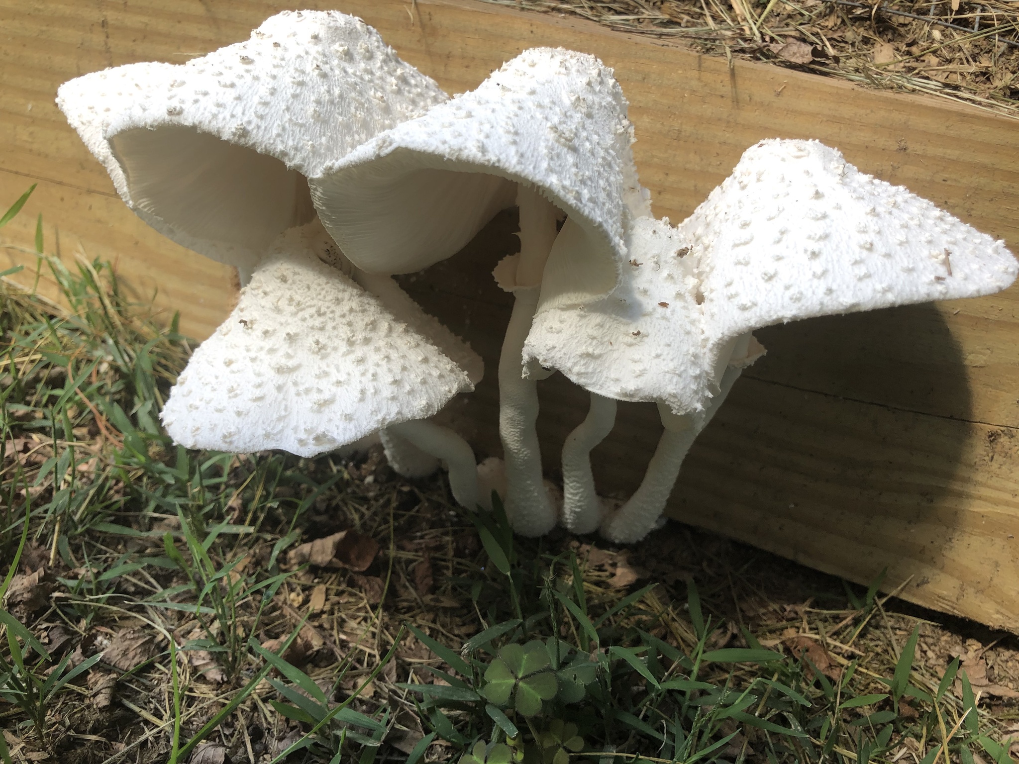
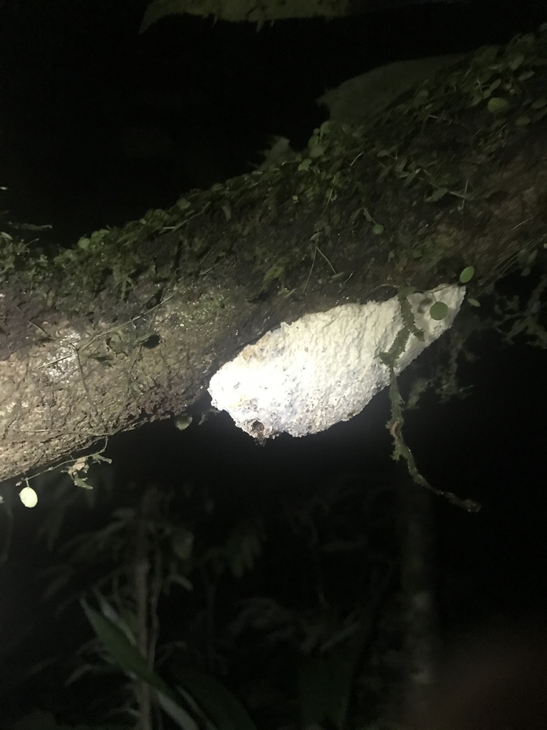
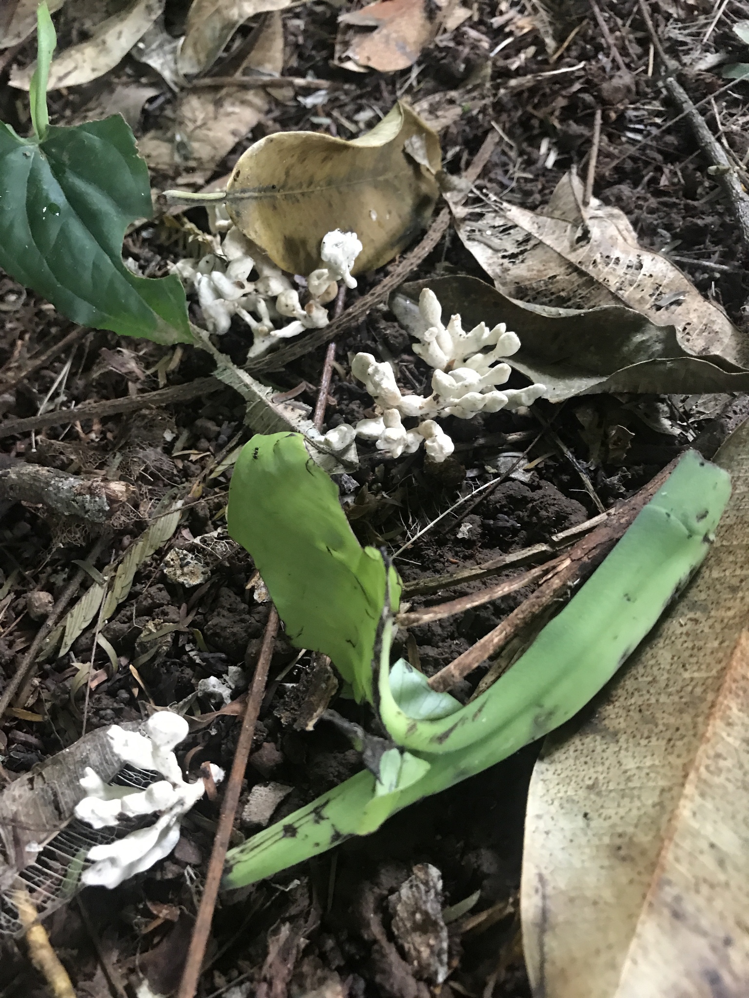
In nature Leucocoprinus cretaceus is a very versatile saprotroph and is observed growing on trees, fallen wood, amongst woodchips, from termite tests, in plant pots, from compost piles or in compost bins or just straight from the ground. There are also numerous observations of it growing in houses from walls, floors, furniture and carpets (some of which are collected here) and whilst it would be inadvisable to eat any mushrooms grown on such structures it does point to a real versatility in their growth habits.

It is therefore likely that they will perform well on many different substrates in captivity and whilst it will take much more testing to determine the optimal substrate they have so far grown well on basically everything that has been inoculated. Potting soil and coir perform well and the addition of leaf matter from used tea or fallen leaves from chillies and tomatoes appears to greatly improve growth though these nutritious substrates also result in the production of far greater numbers of sclerotia.

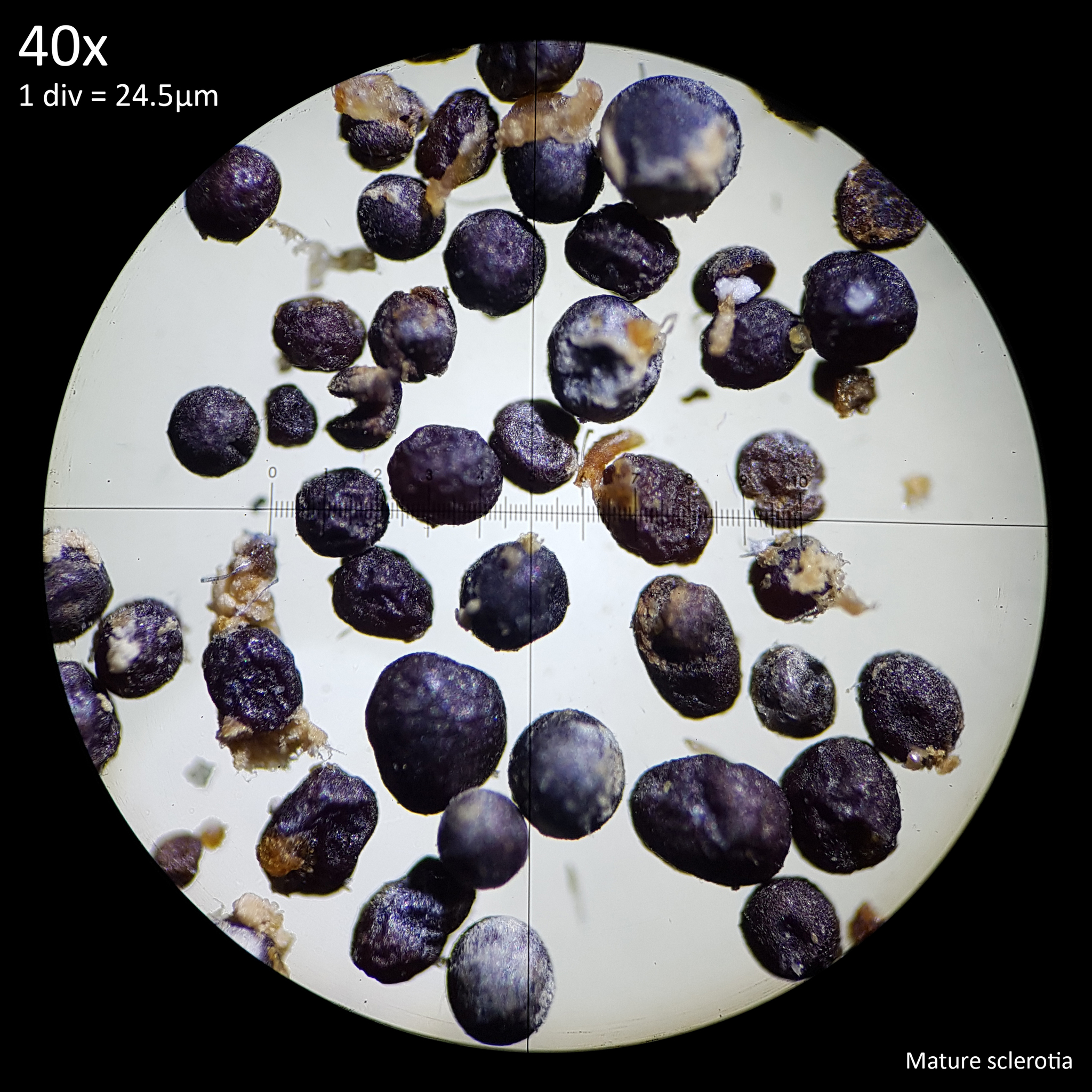
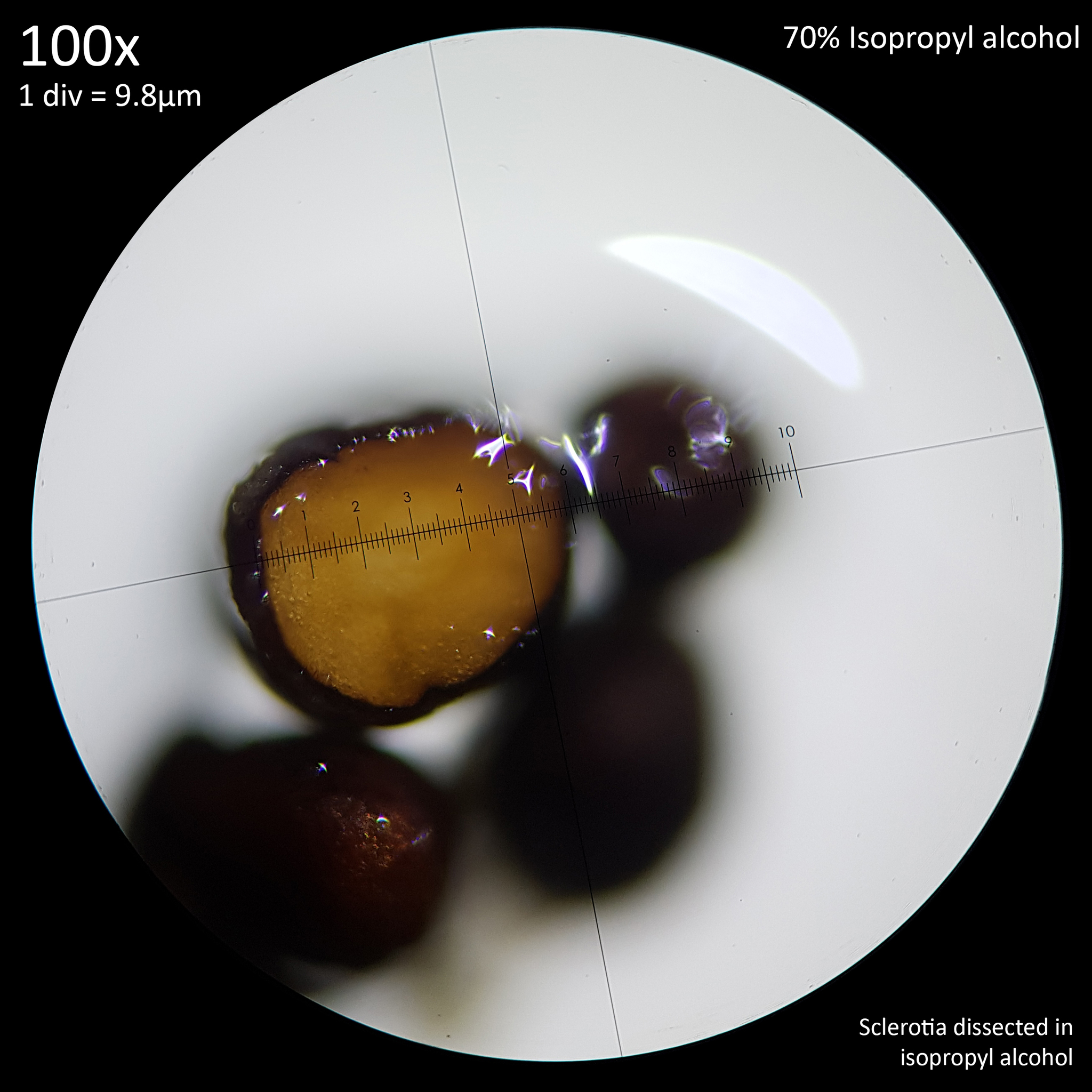
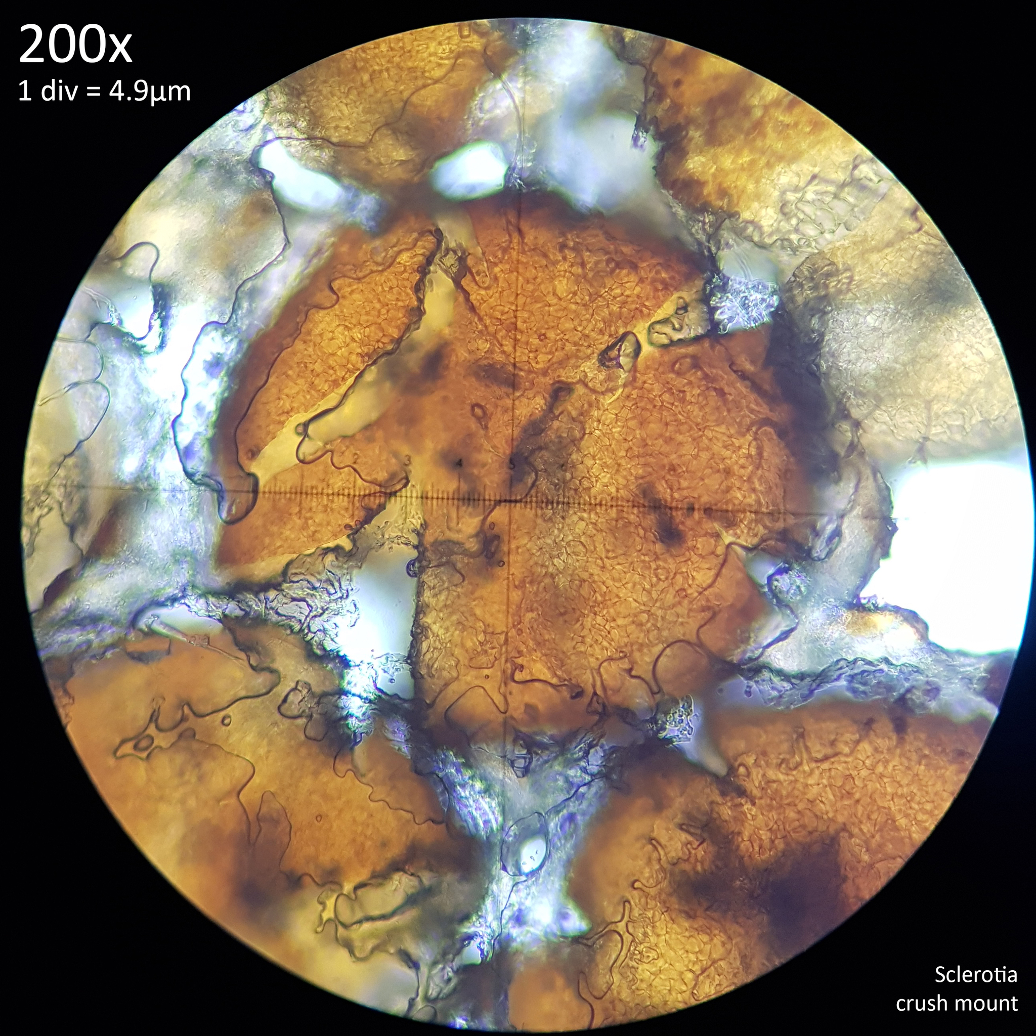
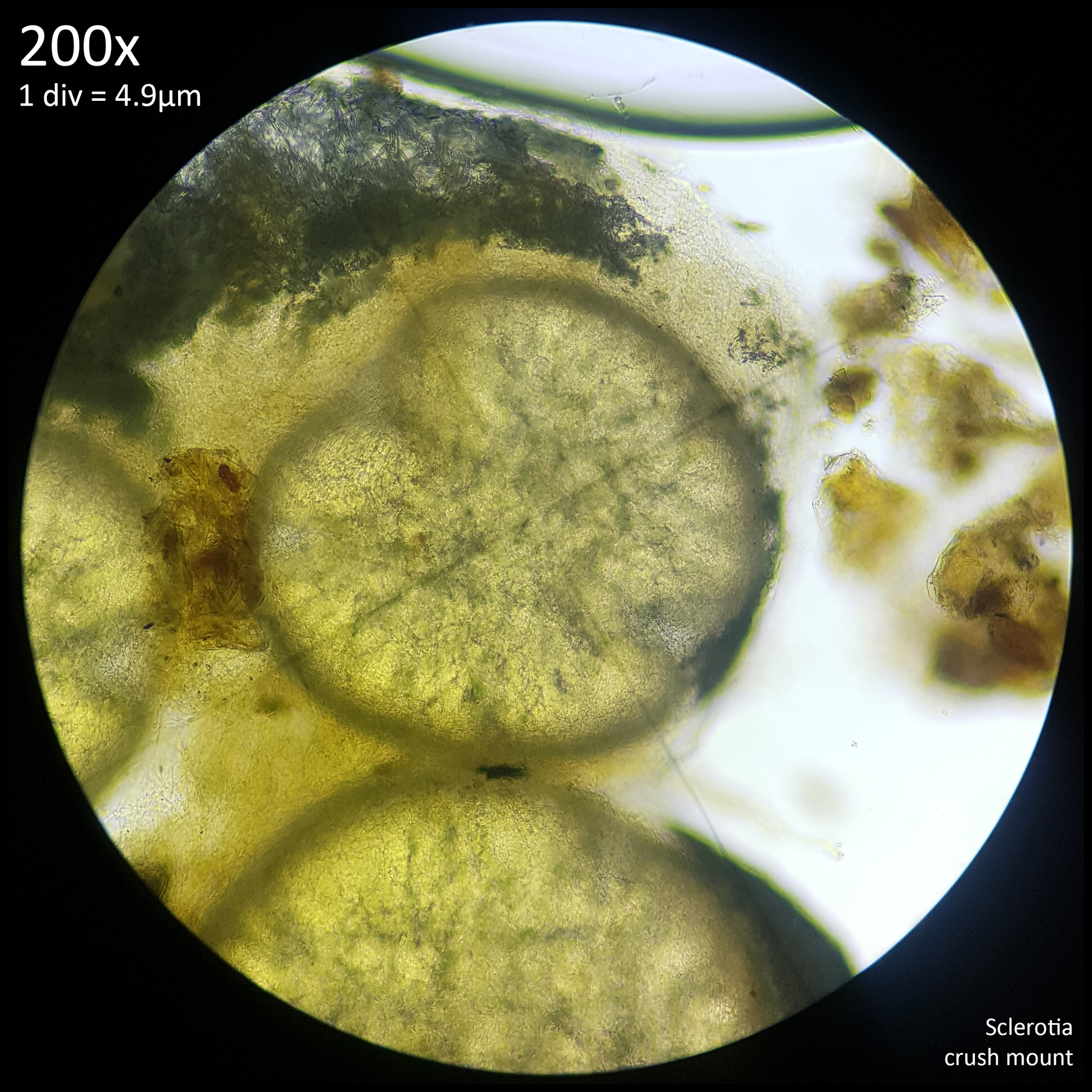
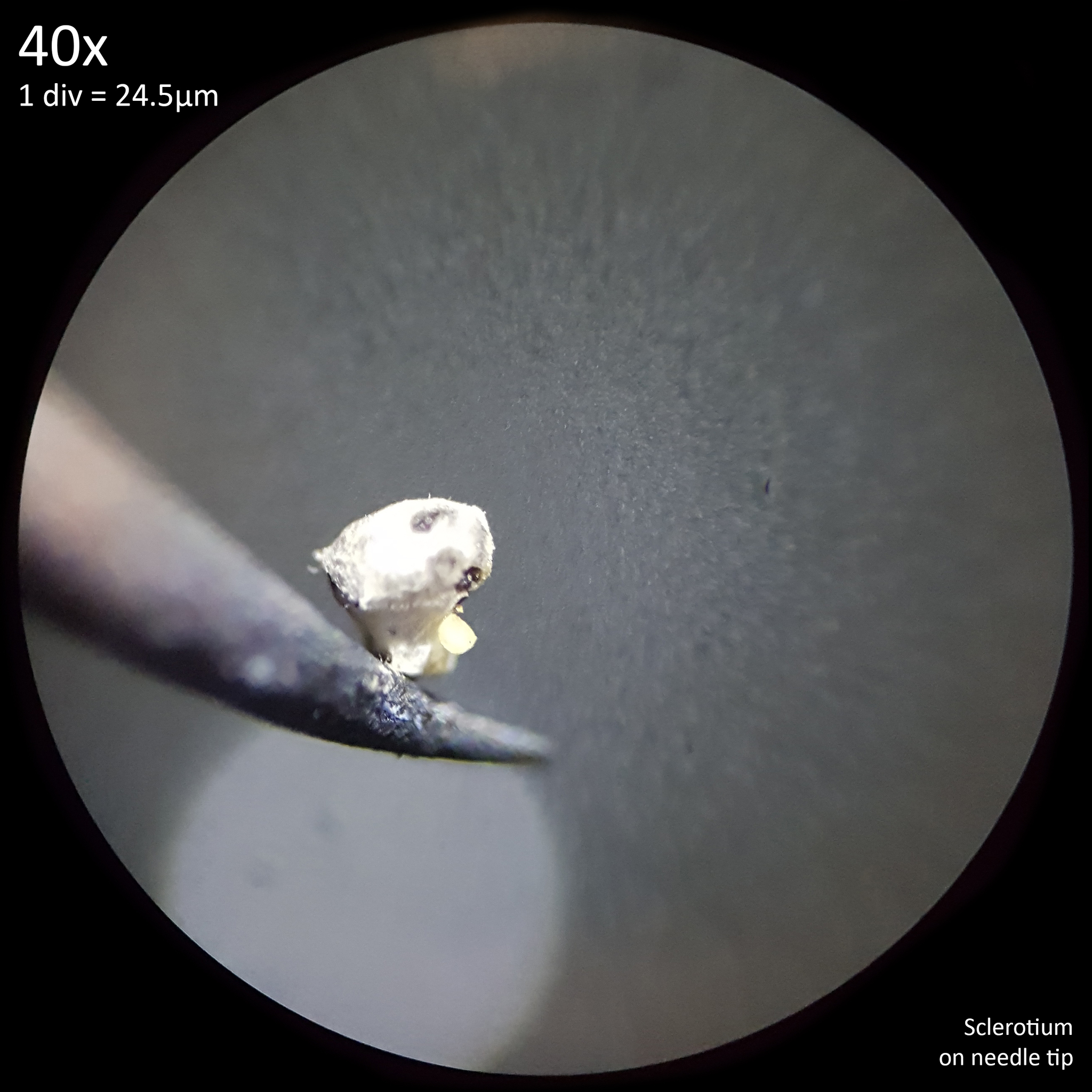
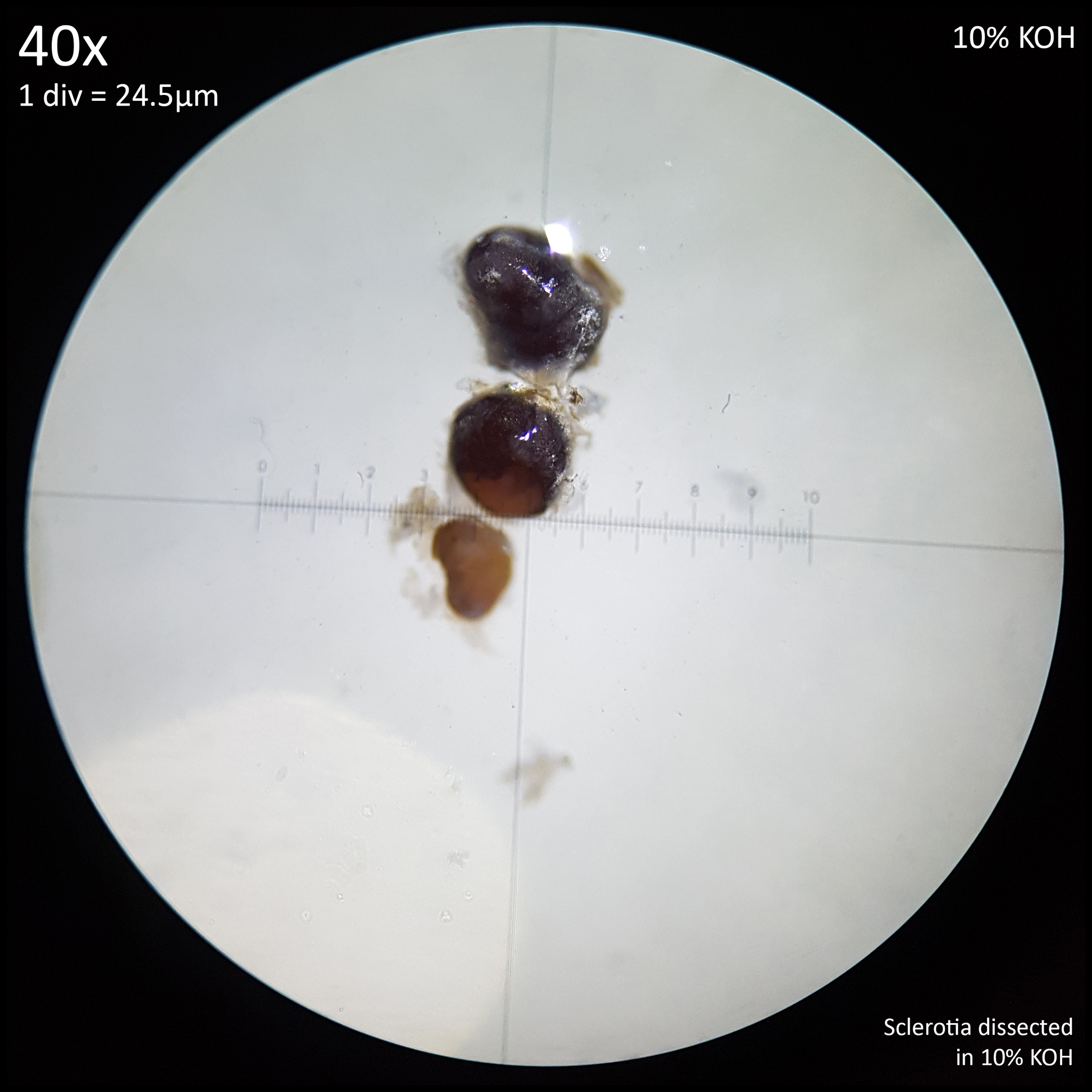
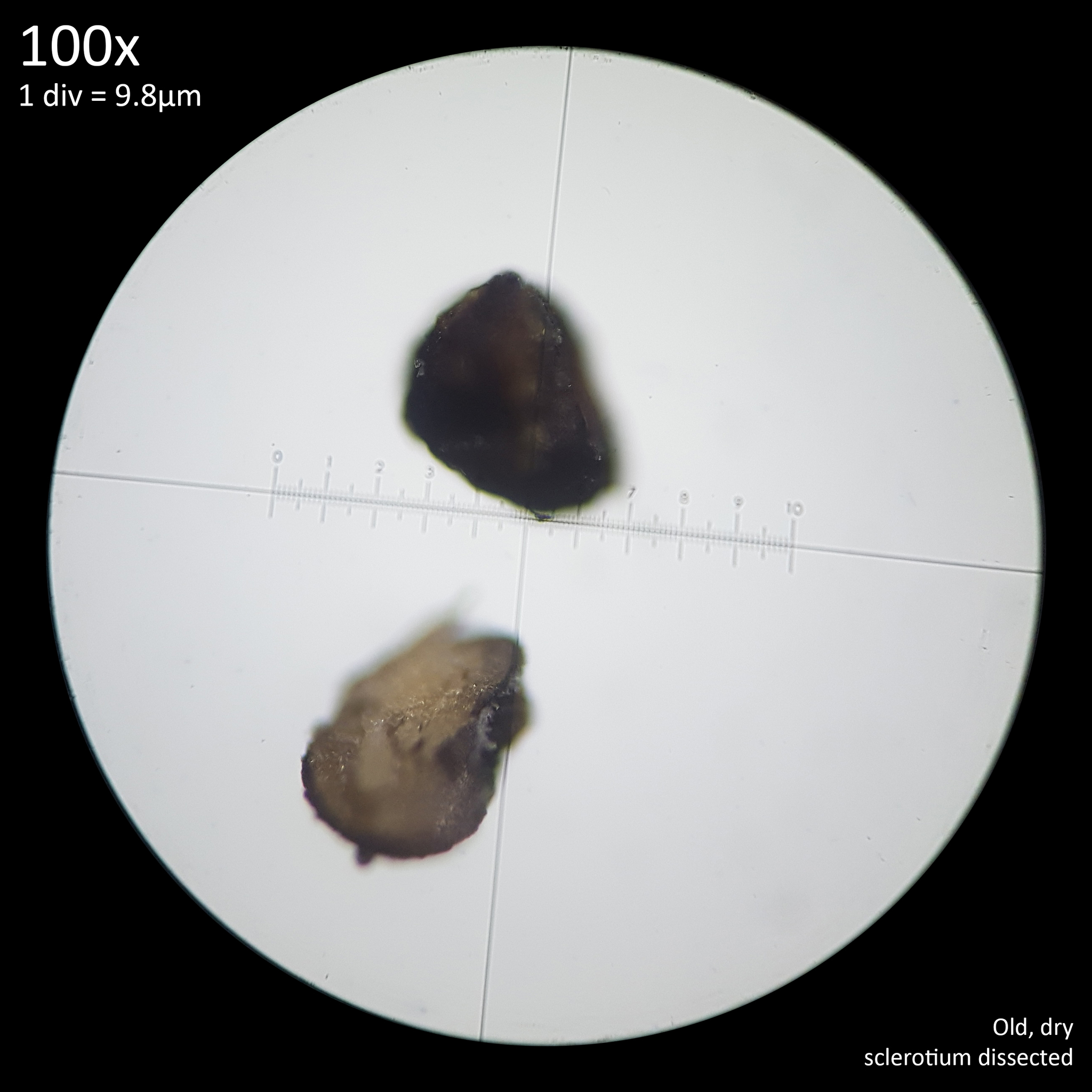
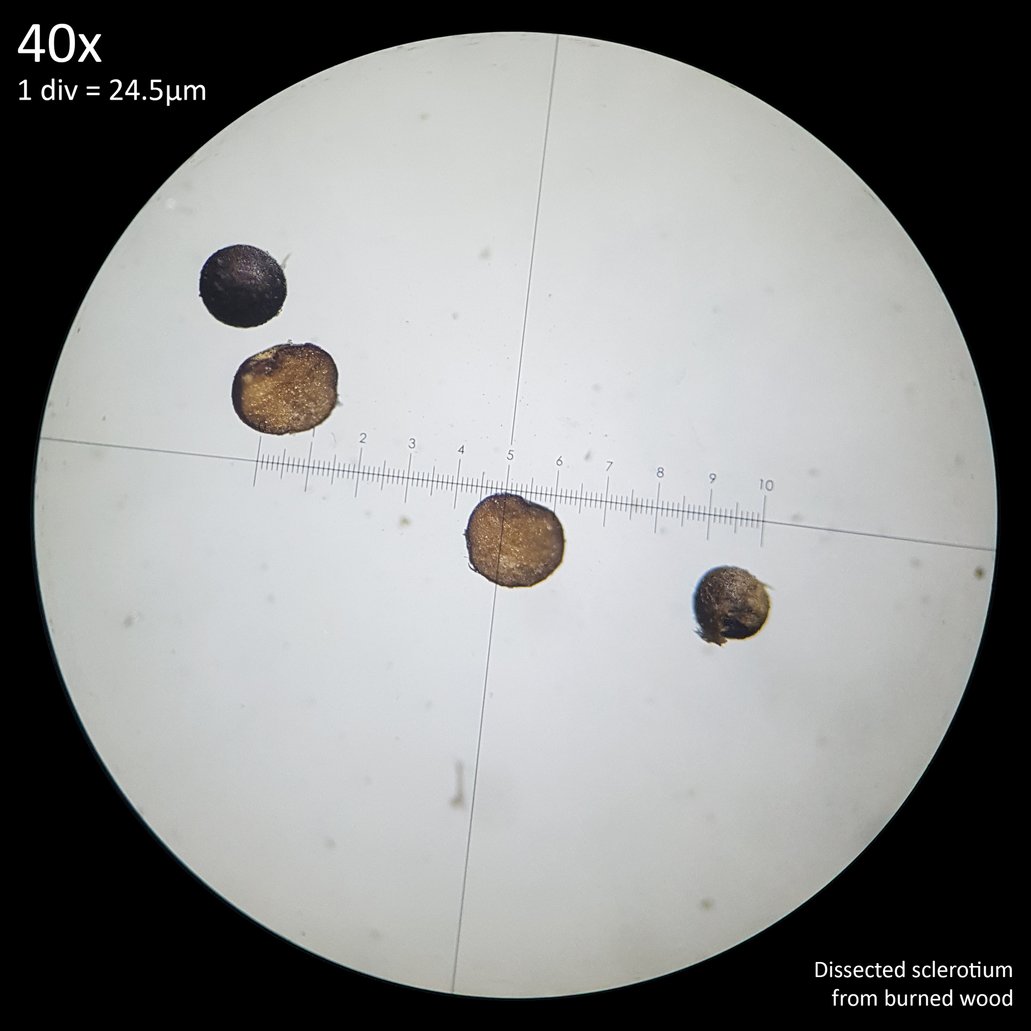
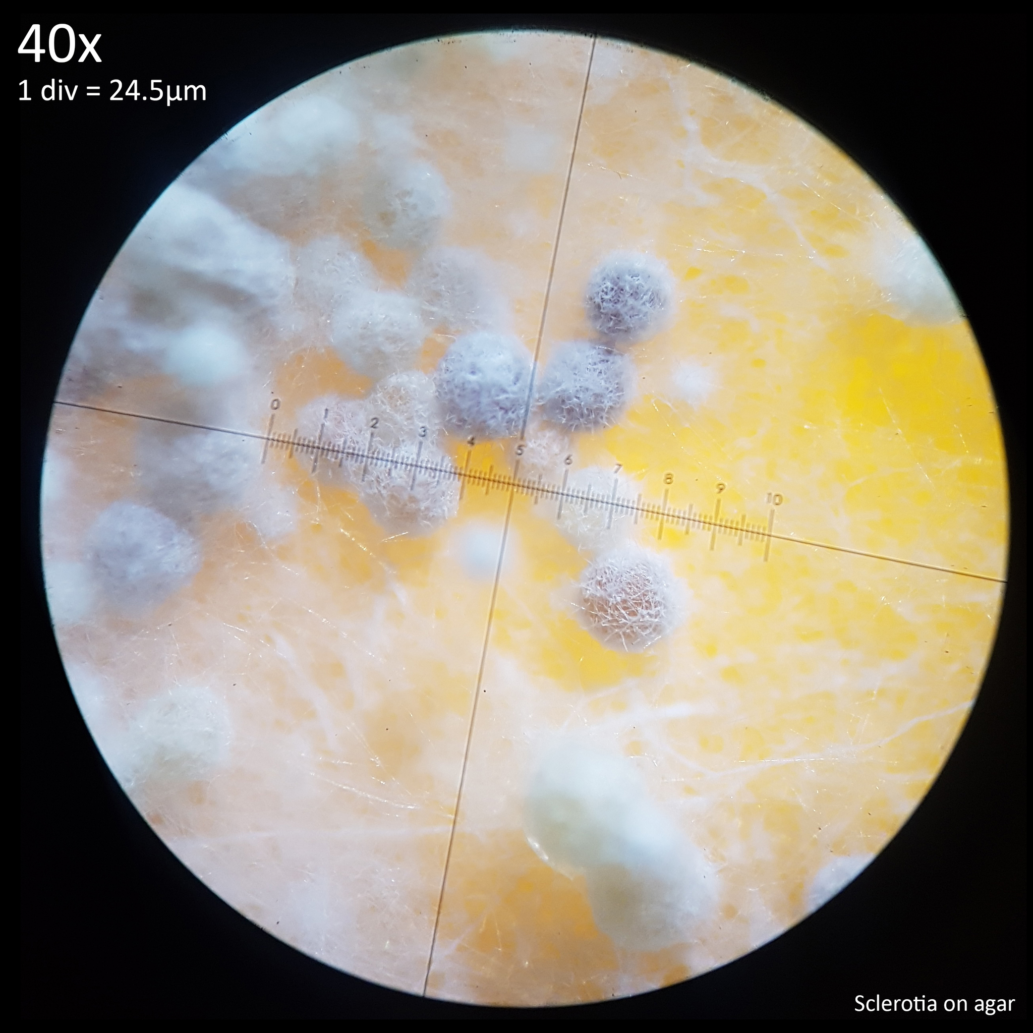
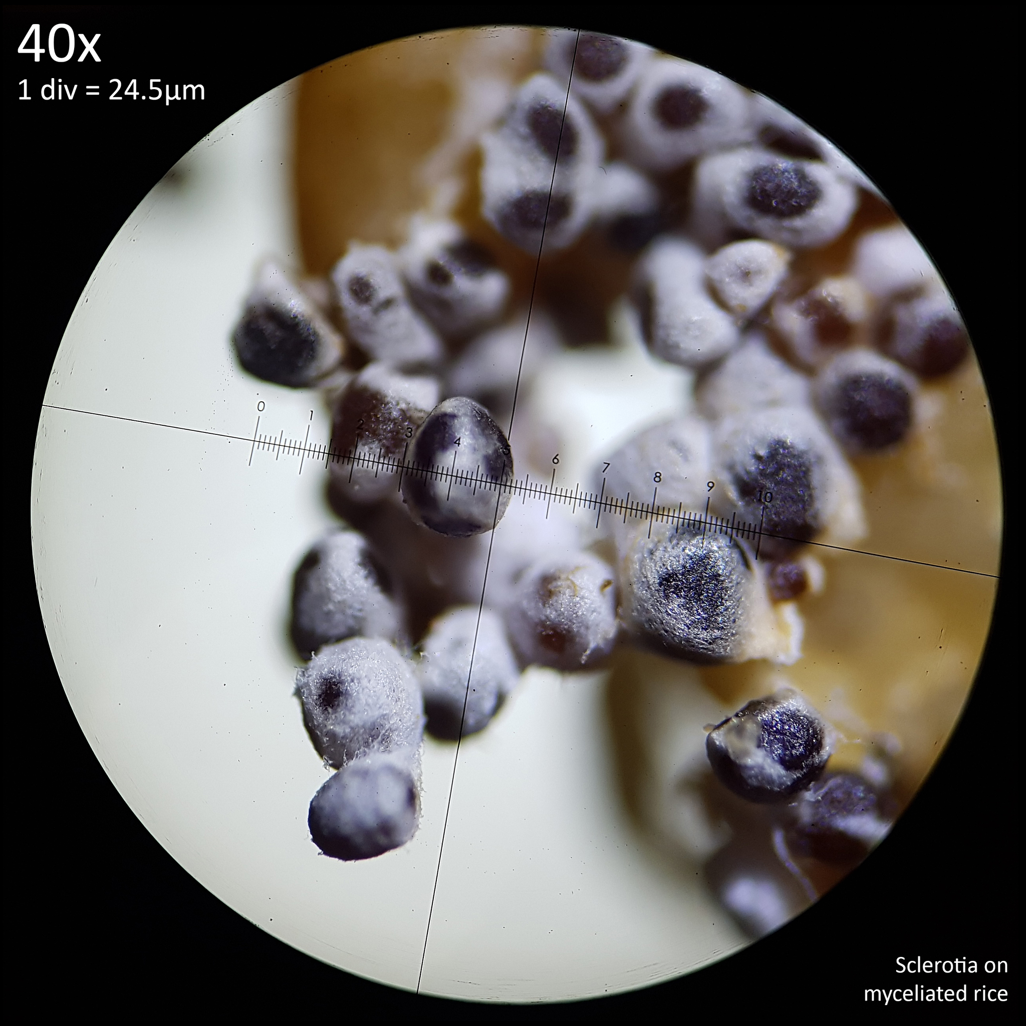
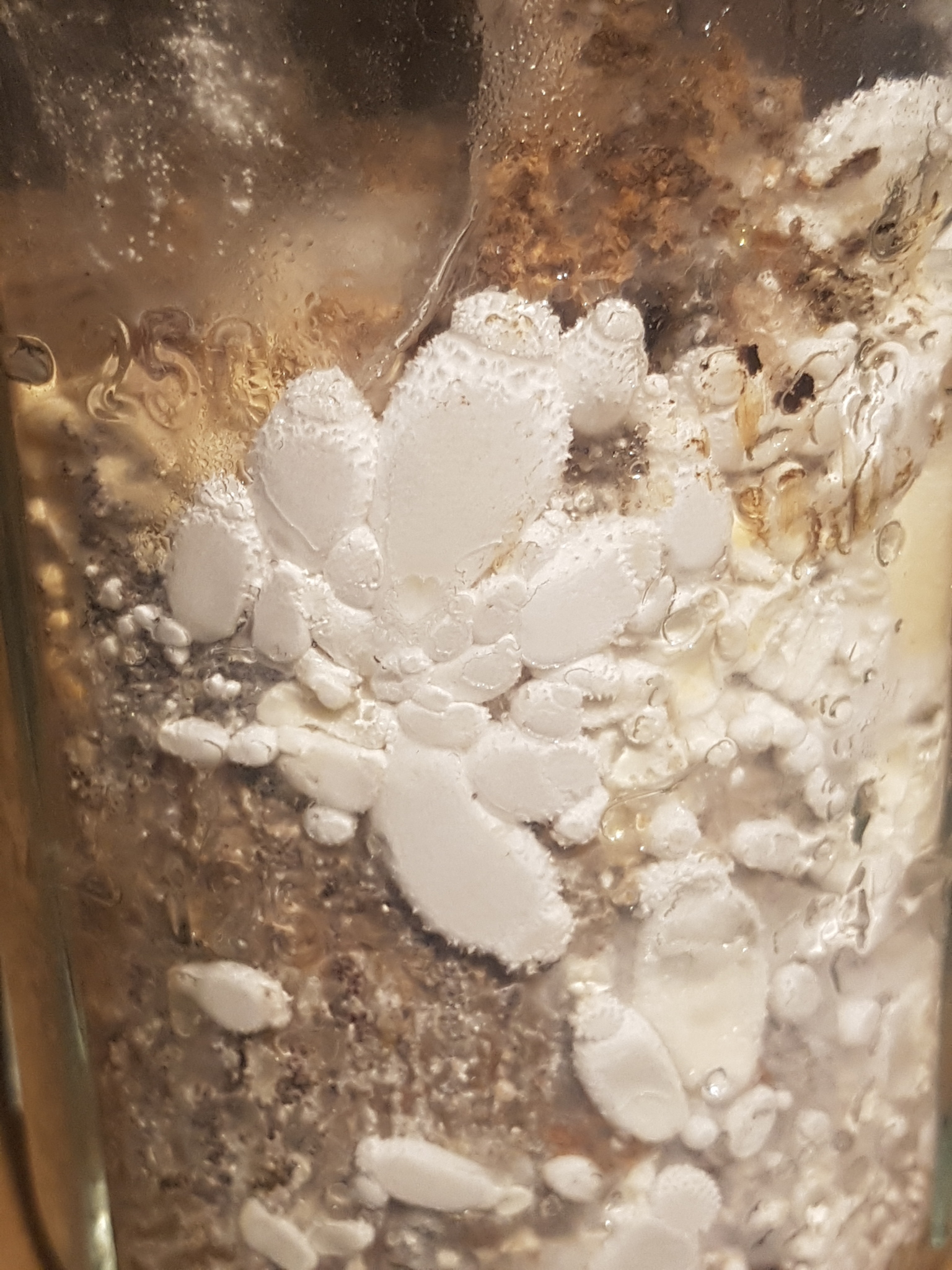
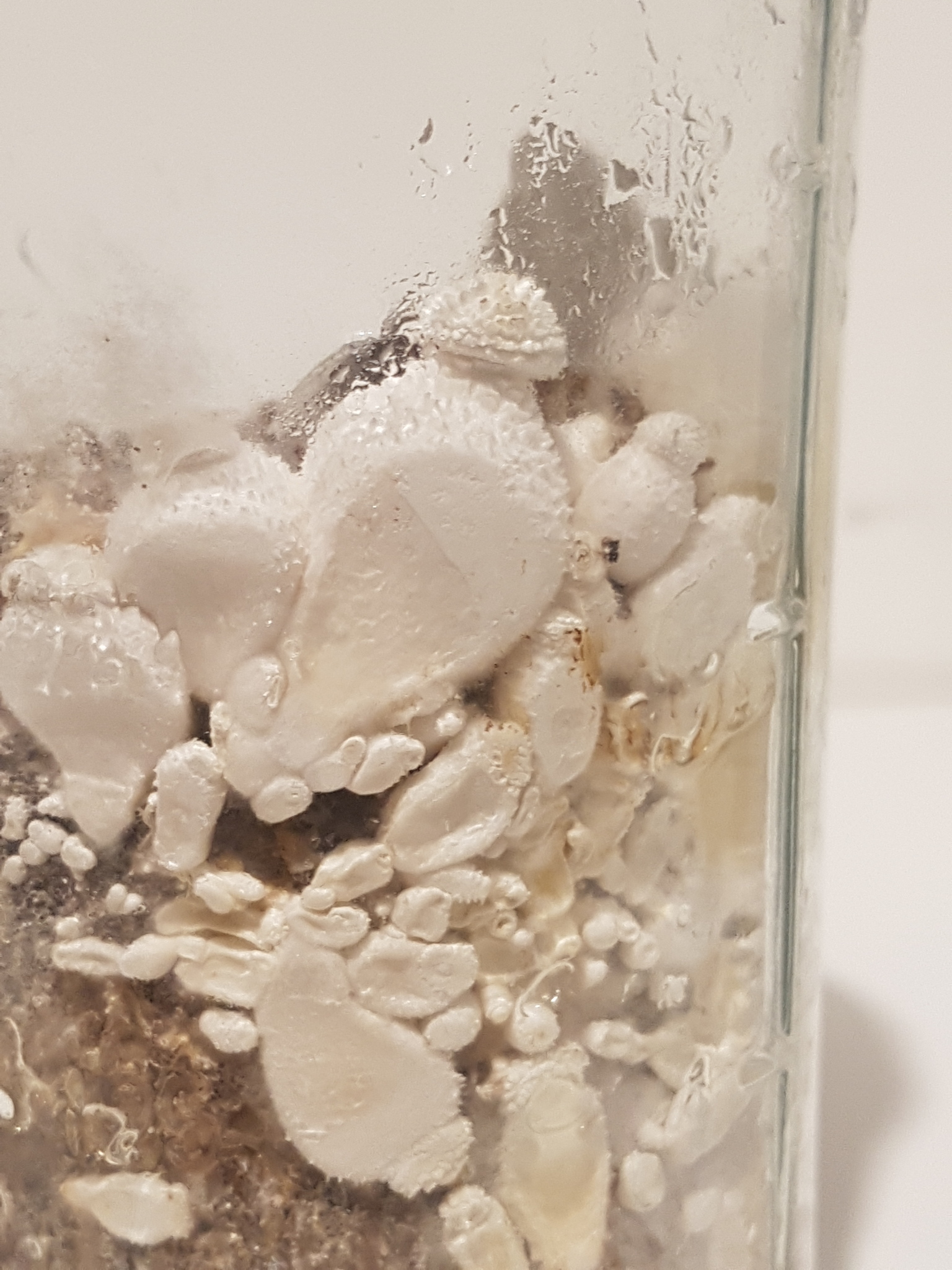
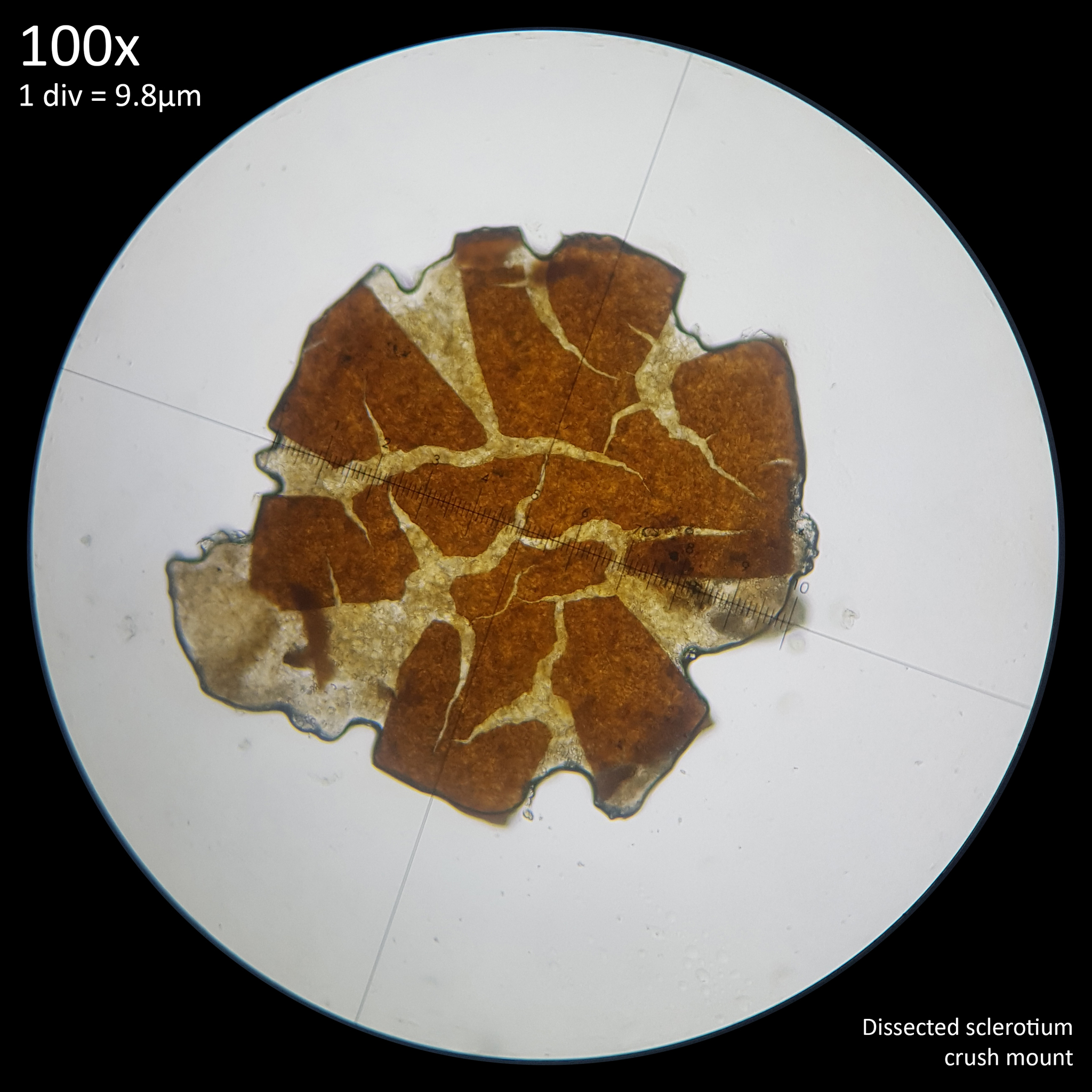
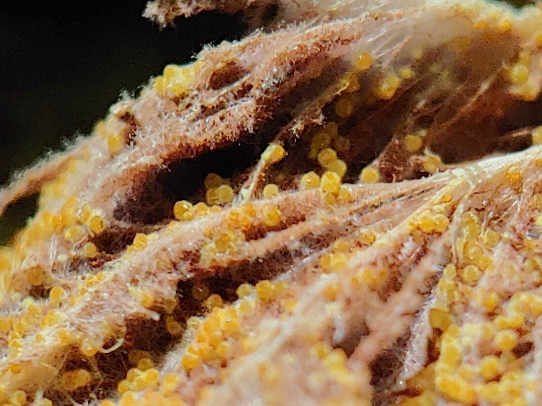
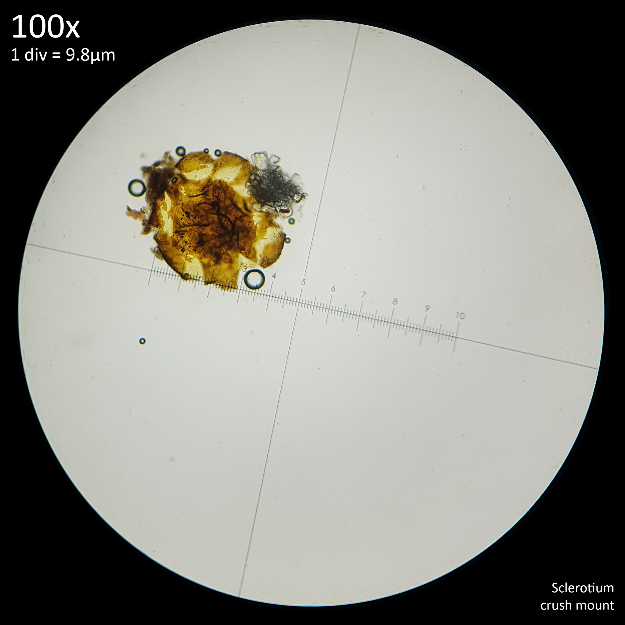
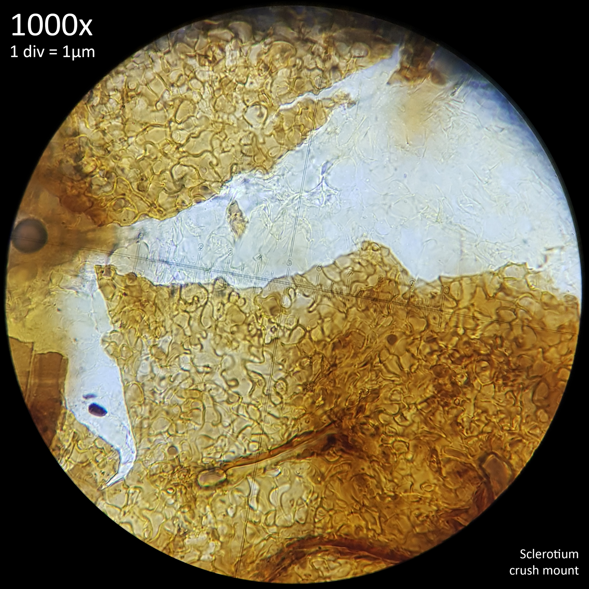
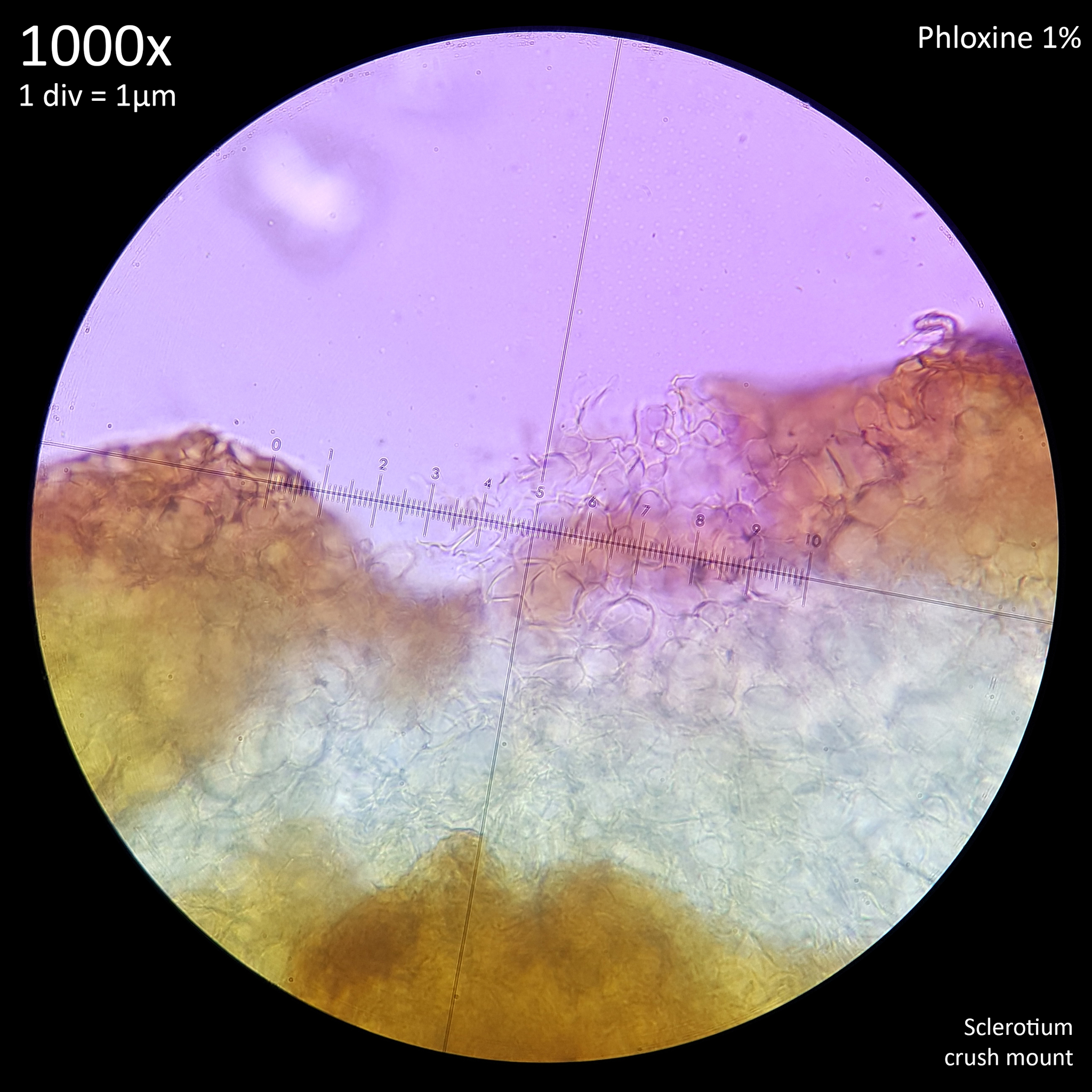
Note that is it important to recognise the role sclerotia play in the growth cycle and the edibility of these hard, sand grain like structures has not been assessed with only the mushrooms consumed. In each case detailed below in which mushrooms were consumed time was first taken to remove the substrate and sclerotia from the base. Sclerotia can stick to the exterior of the mushrooms and in some cases are embedded in the surface of the stem base although dissection has so far not revealed any deeper inside the mushrooms. As the sclerotia are very hard and have shown some resistance to both heat and chemicals it is not yet clear how digestible they would be or if cooking would be reliable at rendering them inert. An additional concern is that the sclerotia appear to serve as a distribution method with new mycelium being able to grow very easily and rapidly from the displaced sclerotia when placed in a humid environment. Even if they are stuck to the inside of a glass jar and not in contact with the substrate mycelium will grow out from them. The sclerotia are the perfect size to get stuck in the gum line or in a gap between teeth and whilst it seems unlikely that they would manage to grow in this environment without being suppressed by the immune system it is probably best to avoid putting this to the test until more is known. Unlike plant seeds which would absorb water and soften over time if lodged between teeth the sclerotia appear to remain hard and unchanged even if soaked in water for a prolonged duration.


5.1 Observations

Fig. 12 - First mushroom harvested

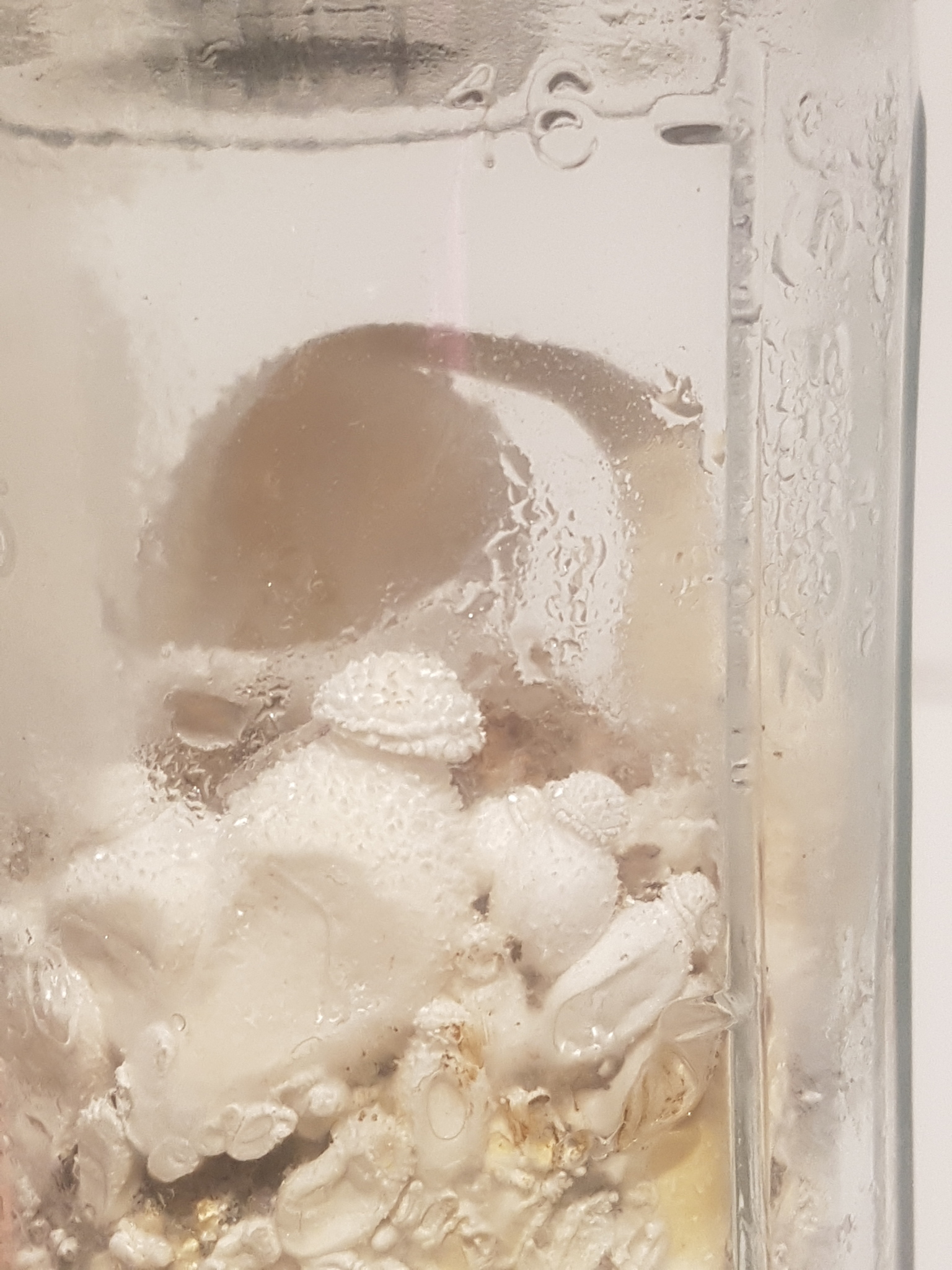
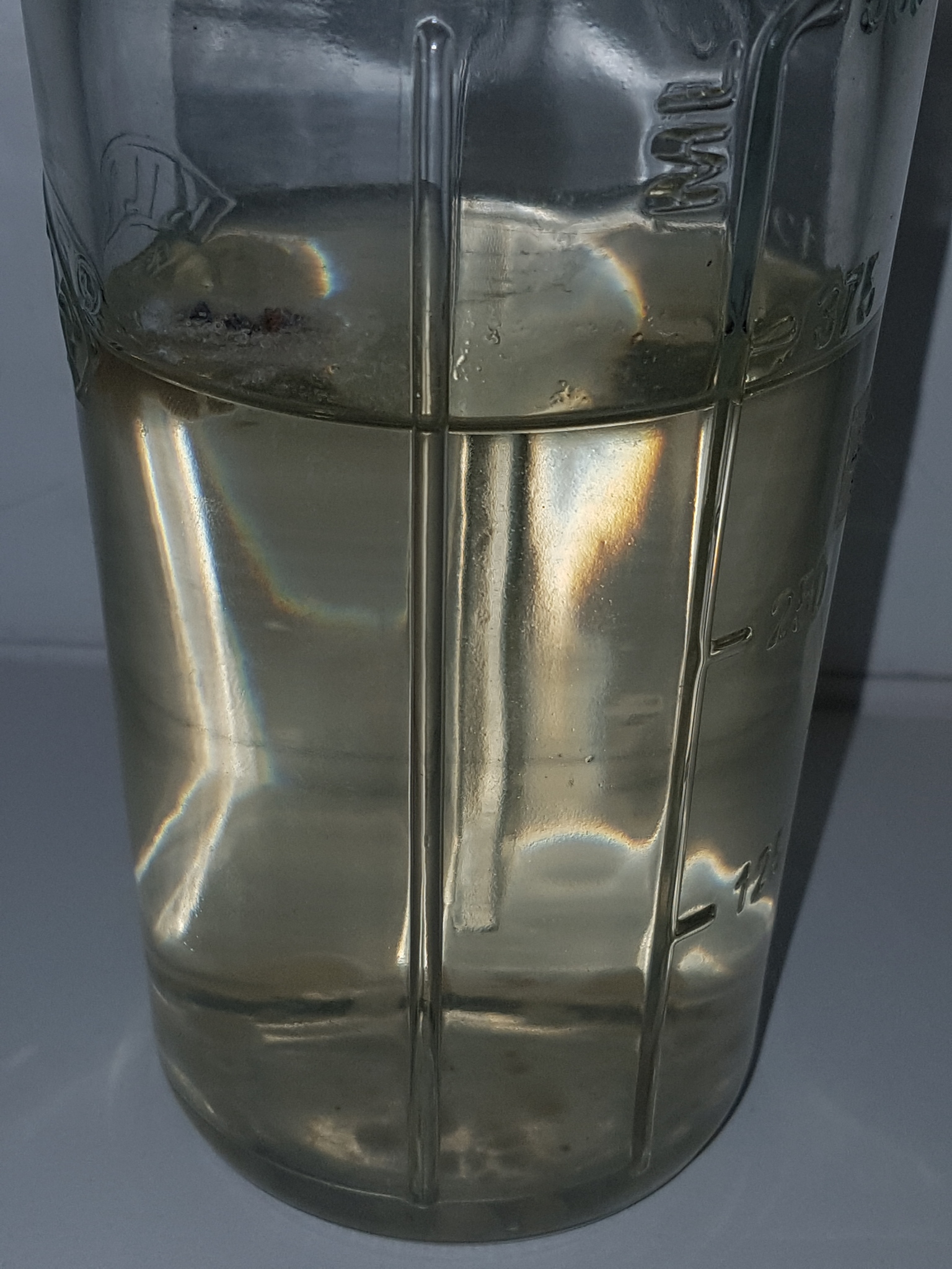
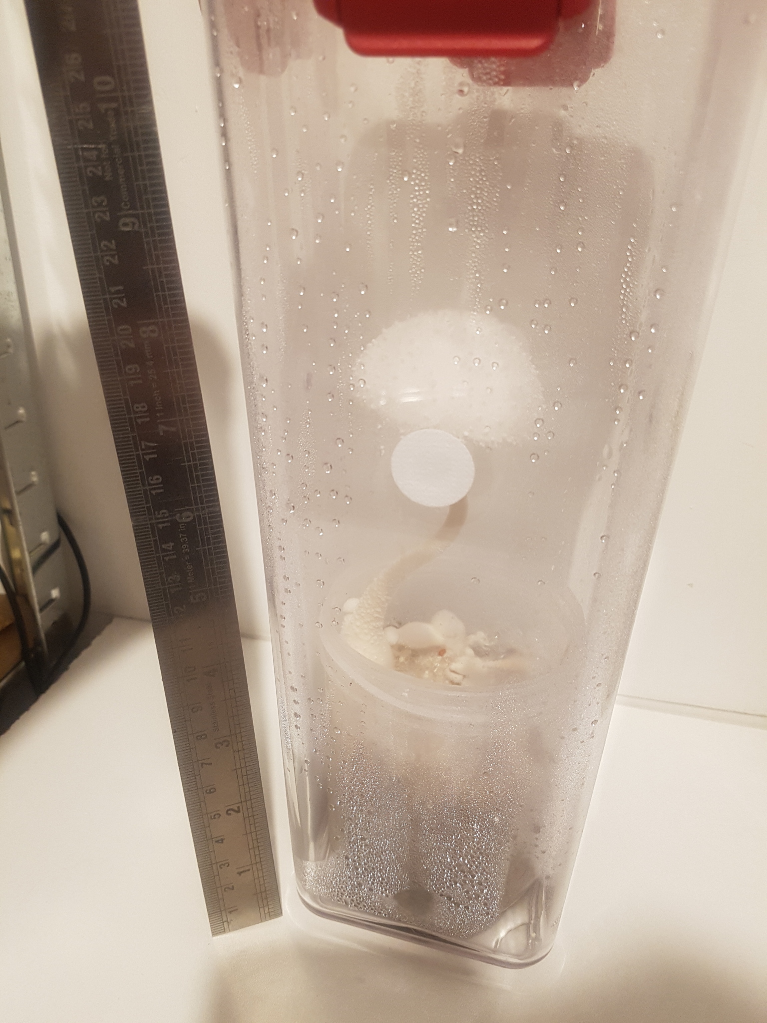
On 11/08/23 one mushroom weighing ~5.4g was harvested from a colonised jar of enriched rice used for the sclerotia experiment. The substrate was comprised of 75g brown rice, 0.75g yeast extract, 0.75g soluble starch (potato), 0.75g gypsum, 20g hardwood fuel pellets (oak), 105ml water in a tall, 500ml mason jar, filled about half full with a 0.3µm PTFE filter disc on the lid for airflow. The jar was inoculated from colonised agar 45 days prior to harvesting the mushroom but was only initially intended to test for contamination since this substrate had been prepared and sterilised for a previous experiment some months prior but went unused and subsequently appeared dry. The excellent growth therefore came as a surprise. The day by day growth documenting the development of the first primordia after harvesting sclerotia is displayed in figures 19a-19h in 'A preliminary investigation of sclerotia in Leucocoprinus cretaceus'.

The cap had opened overnight and had developed a brownish yellow centre. Upon opening the jar to harvest, a strong and pungent, though not unpleasant fungal smell was immediately noticeable however the mushroom itself did not smell so distinctly. The mature mushroom has a pleasant fungal smell but it is not overly noticeable compared to the smell of the mycelium and the younger pins which are far stronger. The immature mushrooms individually do not smell that strongly but when clustered together it is quite noticeable. When the stem of the mature mushroom was cut in half the smell was slightly stronger and more pleasant. The base of the stem was removed and kept for study as it came away with some substrate and many sclerotia clustered around it. After leaving it in the open air for a couple hours the smell seemed faintly similar to that of cooked Grifola frondosa but still with a slight chemical undertone. This however was only noticed after cooking the mushroom and so may have been biased by the whole house smelling like this. When left longer to dry in the open air there was no smell to the stem or base with the mycelium and sclerotia on the substrate. A section of the cap amounting to approximately a quarter of the total cap was left to spore print in a sealed tub and remained left sealed for some days after. Upon opening this no significant smell was noted.

The taste of the mushroom raw was likewise not unpleasant though not overly distinctive. After spitting it out the aftertaste was more noticeable and had a slightly chemical or floral taste which was hard to place. The taste of the cap was perhaps slightly less pleasant than that of the stem with a more noticeable chemical taste however it is the texture of the cap that makes it less desirable to taste as it feels like rolling a piece of cotton wool around in your mouth. It is unsubstantial and cottony in texture much like the description found in many Leucocoprinus species.

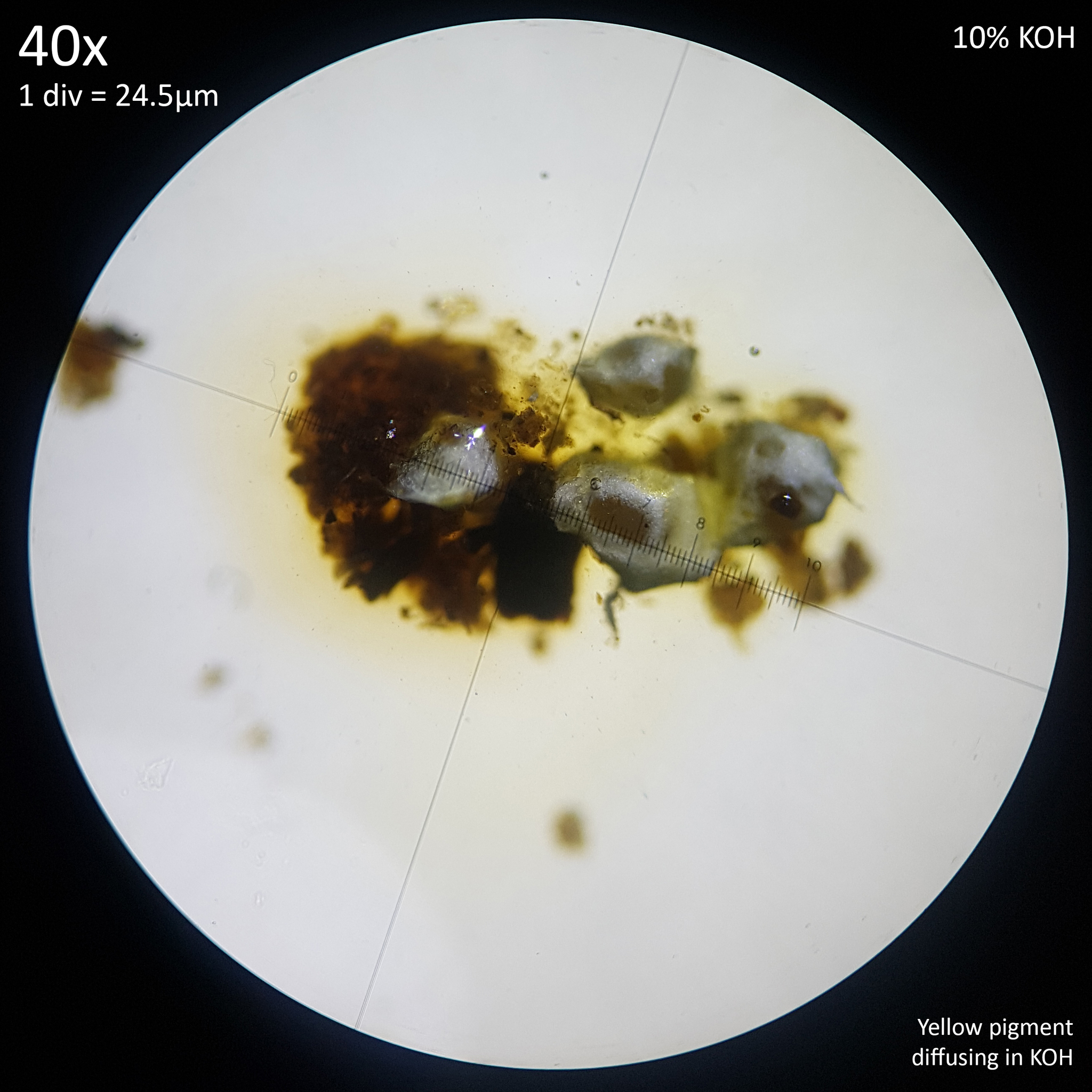
The cap falls apart relatively easily when handled and is fairly fragile with thin flesh whereas the stem is firm and rigid. The base of the stem is solid and quite firm, not compressing much at all when squeezed however when a piece was left for some hours it became soft and easily compressed. Higher up the stem is pithy inside, though still quite firm and felt as though it could be too tough to eat. On squeezing the stem slightly it did not yield greatly but a yellowish or brownish colour similar to the centre of the cap is produced, however the reaction is not instant and not very distinctive. This colouration was already present in some places of the stem before it was harvested but less noticeable beneath the powdery white scales, which easily displace and stain the fingers on touching. So squeezing it also removed the scales and increased the visibility of this colouration. When the base of the stem was cut no discolouration was noted but upon cutting the thinner stem higher up a yellowing occurred on the outer surface but stopped 1mm or less into the pithy inner flesh, which did not discolour. A section of this inner flesh was tested with KOH to see if this would accelerate discolouration but it did not. No reaction to KOH or FeSO4 was observed on any part of the mushroom.


5.2 Cooking

Please note that consuming this mushroom was only undertaken after extensive reading of the taxonomic history and literature on this species and others in order to determine that it would be safe. Toxic species in the Agaricaceae family are generally only gastrointestinal in nature, if Lepiota species are excluded and taken as belonging to Verrucosporaceae (as iNaturalist has them in opposition to Species Fungorum and Mycobank which still classify them as Agaricaceae). There are some case reports suggesting fatal poisoning by Chlorophyllum molybdites in small children and dogs but no such cases have been reported in adults. I have seen no fatal reports for anything else in the family and therefore it did not seem likely that there would be any risk of serious toxicity. The multiple mentions of edibility in the literature for this species also served to dispel fears but only after sufficiently interrogating each source to try and confirm which species they were actually talking about - a vital step given the numerous confusions found in the taxonomy. The suggestions of edibility would not have been enough to give me confidence to try eating it if there was anything seriously dangerous in the family.

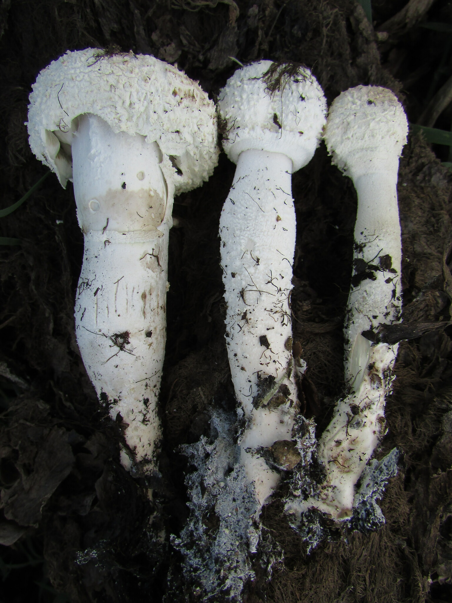
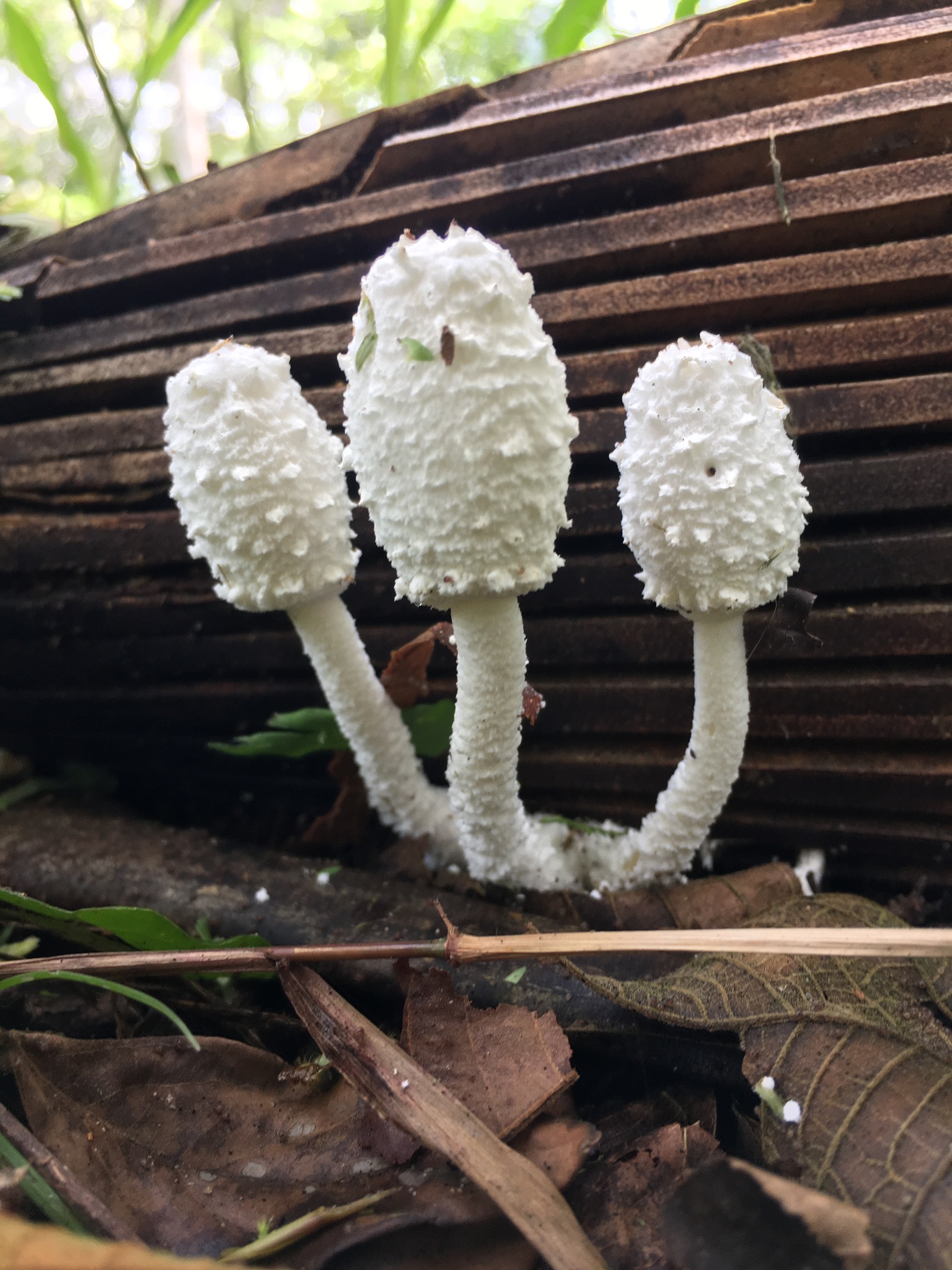
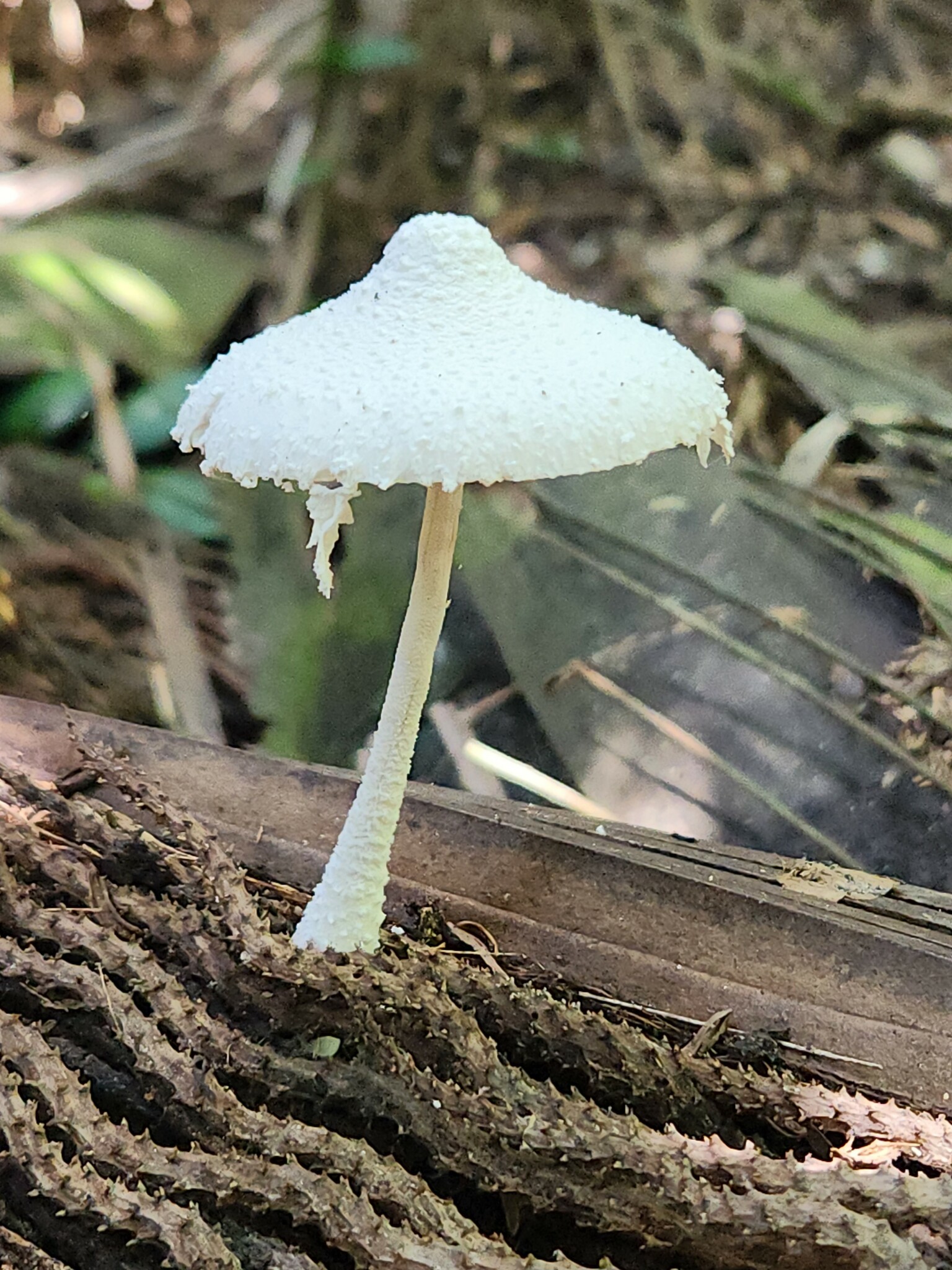
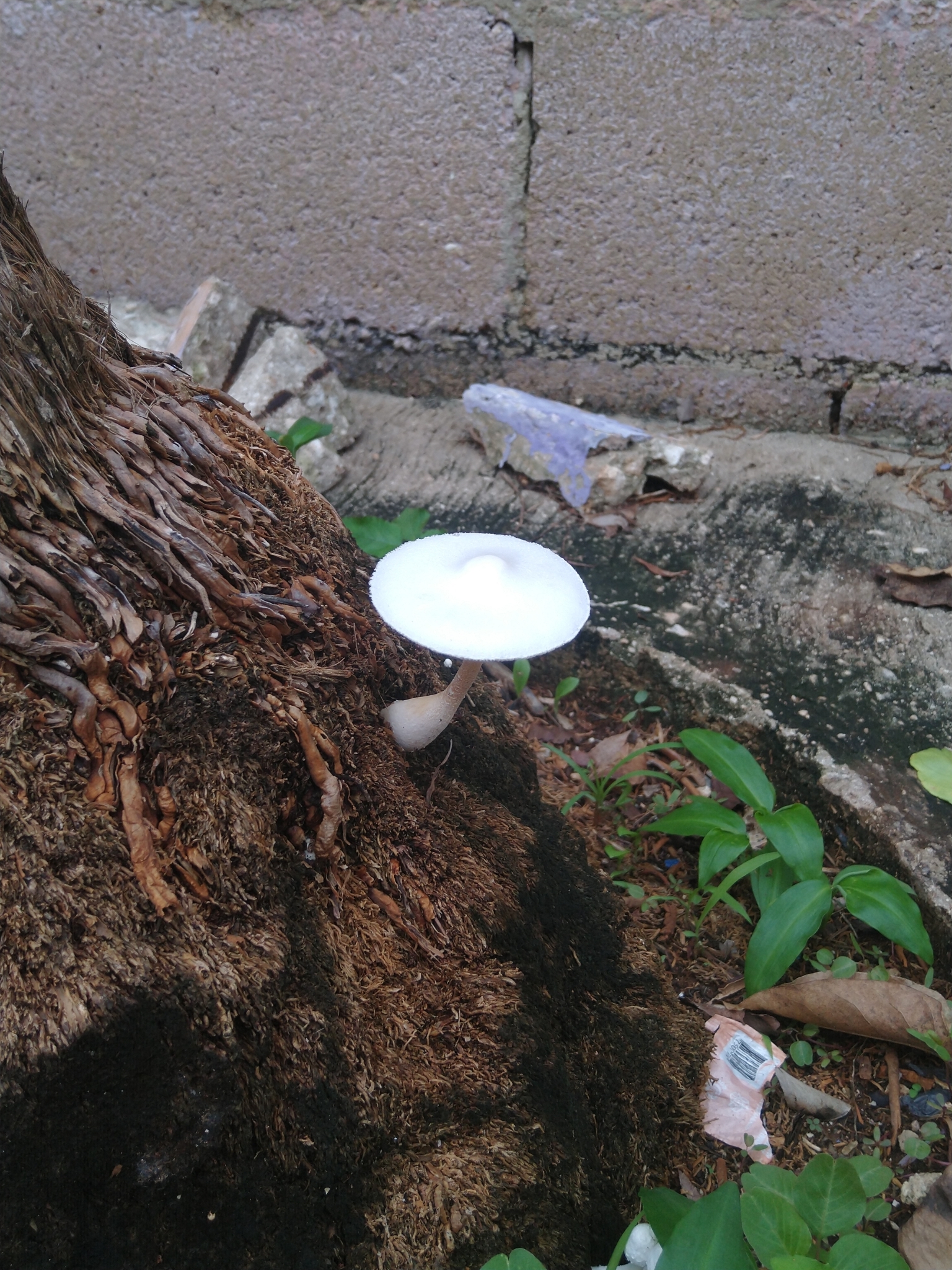
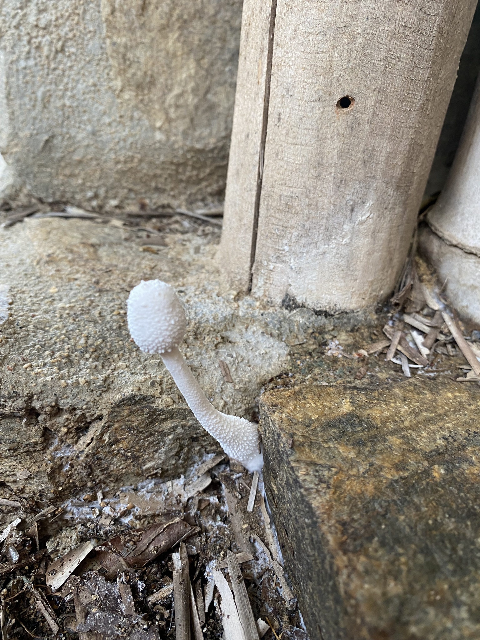
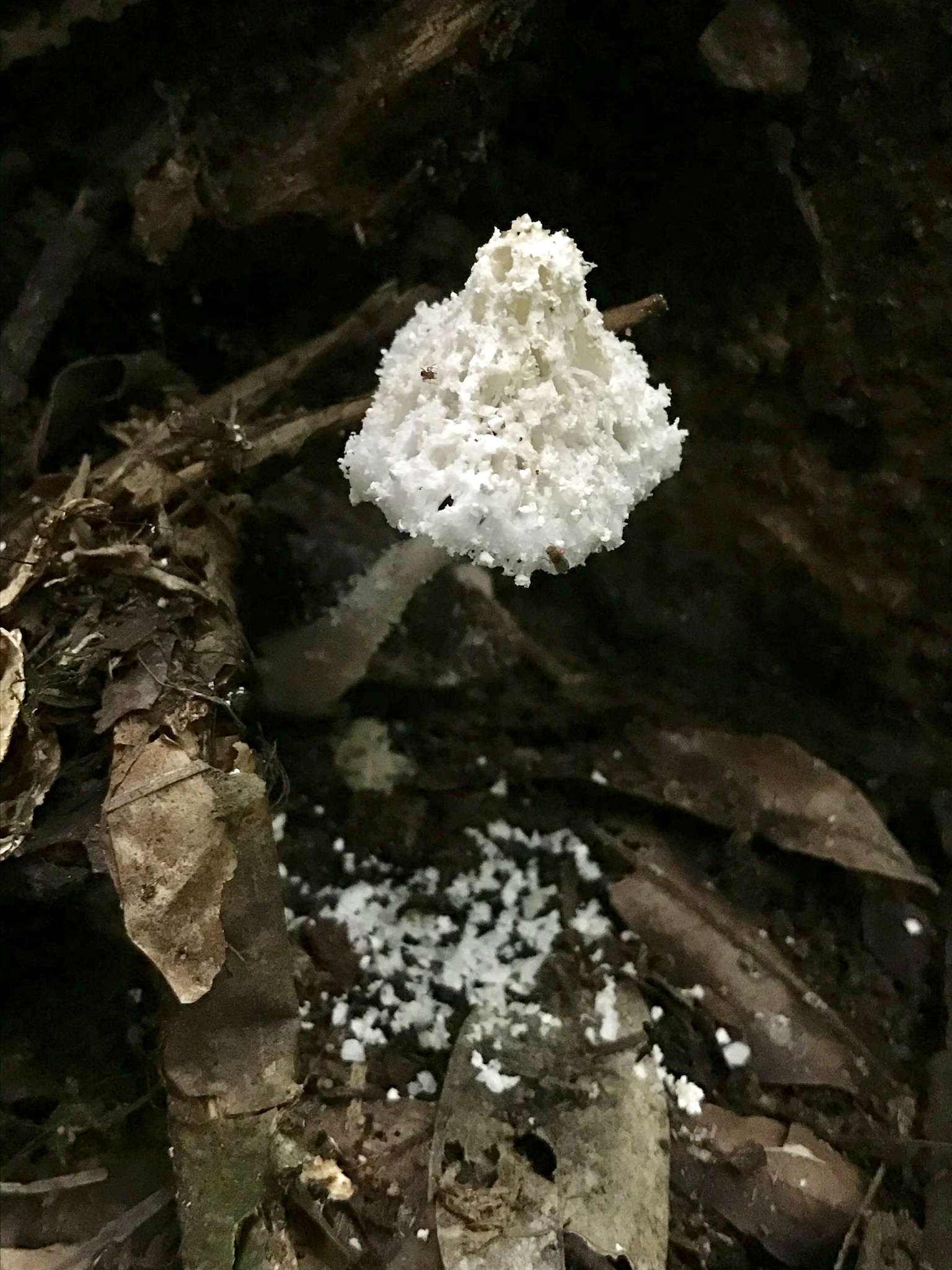
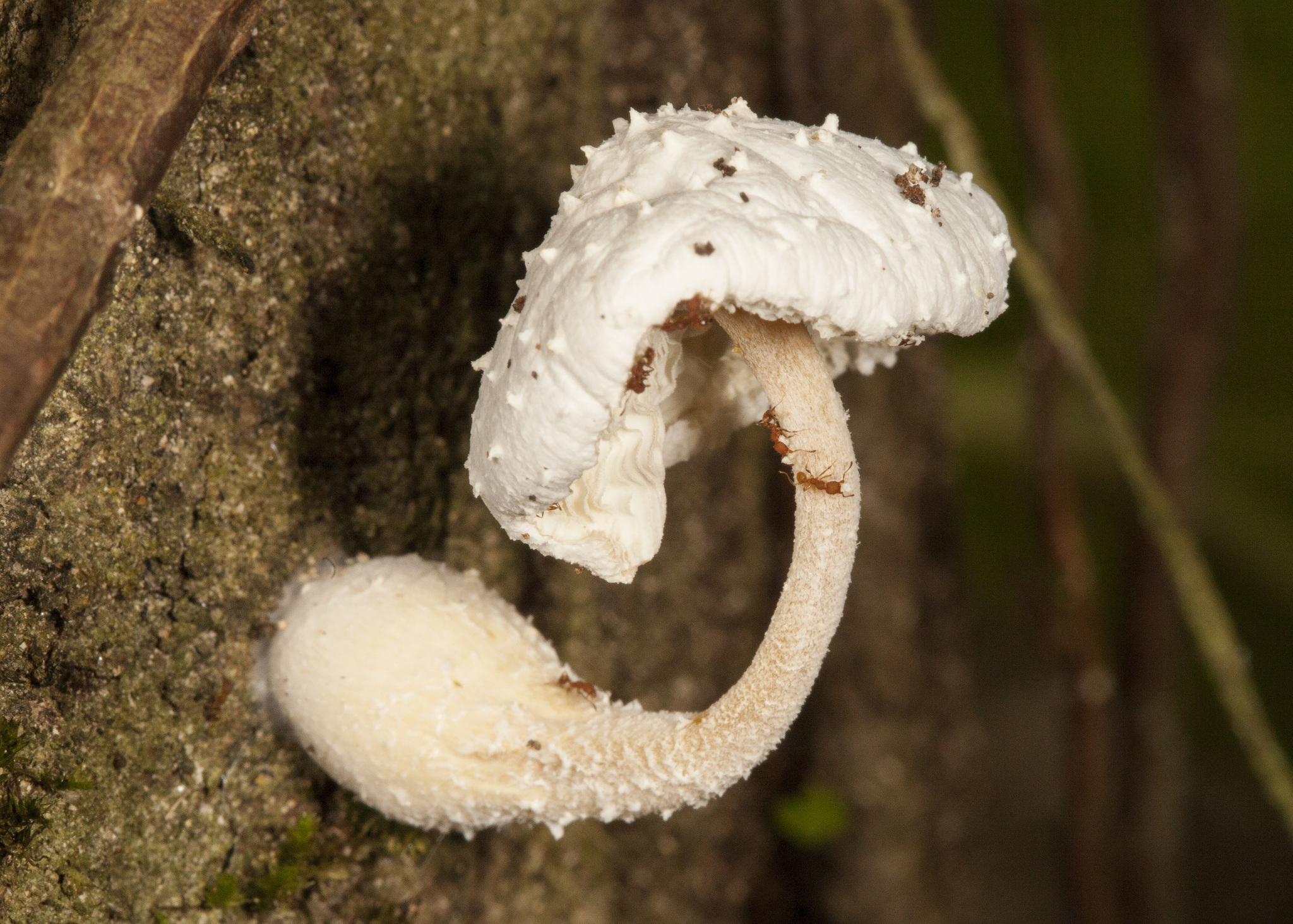
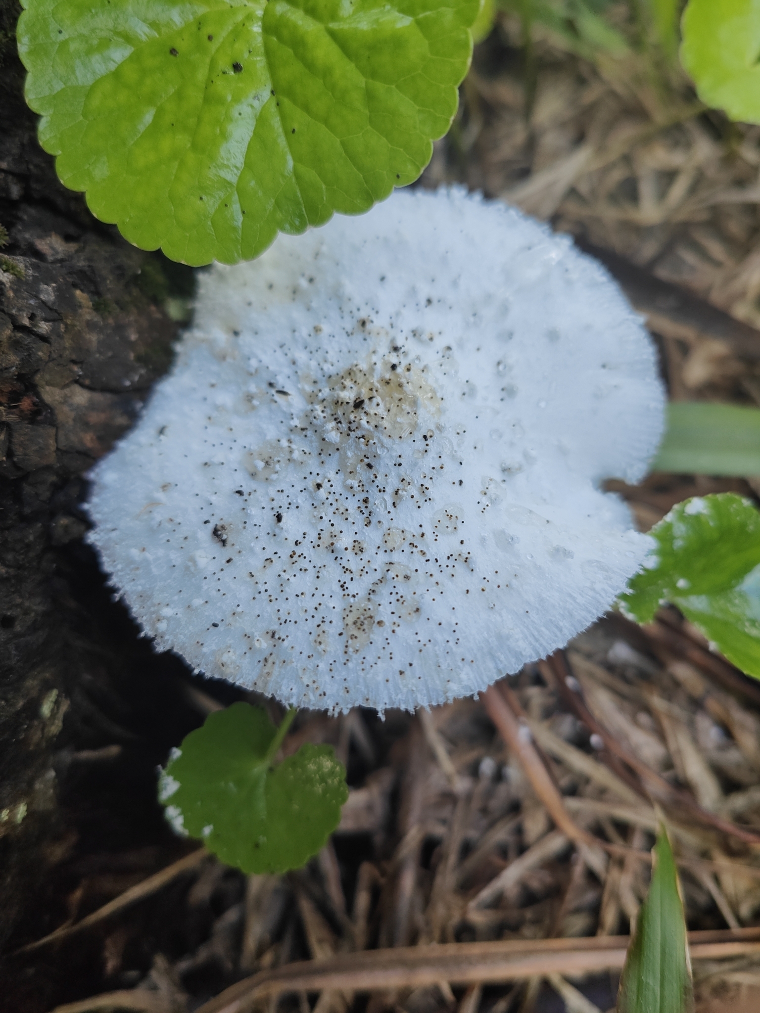
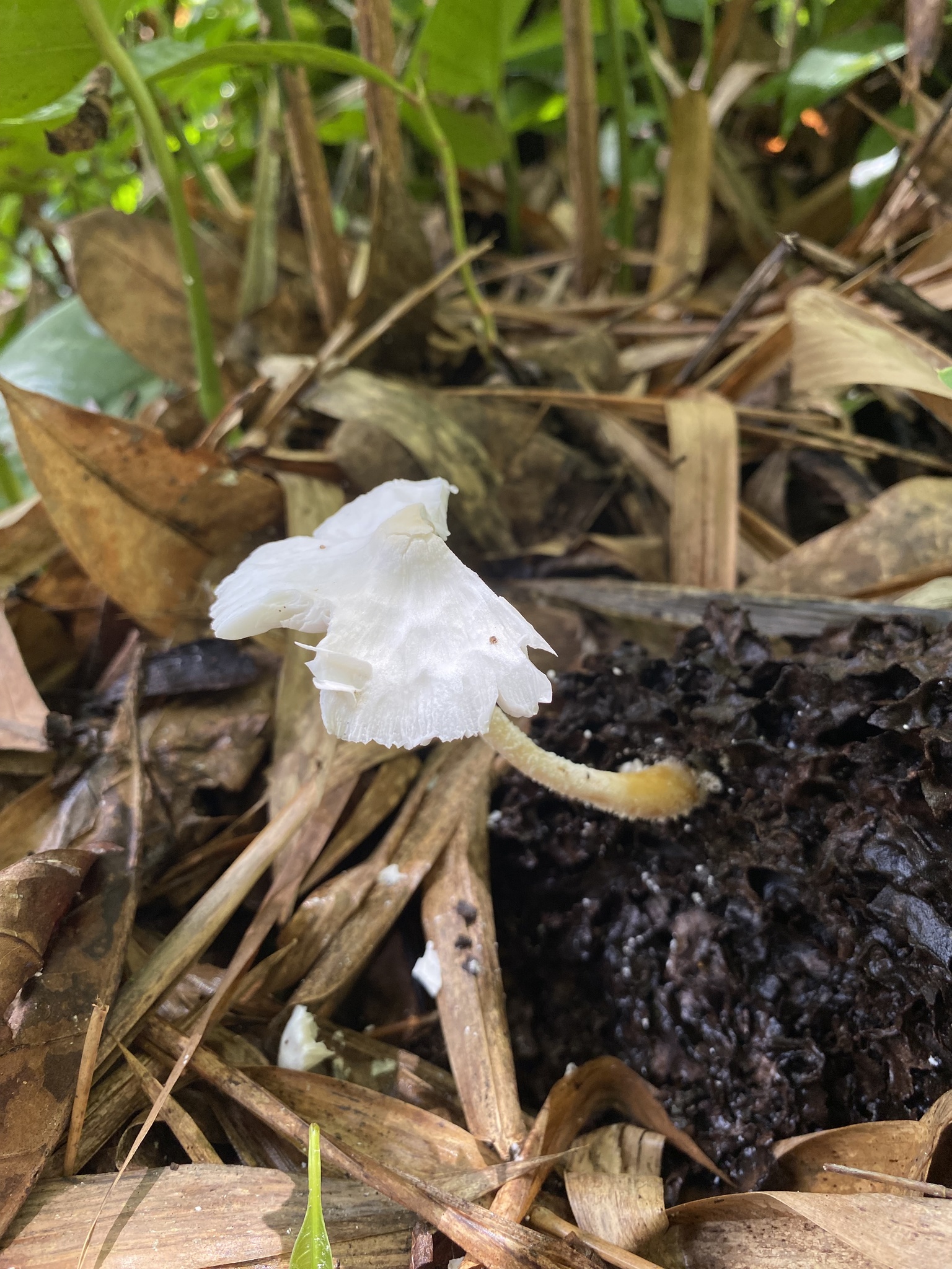
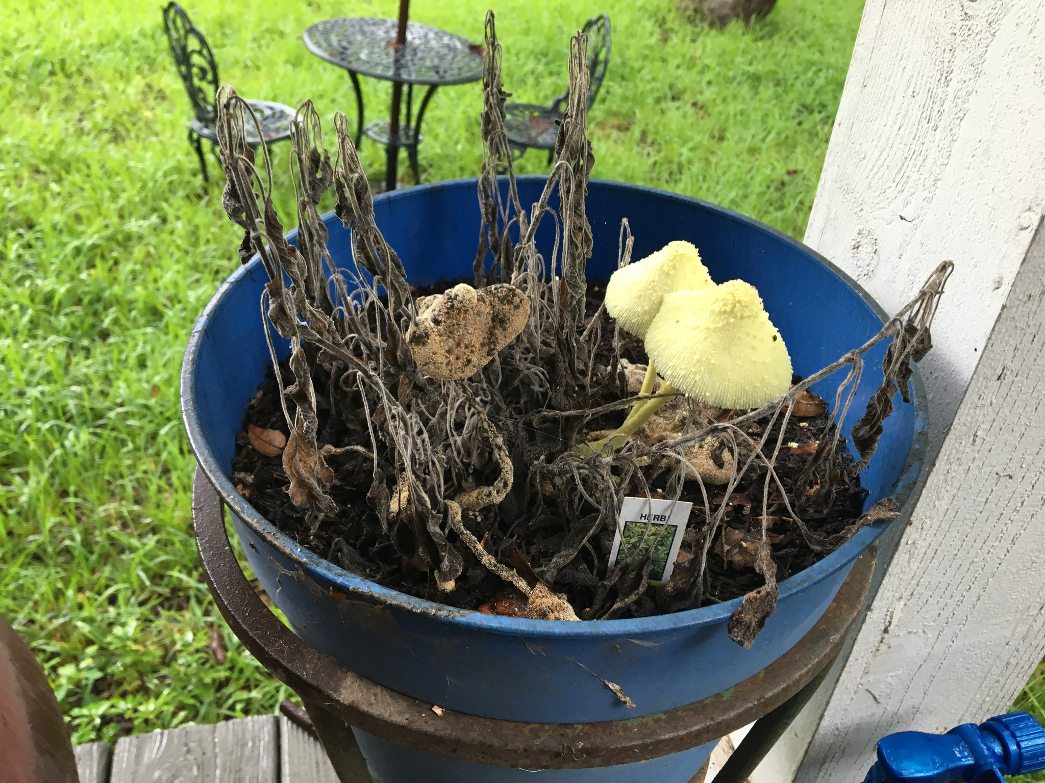
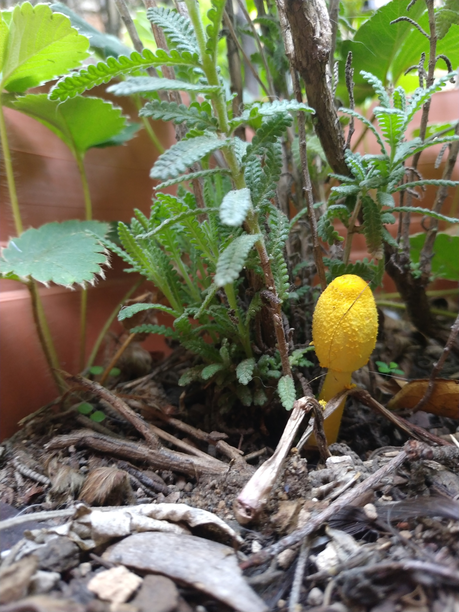
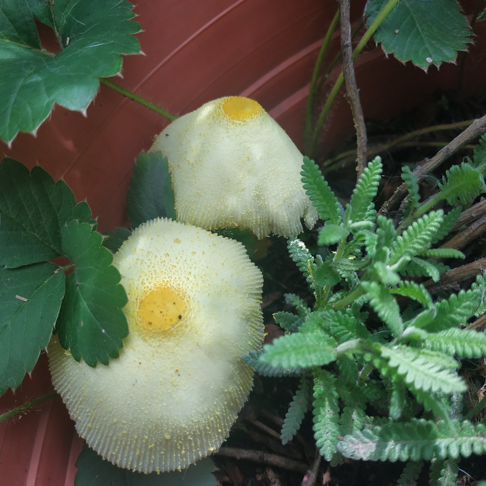
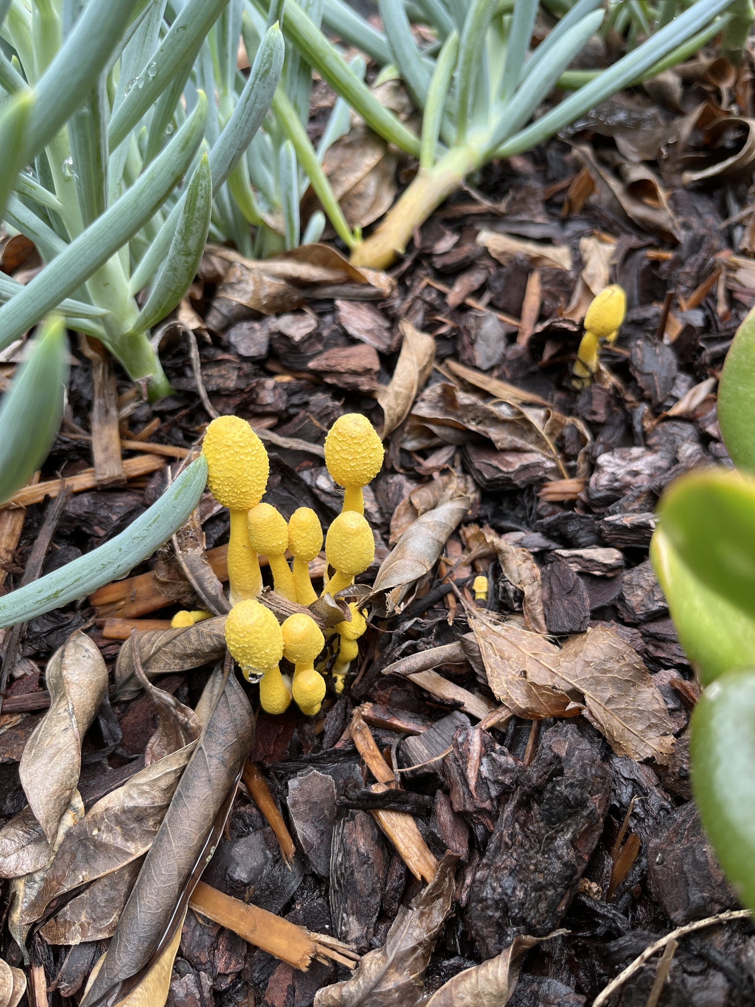
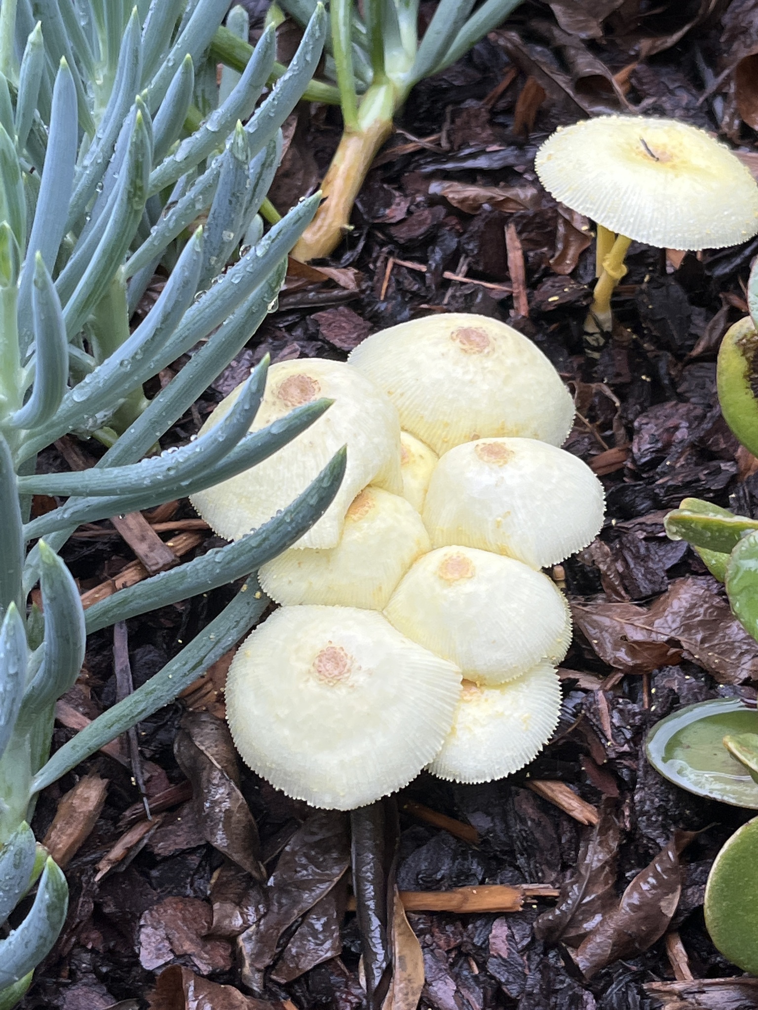
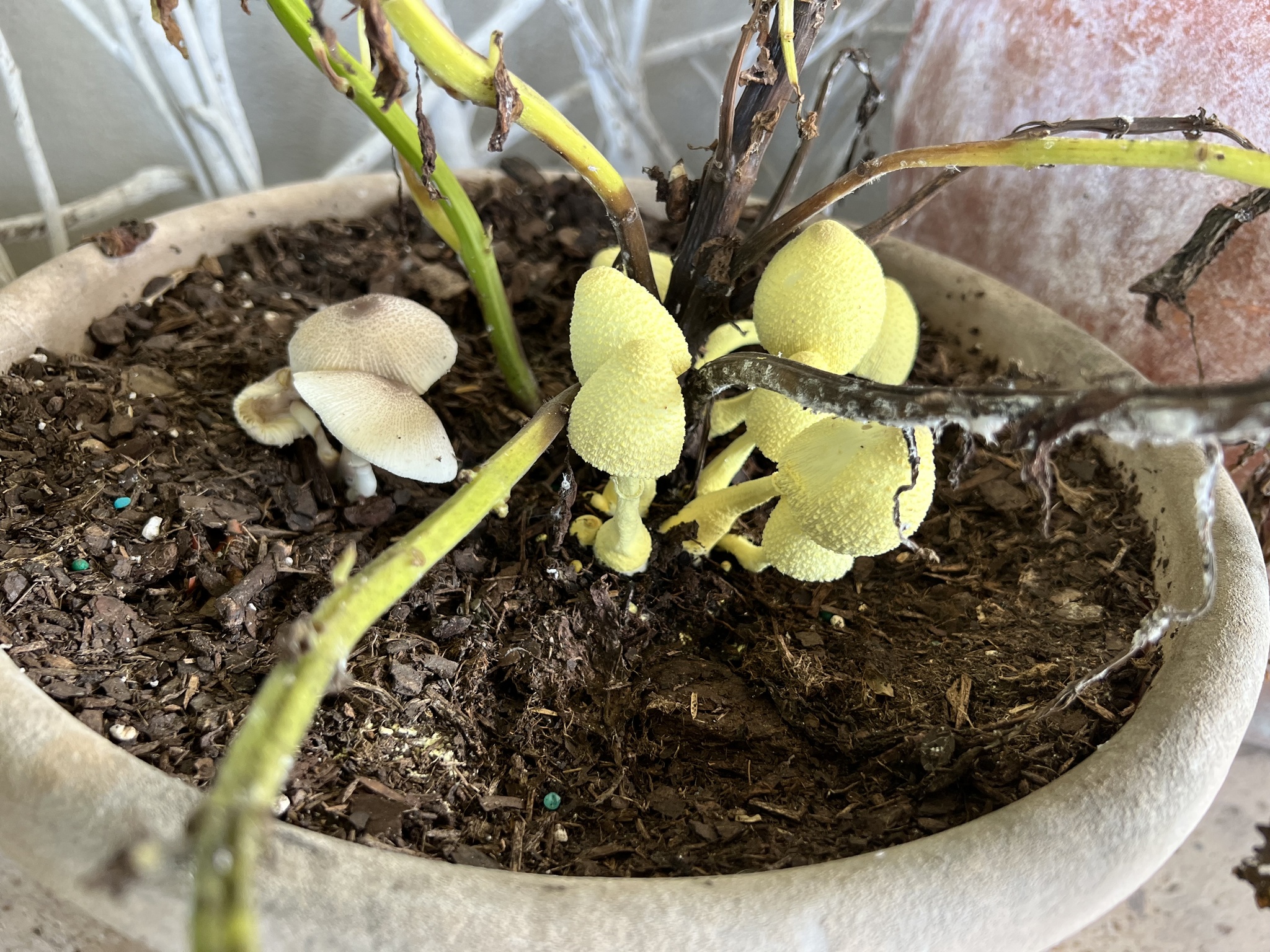
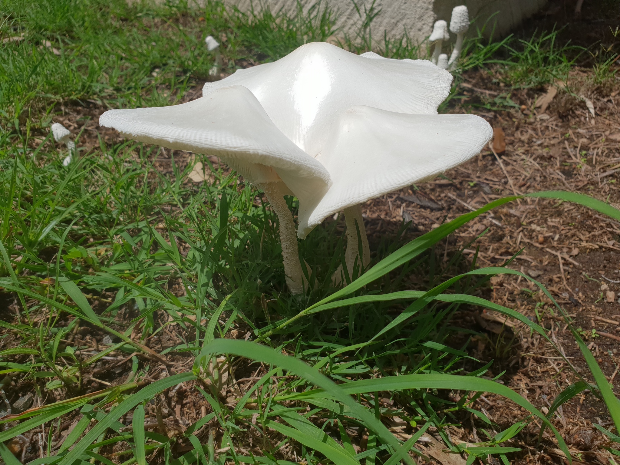
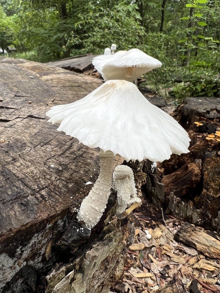
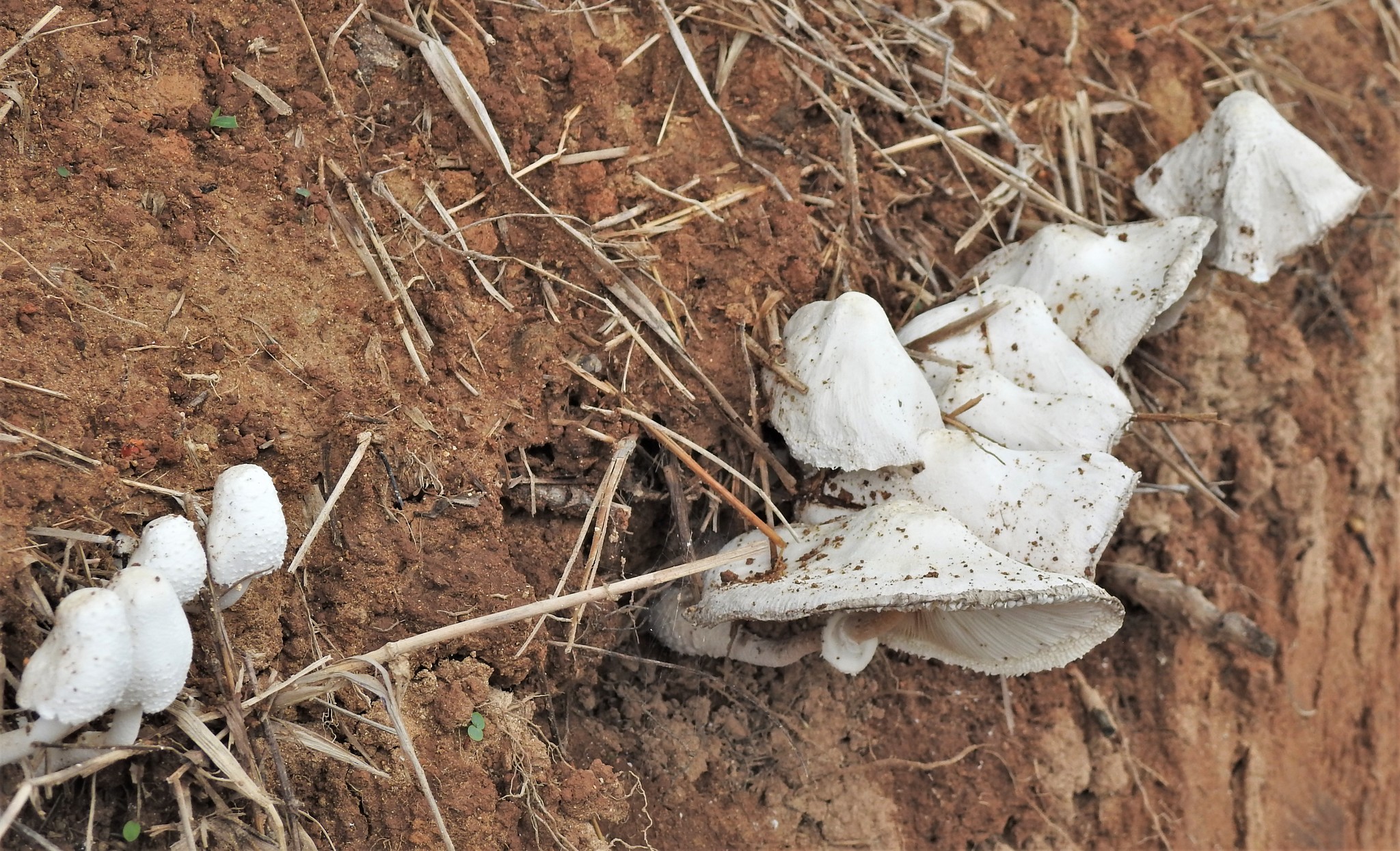
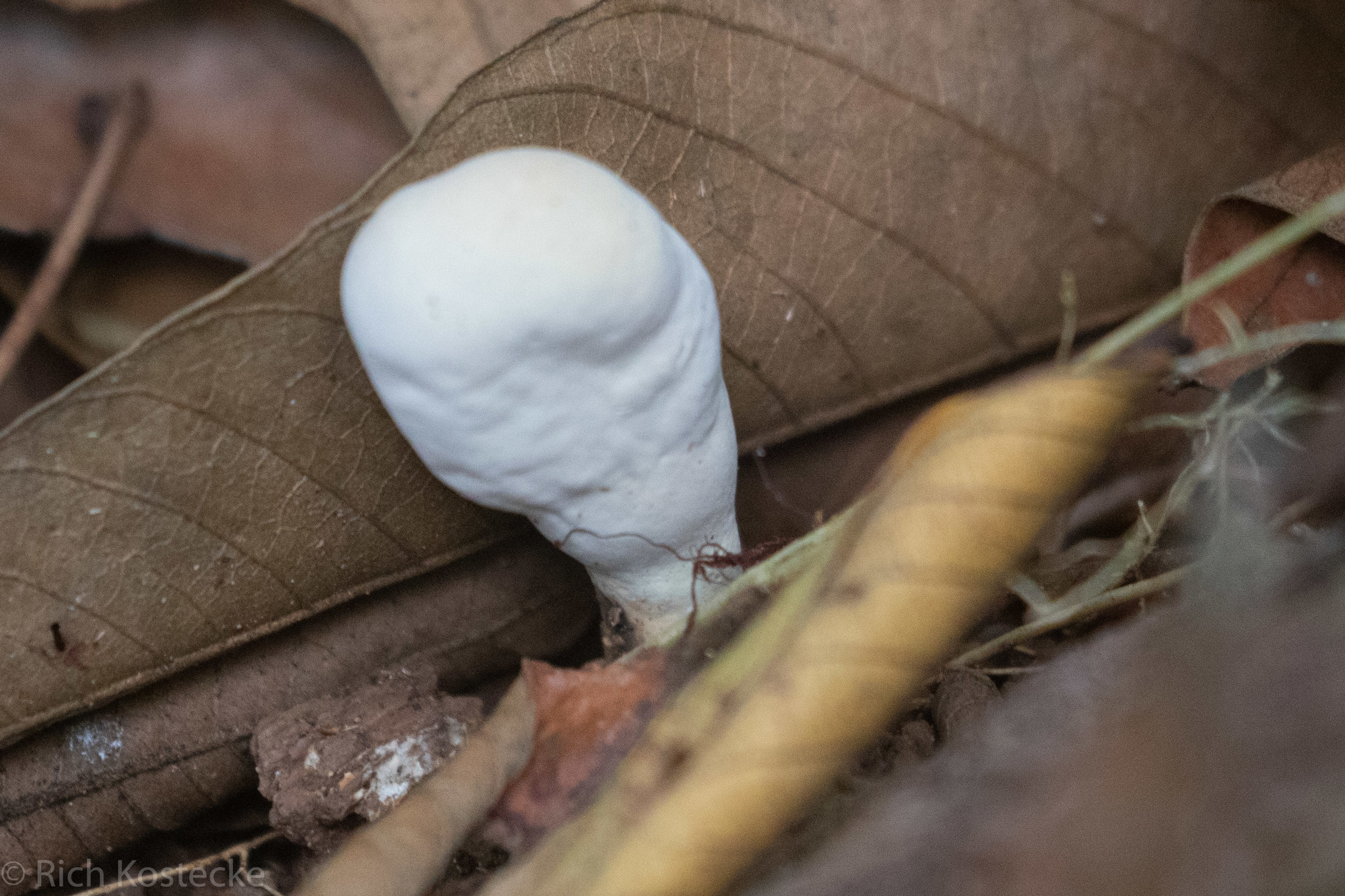
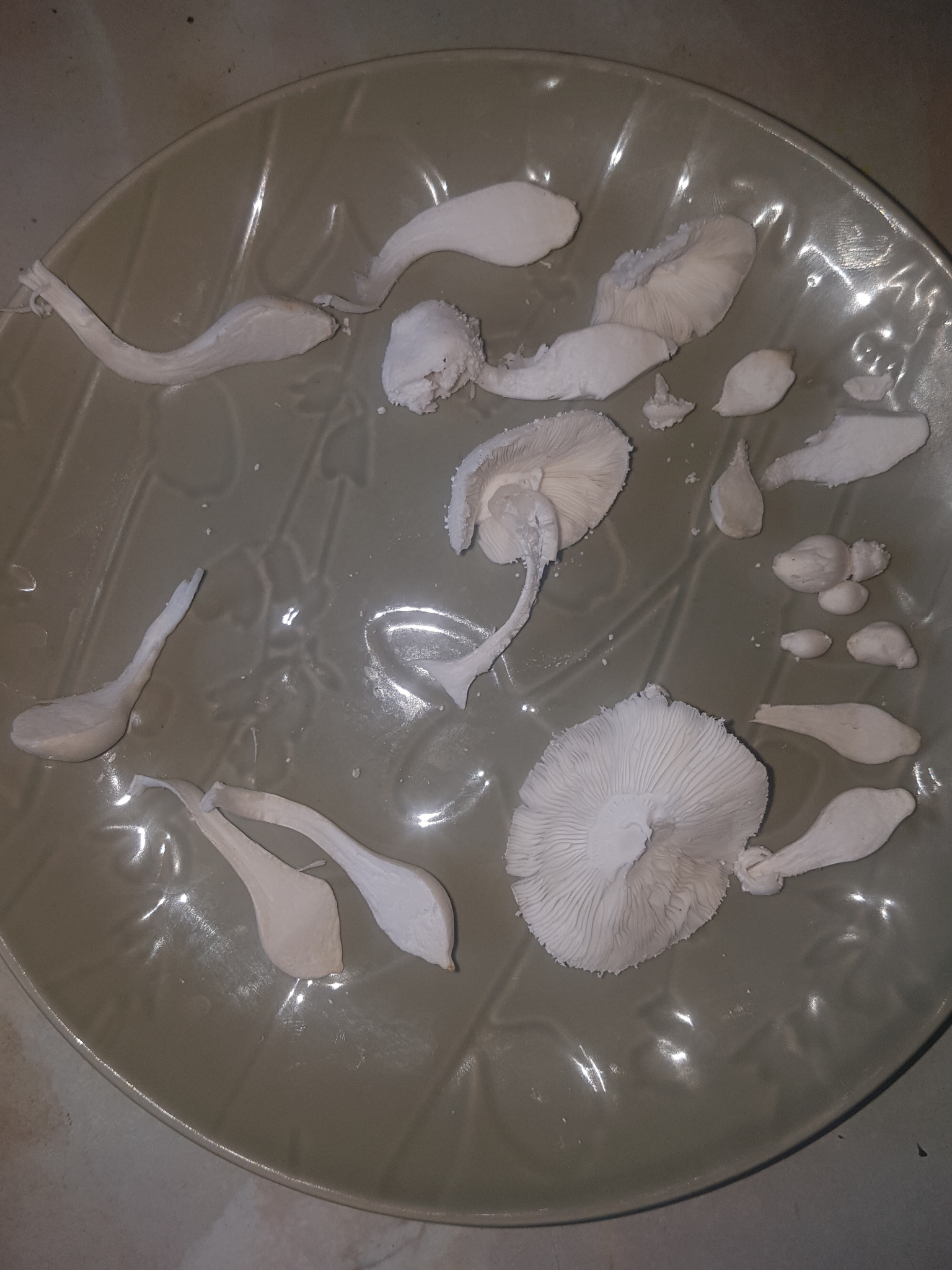
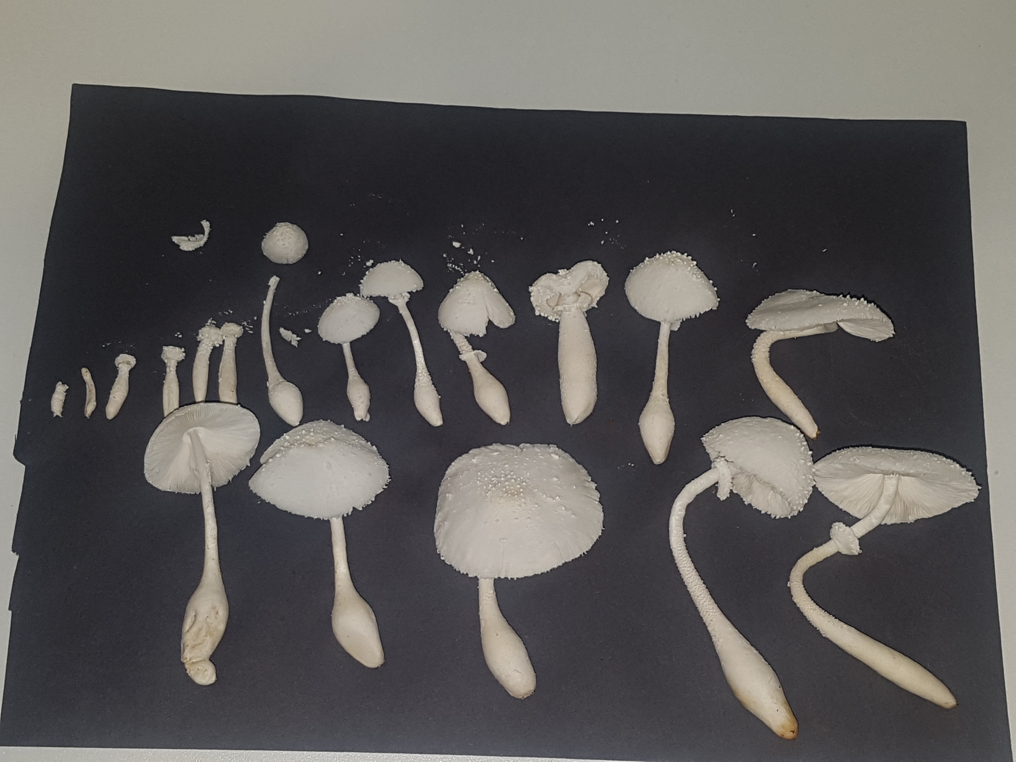
The species identity was confirmed via microscopy as well as observations of the intact mushrooms whilst growing them. In the wild L. cretaceus can lose many of its distinctive identification features due to rain and damage as can be seen here. There are numerous Leucocoprinus and Leucoagaricus species for which it could be mistaken and for which the edibility is still unknown. More seriously it would not be inconceivable for inexperienced foragers to confuse this species with some of the deadly Lepiota or Amanita species. Species in Amanita Sect. Roanokenses get mistaken for L. cretaceus regularly and some of those can be seriously toxic.

Whilst consuming the cultured mushrooms has not caused me any issues I would not suggest that this species is suitable for foraging due to the short period of time in which they are fresh and the risk of confusion.
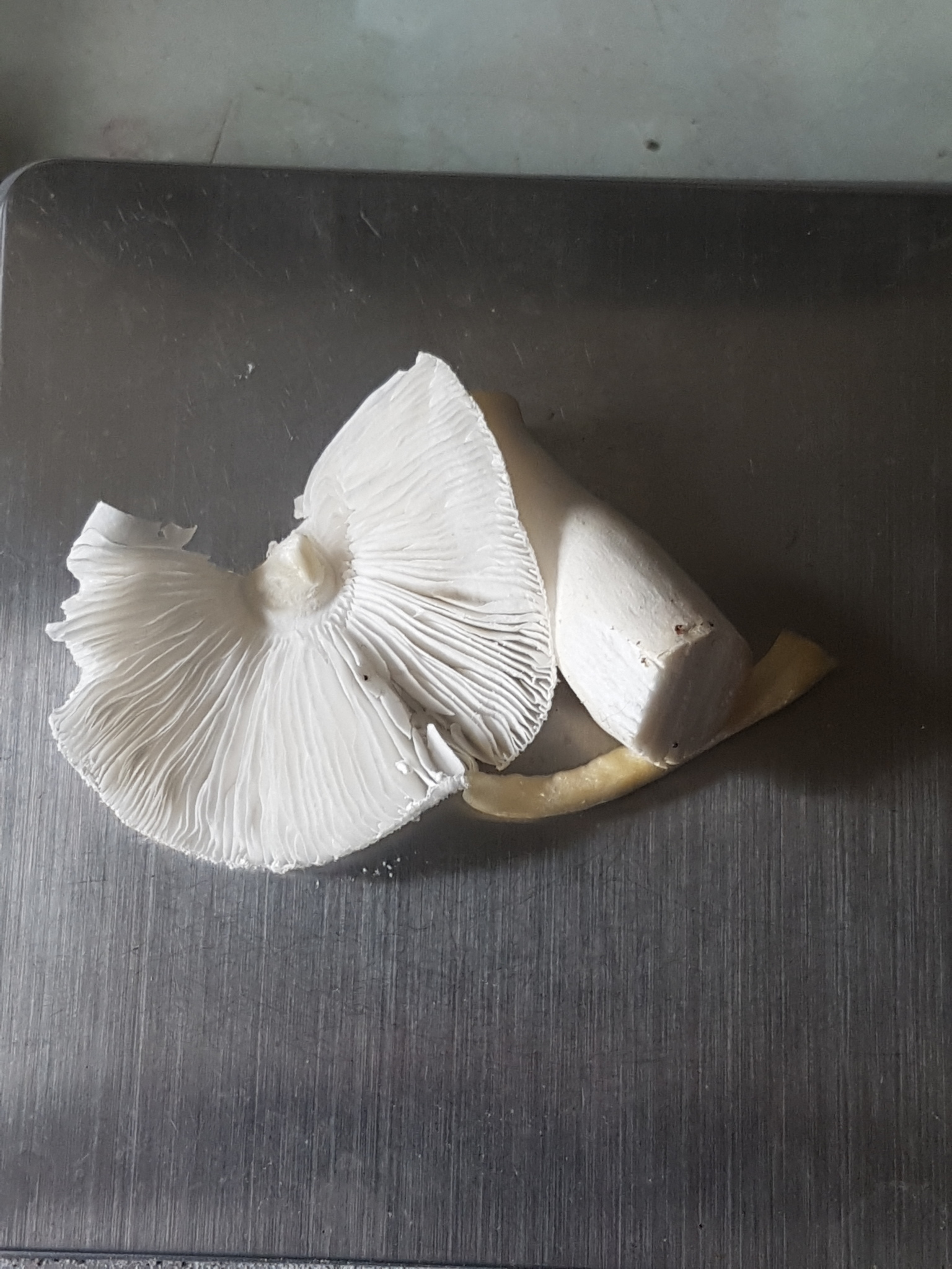
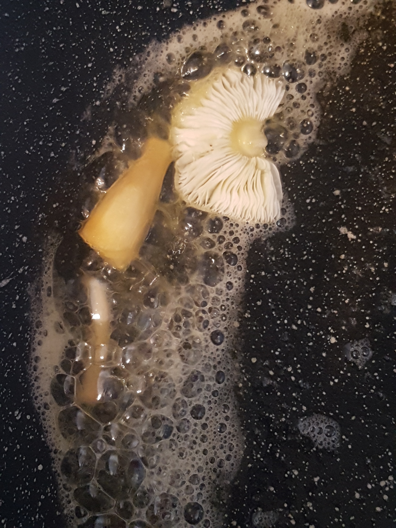
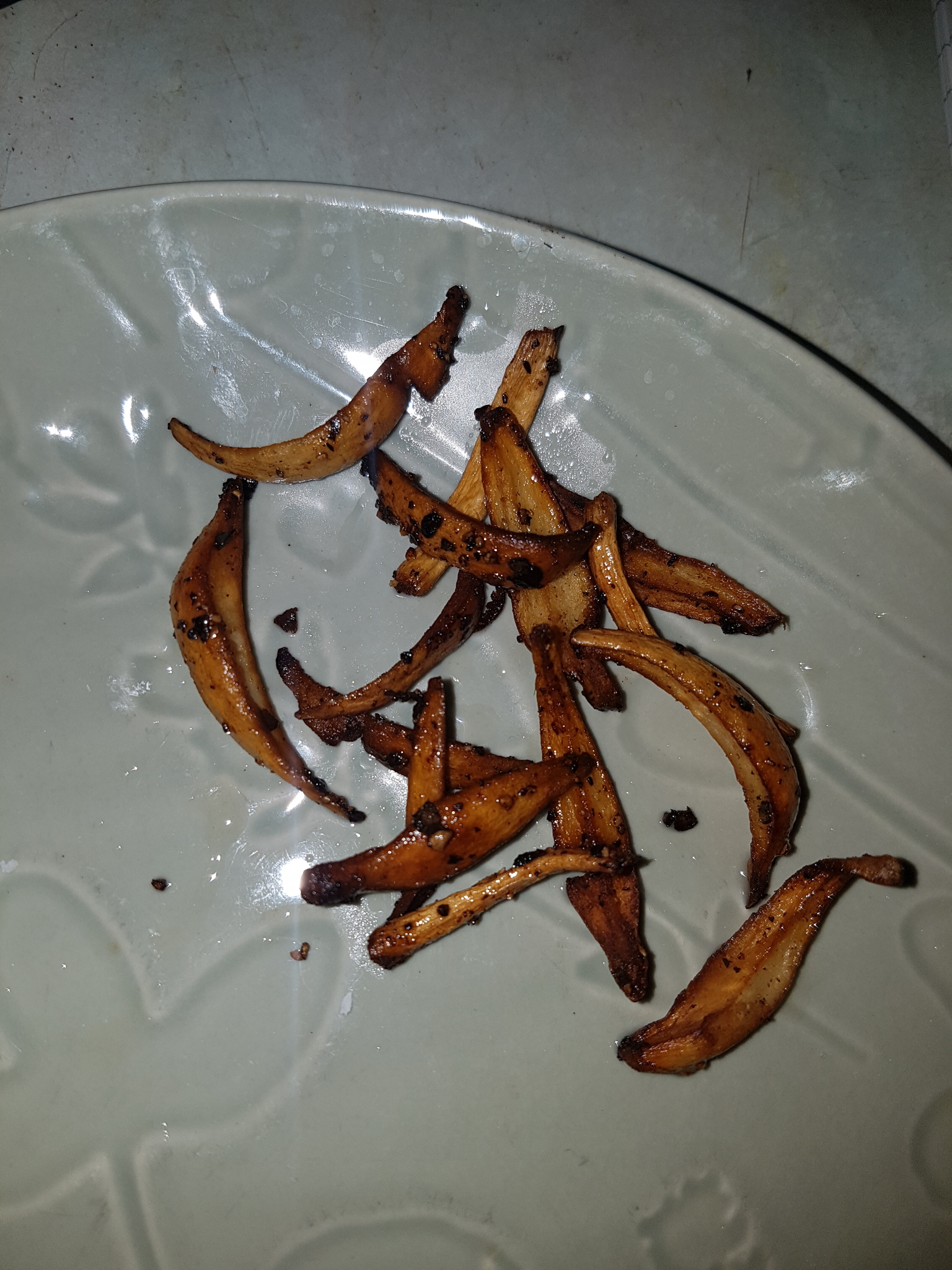

Fig. 13a - Before cooking Fig. 13b - During cooking Fig. 13c - After cooking

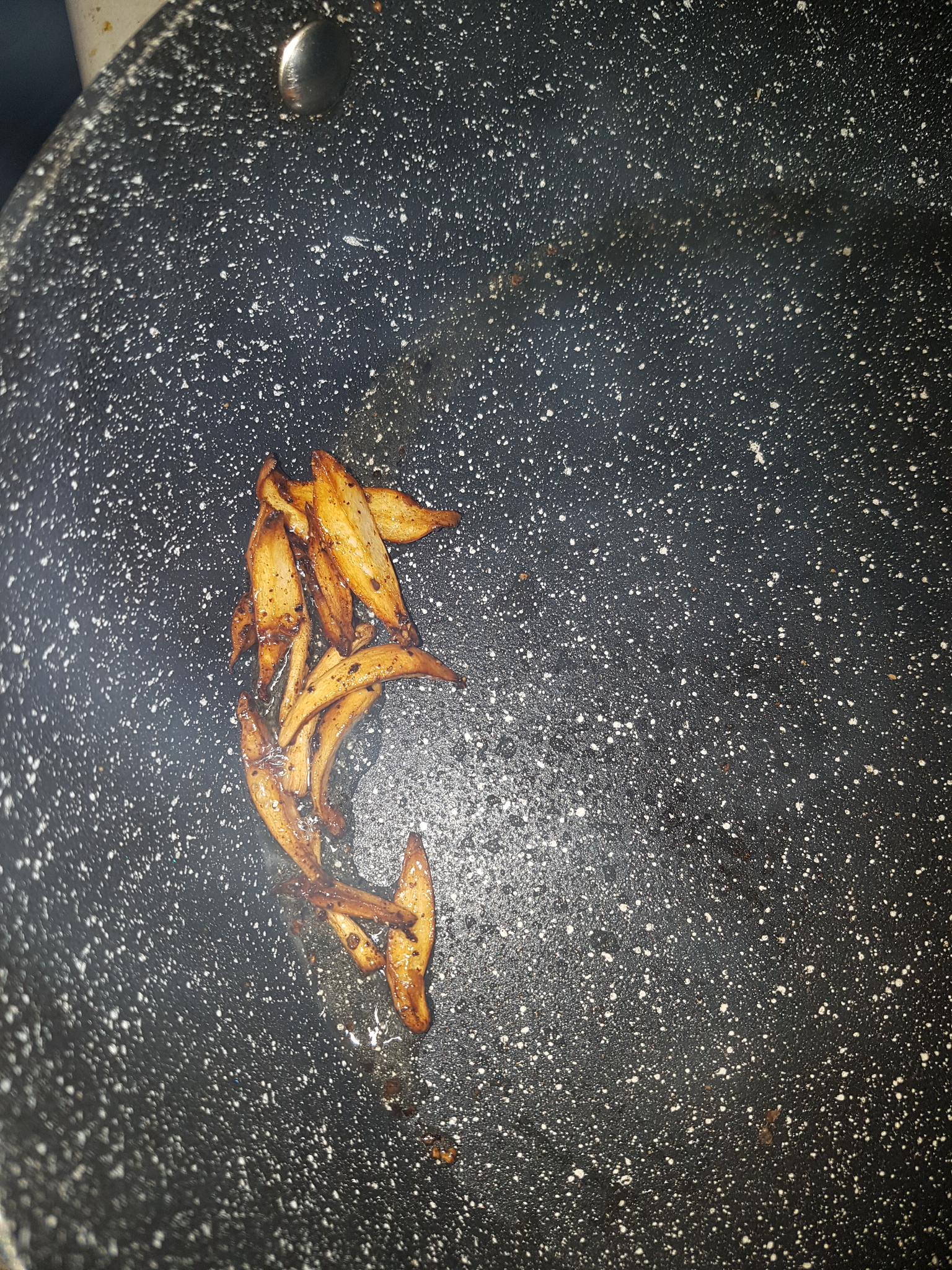
An hour after harvesting, 1g of cap and 2.4g of stem was cooked by sauteing in a pan with butter, without any seasoning. This amounted to most of the cap besides a section kept to study and most of the stem besides the very bottom of the base and some small pieces that were tested.

After browning the butter slightly the cooking time was approximately 5 minutes on a low heat in a stone coated pan. The mushroom was initially divided into three pieces - the cap, middle of stem and base of stem but as the base was thick and did not shrivel when cooked it was cut in half during cooking to brown both sides. Surprisingly the mushroom did not shrivel and diminish as much as some others do when cooked and it browned quickly. The cap reduced in size slightly but the stem pieces retained their consistency well. The smell during cooking was pleasant and persisted in the house for a few hours afterwards being somewhat reminiscent of the smell of fried chicken or similarly chicken-like as the smell of cooking chicken of the woods and hen of the woods, though the smell of the butter itself may have influenced this comparison.

The thinner, middle piece of the stem was tasted first and was surprisingly delicious. It was meaty and flavourful and somewhat reminiscent of chicken of the woods with a texture that was neither too tough, crispy, chewy, soggy or insubstantial. It is close to or slightly stronger than Pioppino, Cyclocybe aegerita in taste but with a slightly firmer, meatier texture which made it more pleasant. The cap was not as great as it had browned and cooked more than the stem and so had a slightly burnt taste, a little like burnt toast - though it did not look as if it was burnt and was not crispy. It was not unpleasant as such but the texture was still cottony and relatively insubstantial. A slight hint of the chemical taste that the cap possessed when raw was noticeable but was not enough to make it unpleasant. It may be necessary to start cooking the stems first and add the caps after a minute or two in order to prevent overcooking, or better yet harvest younger mushrooms before the cap opens. The two pieces of the base of the stem did not taste as strong as the middle section though were still very pleasant. With their greater girth they had not cooked through as much even when cut in half so were softer inside and similar in taste and consistency to cooked chicken of the woods.

Mattirolo makes the comparison to Pelliccione - Macrolepiota procera but from his description it is not certain if he is comparing the taste or only the appearance of the gills. As I have only ever found Macrolepiota procera in protected woodland on a nature reserve and hence have not picked and eaten them I cannot make this comparison however I do know that the stems are often regarded as too tough to eat and are discarded. So it interesting that with Leucocoprinus cretaceus the stem texture is actually very pleasant and frankly perfect for consumption. I am not a huge fan of the texture of many cooked mushrooms which can often be too soggy or overly crispy but I thoroughly enjoyed sampling this mushroom. It seemed sensible to consume only a small amount to test for any gastrointestinal symptoms but frankly if more than one had grown I would have been tempted to eat them all since the taste left me wanting more. No symptoms occurred as a result of eating this mushroom and there was no reaction to alcohol.

The best time to harvest these for consumption may be before the cap opens or even before the stem has substantially grown. After harvesting and eating this mushroom the immature mushrooms remaining in the jar aborted and gradually diminished however many new primordia grew afterwards. Aborted mushrooms appear to desiccate themselves quite effectively even whilst embedded in a moist substrate.


Fig. 14a - After harvesting Fig. 14b - After cutting Fig. 14c - During cooking

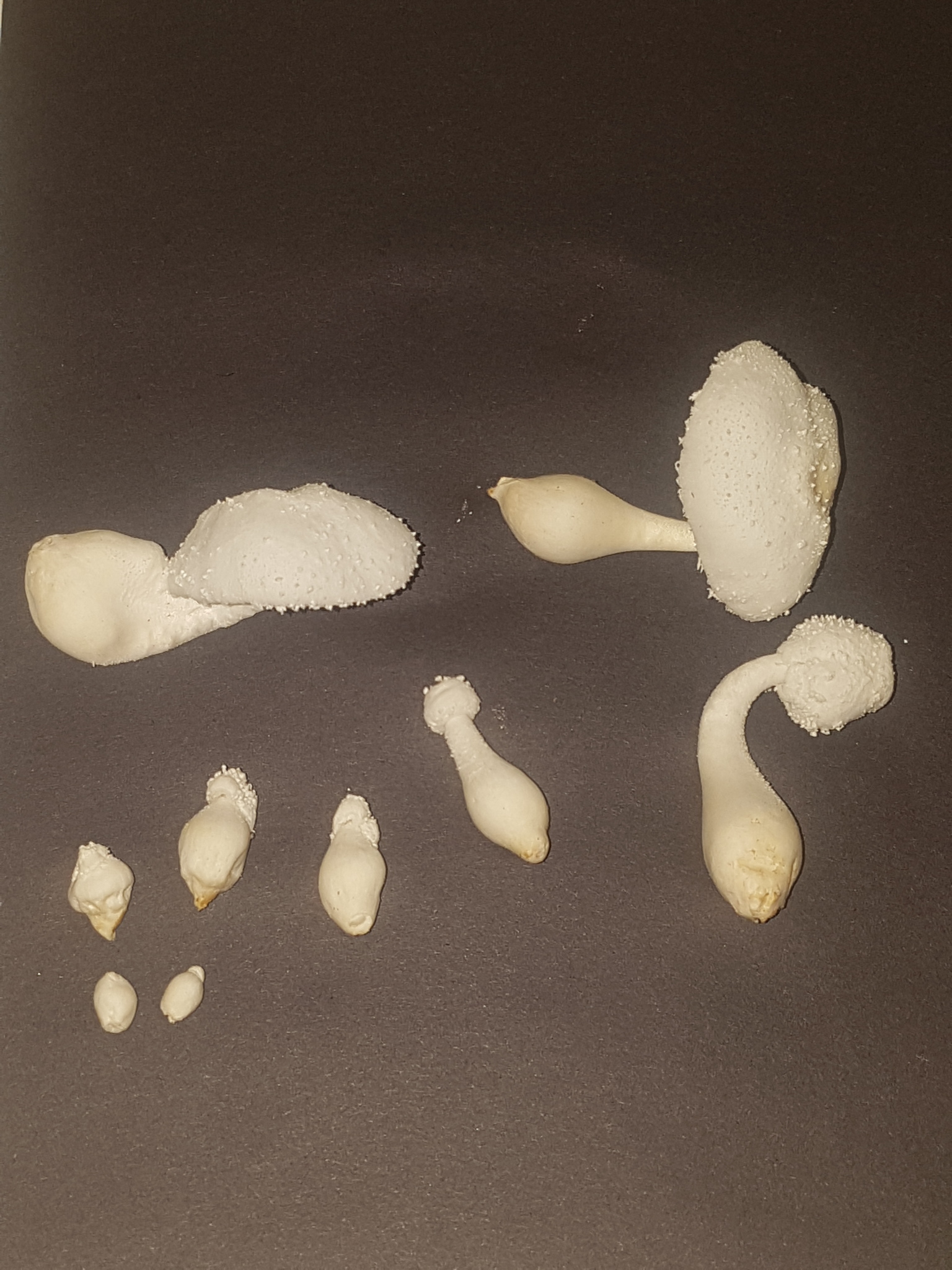
On 15/08/23 more mushrooms were harvested from the same jar with a total fresh weight of 10.78g. This represented one large mature mushroom with the cap open, one of a similar size with a closed cap, a smaller less mature one with a closed cap, two conjoined mushrooms with the base and cap attached but individual stems noticeable and then a few small, undeveloped pins that came attached to others.

One mature mushroom cap and stem was sauteed in sunflower oil for ~2-3 minutes without seasoning. The whole stem was cut down the middle from top to bottom and cooked in two pieces and the cap was cut in two. The stem was again pleasant, though was better in butter and the cap still had the slight burnt taste. It was not unpleasant but again reinforced the thought that they are probably best harvested before the cap opens.

The remaining mushrooms were sauteed in butter for 4-5 minutes without seasoning. The unopened caps proved far better than the open ones and were quite reasonable to eat. In both taste and texture they were preferable. The stems were again very pleasant with the thinner portion becoming more crisped than the base, which had soft flesh within. The small immature mushrooms were cut in half down the middle or cooked whole. Subsequently they cooked faster than the larger ones and browned more but did not burn. The taste was not substantially different to the mature stems but no off taste or texture from the immature cap was noticed.
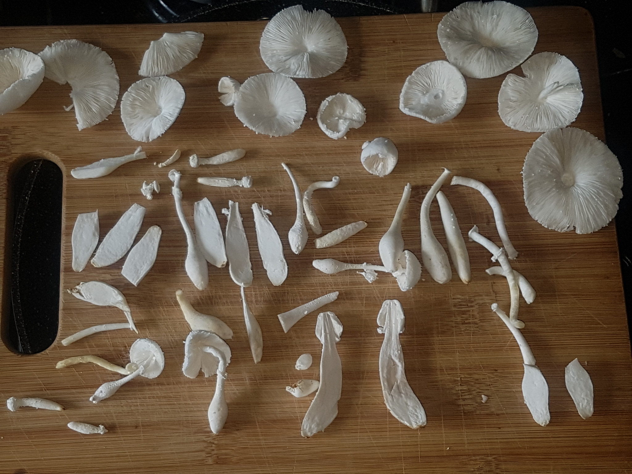
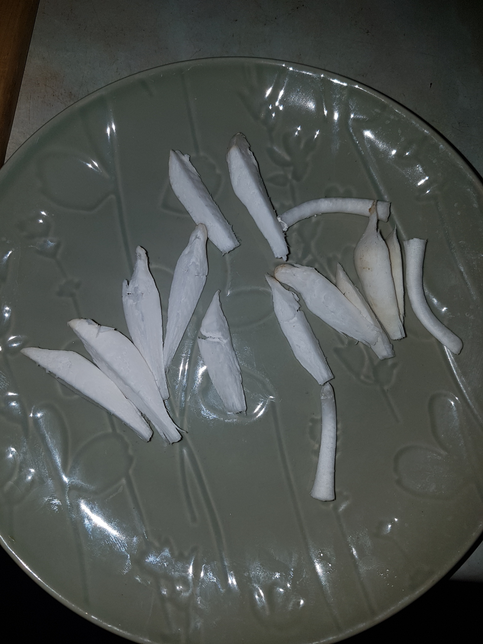

Fig. 15a - After harvesting Fig. 15b - After cutting

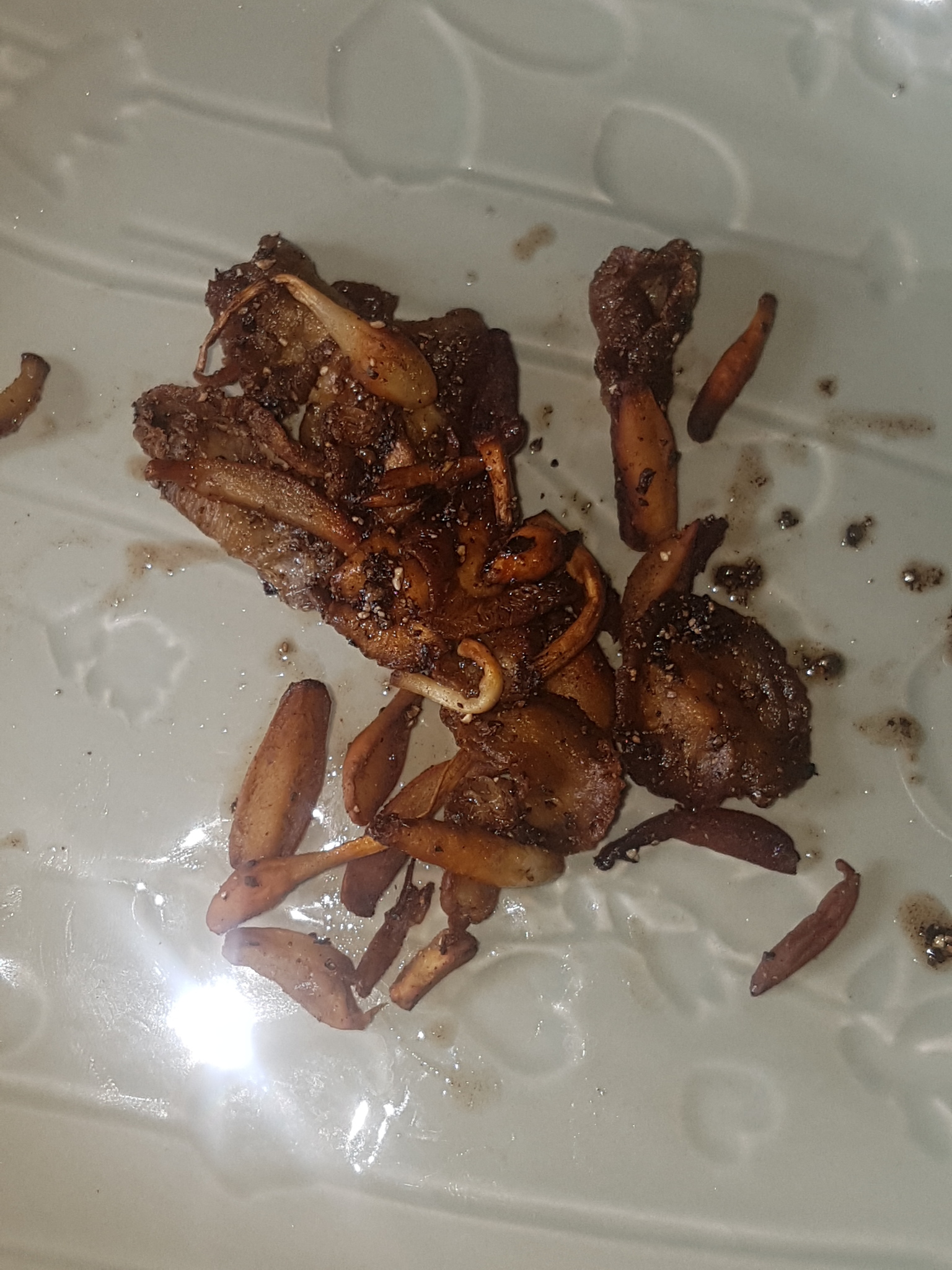
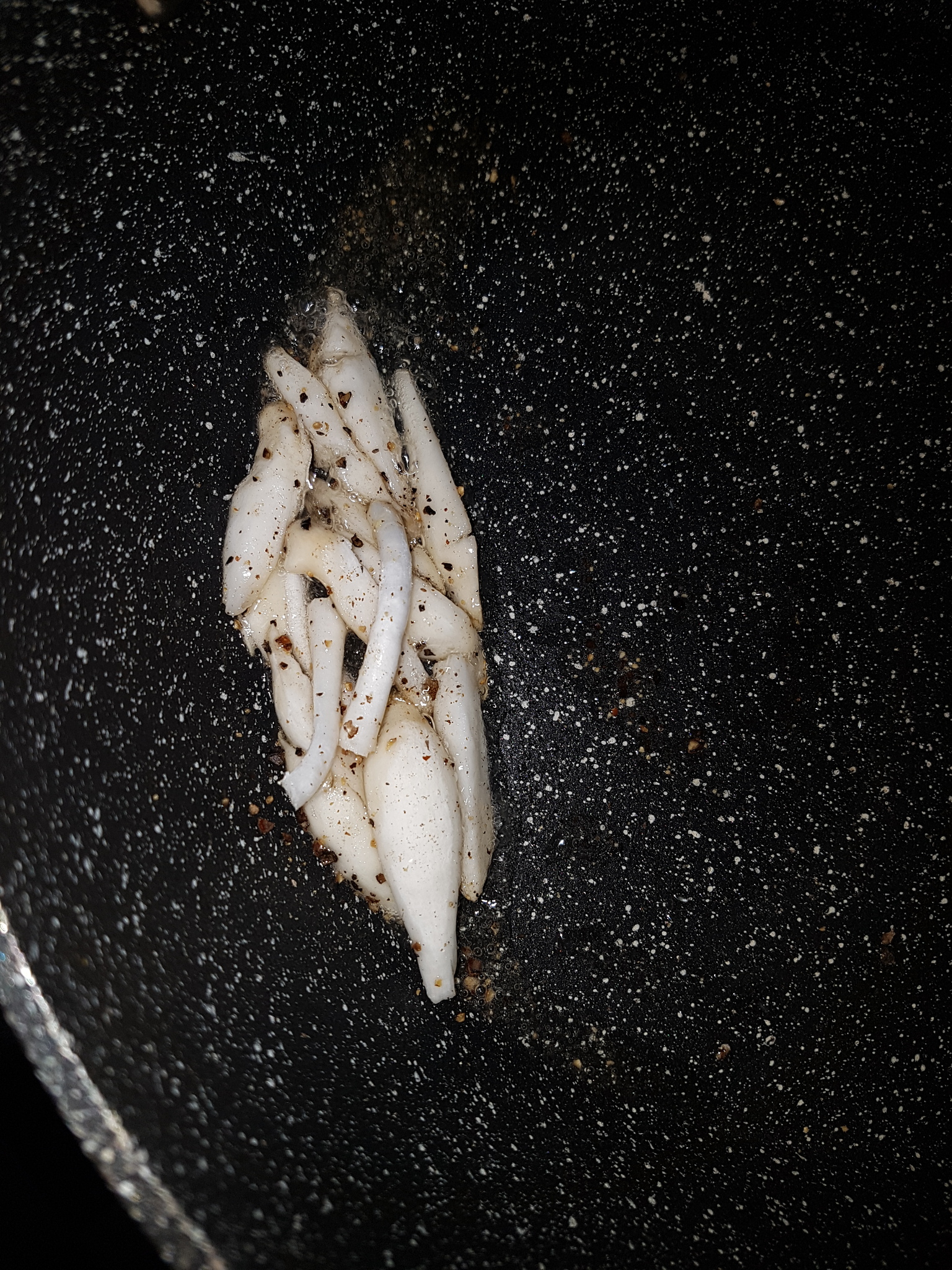
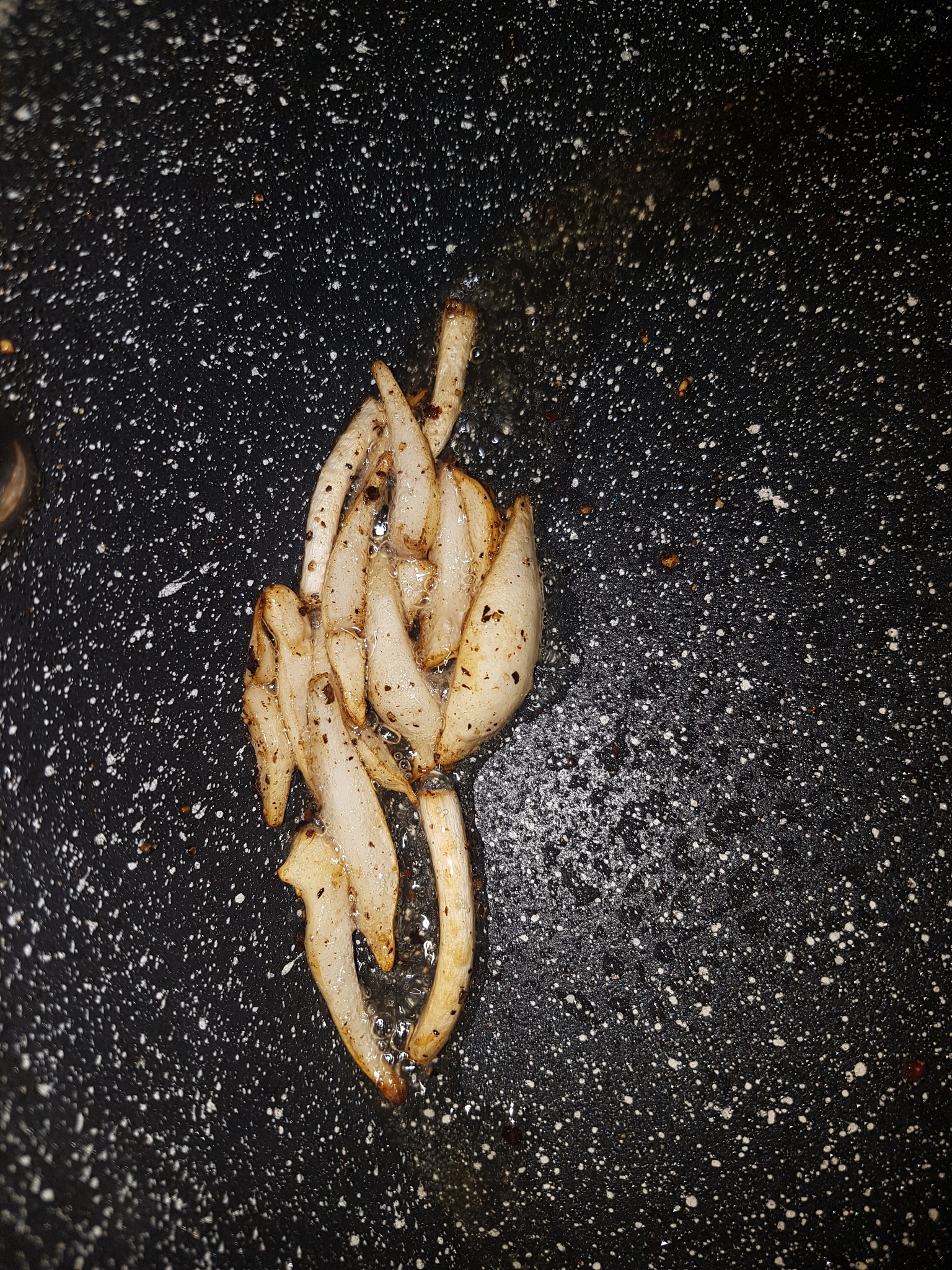
On 01/09/23 two dozen mushrooms and some immature pins weighing a total of 35.87g fresh were cooked and consumed. Mushrooms were harvested from several jars over a two day period with some stored in the fridge overnight. They were grown in jars containing rice, potting soil or a potting soil and coir mix. Some smaller ones were grown in a small container full of only wheat bran and water which had been intended to test sclerotia growth but resulted in mushrooms nonetheless.

The mushrooms were divided into two portions with the first sauteed in just butter and the second sauteed in butter and seasoned with black pepper. Each portion contained a mix of mushroom caps and stems in different stages of maturity. The stems were cut into sections and added to the pan first with the caps added a minute or two later once the stems had started to brown. The caps of the young mushrooms that had either yet to open or only recently opened were vastly preferable to the older ones and when cooked with pepper actually tasted even better than the stems, though perhaps this was just the result of them absorbing a great deal of butter. Some that were overcooked or older still had a slight unpleasant bitterness and the texture was still not ideal with any of the ones that had expanded though they were still tolerable and not a big problem. With caps that remained closed or had only just started to open there was a meatier texture and they cooked quite well. As a result the taste was surprisingly variable between the different parts at different stages of maturity.
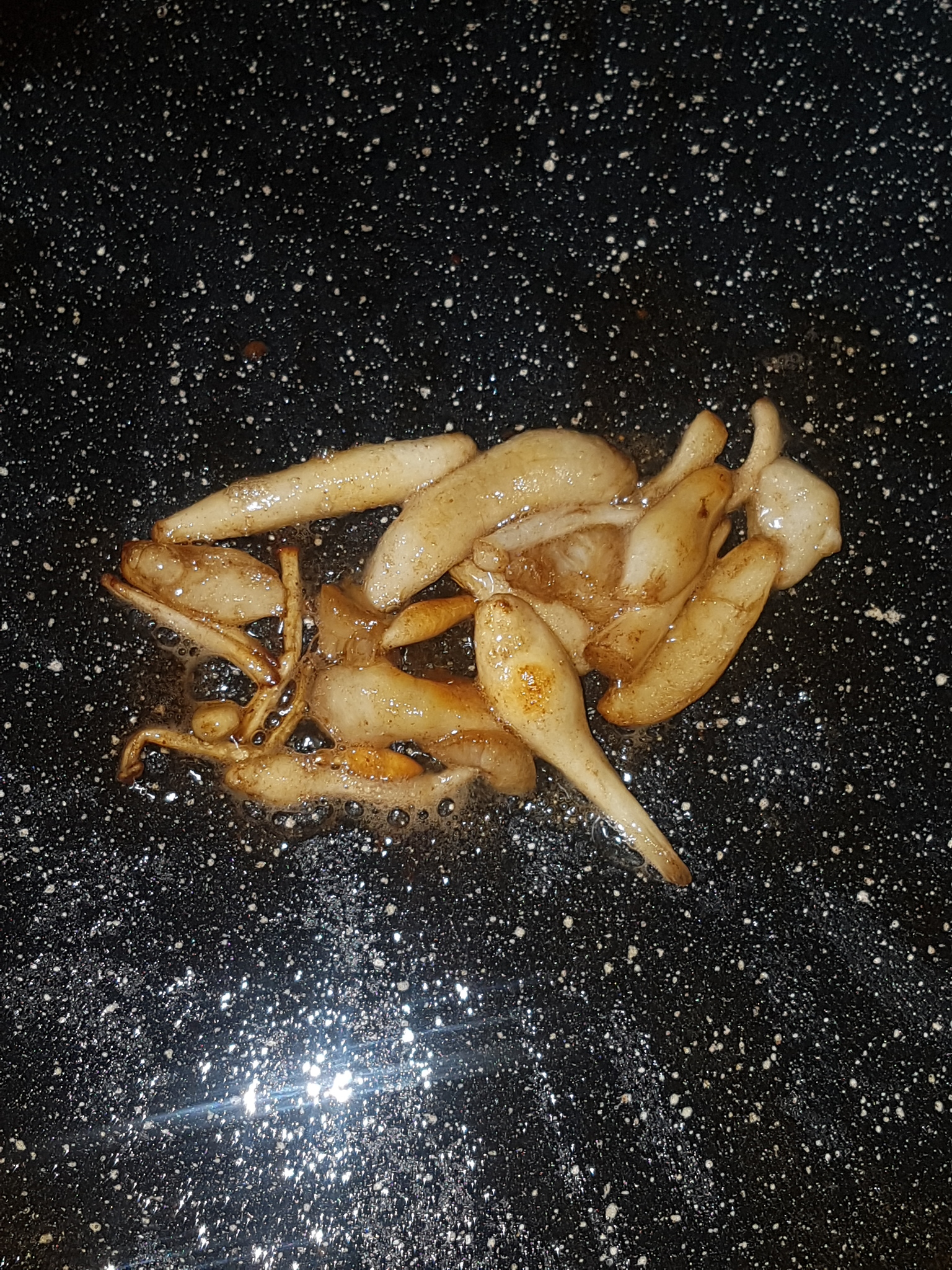
Fig. 15c - During cooking Fig. 15d - During cooking Fig. 15e - After cooking

No issues arose after eating this larger serving. The interior flesh in some thicker pieces of stem that had only been bisected was not perfectly cooked and so it is probably better to cut into quarters. The ideal time for harvesting is probably when pins have started to enlarge but before the caps have begun to open. If mature caps are to be cooked they require less time than the stems so as to be palatable. One mushroom that formed had a stem that was wider for a greater portion of the length and grew to around finger size with a cap that was small and unopened and this proved ideal for consumption. Similar has been observed multiple times but it is not yet clear if this is the result of chance environmental conditions or whether this trait can be reproduced via cloning.

Experimentation with consumption of the mushrooms was paused at this time due to unusual symptoms of nasal congestion which occurred and persisted in the weeks following. It was severe enough to cause constant sneezing and at times a complete loss of smell such that it was not possible to distinguish isopropyl alcohol or vinegar from water by smelling a tub held right under the nose. Whilst it seemed unlikely that the mushrooms were responsible for these peculiar symptoms they could not be ruled out as the cause and so further consumption had to be halted. The very strong and unusual smell of the substrate and sclerotia was also considered as the cause and general hypochondria ensued due to a family history of nasal polyps causing anosmia.

However the culprit appeared to be contamination of the grow room with mites, probably Tyrophagus puterescents which in some places had left significant quantities of dusty material behind which was triggering a major allergic response. Symptoms quickly disappeared when walking outside and escalated when entering the grow room. Some of the mushrooms consumed were also collected from substrates that were likely contaminated by the mites. Once the infested material was removed, the debris cleaned up and more suitably mite-proof containers were employed to prevent further infestation the allergic response diminished. After the symptoms alleviated they did reoccur briefly on a couple of occasions after emptying mite contaminated jars into the compost and so it would appear that exposure to the mite debris was the trigger for the symptoms. During this hiatus any mushrooms harvested were dried for later and on a few occasions some dried stems were added to pizza without issue.
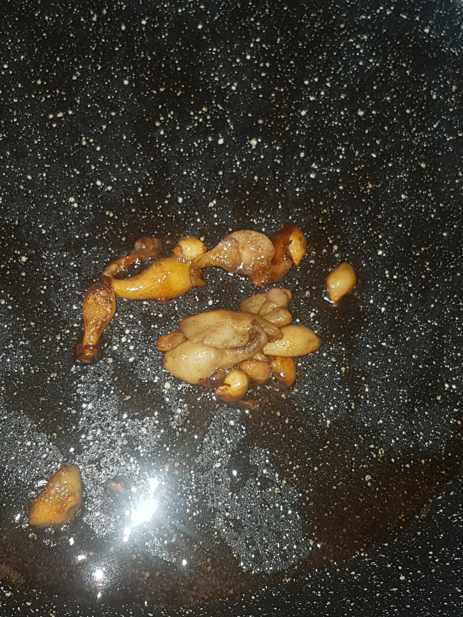
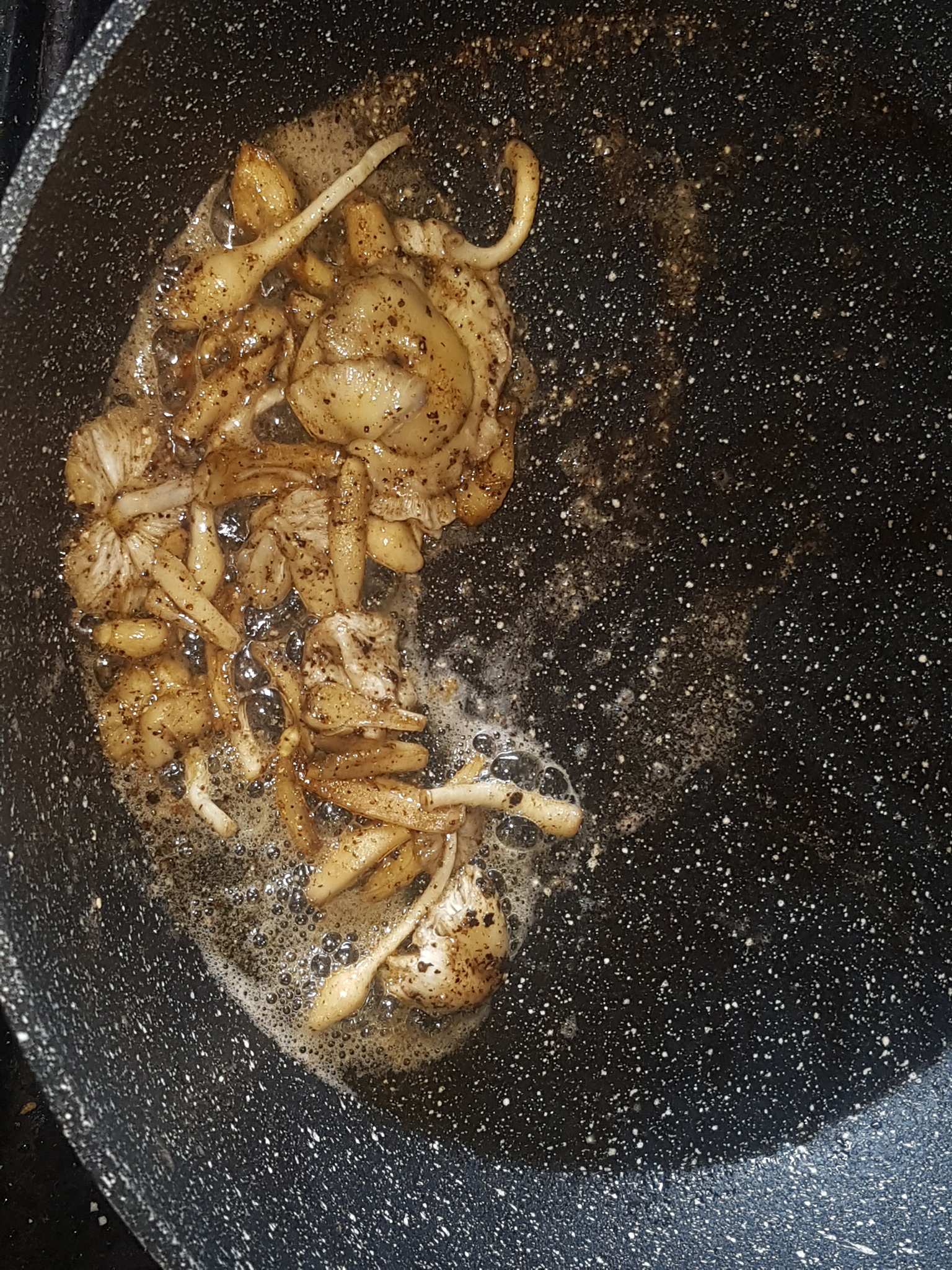

Fig. 16a - 00:36 - Before cooking Fig. 16b - 00:37 - Cooking start Fig. 16c - 00:38 - Cooking Fig. 16d - 00:39 - Cooking Fig. 16e - 00:40 - After cooking

On 31/12/23 three large mushrooms grown on a soil substrate were picked within hours of the caps opening and weighed a total of 20.5g. The caps were removed for spore printing and the masses of sclerotia at the stem base were removed leaving 15g of stems with bases that measured 14.5, 16 and 18mm thick. The stems were cut half way up to separate the thinner top portion of the stem from the thicker base and then the bases were quartered down the centre. All the stem material was then sauteed in sunflower oil with black pepper for approximately 4 minutes. Figures 16a-16e show the browning during cooking with photos taken 1 minute apart. The cooked stems had a pleasant taste and texture with quartering them allowing them to better cook through. No symptoms occurred after consuming them.

The following day a further 7.7g of stems were cooked similarly but with the addition of ground Nigella sativa seeds and Cajun spices which further improved the already good flavour. No symptoms occurred. On 05/01/24 one especially large mushroom was picked which had a total weight of 13.1g of which 8.5g was the stem which had a thickness of 18-19mm at the base. This was basted in sauce and roasted in the oven resulting in a consistency similar to potato wedges with a crispy exterior and softer middle. The next day a 7g mushroom was picked and 4.1g of stem was roasted on a baking tray in sunflower oil with potato wedges. It cooked well and became crispy and firm in around half the time that the potatoes took.

On 21/01/24 dried mushrooms were soaked in water to rehydrate with five dried mushroom stems weighing a total of 1g placed in a cup with approximately 100ml of cold tap water. The stems floated but rehydrated quite readily however they became more soggy and lacked the firmness of the fresh stems. The thick stem bases rehydrated substantially and enlarged to something similar to their original size in three of the five mushrooms but were quite water logged and soggy to the touch. The middle portion of the stem remained quite firm and inflexible similar to when fresh but the apex of the stem was very soggy. Two of the stems remained somewhat shrivelled and had a browner colouration but felt a bit firmer than the others. These may have been specimens that were picked during a different state of maturity or air dried as opposed to placed in the dehydrator. This was not recorded as all dried mushrooms were simply kept together in an air tight container.

The stems were removed from the water after around 10 minutes and weighed 4.2g total which is probably similar to the weight when fresh since these were all small specimens. The sogginess made them awkward to cut and so the smallest were cooked whole. After sauteing in sunflower oil and black pepper for approximately 4 minutes the sogginess was not an issue and they crisped up well. The taste and texture was good and similar to when fresh though perhaps slightly preferable with fresh mushrooms. The dried mushrooms have a very pleasant savoury smell which is noticeable when opening the container but after soaking and cooking this was not noticeable. If larger specimens are to be dried for later cooking it will probably be optimal to cut them first.


5.3 Notes on the Smell

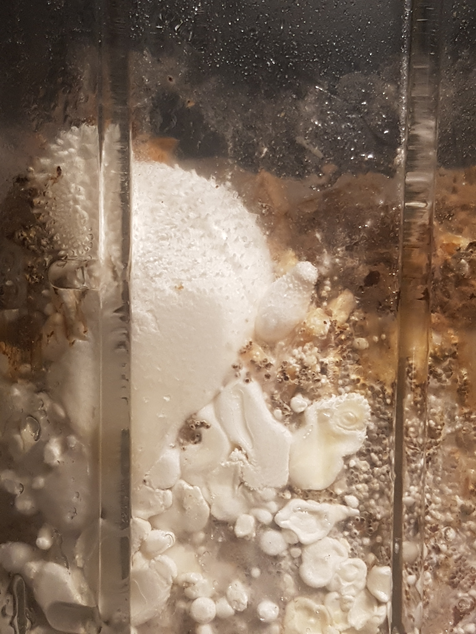
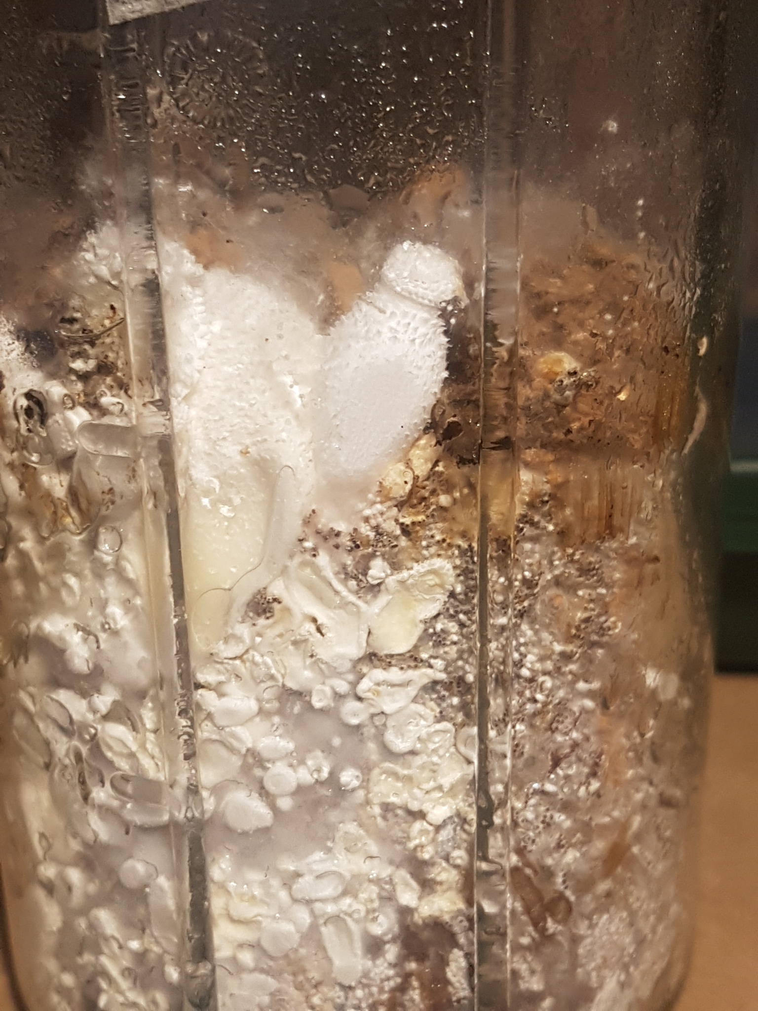
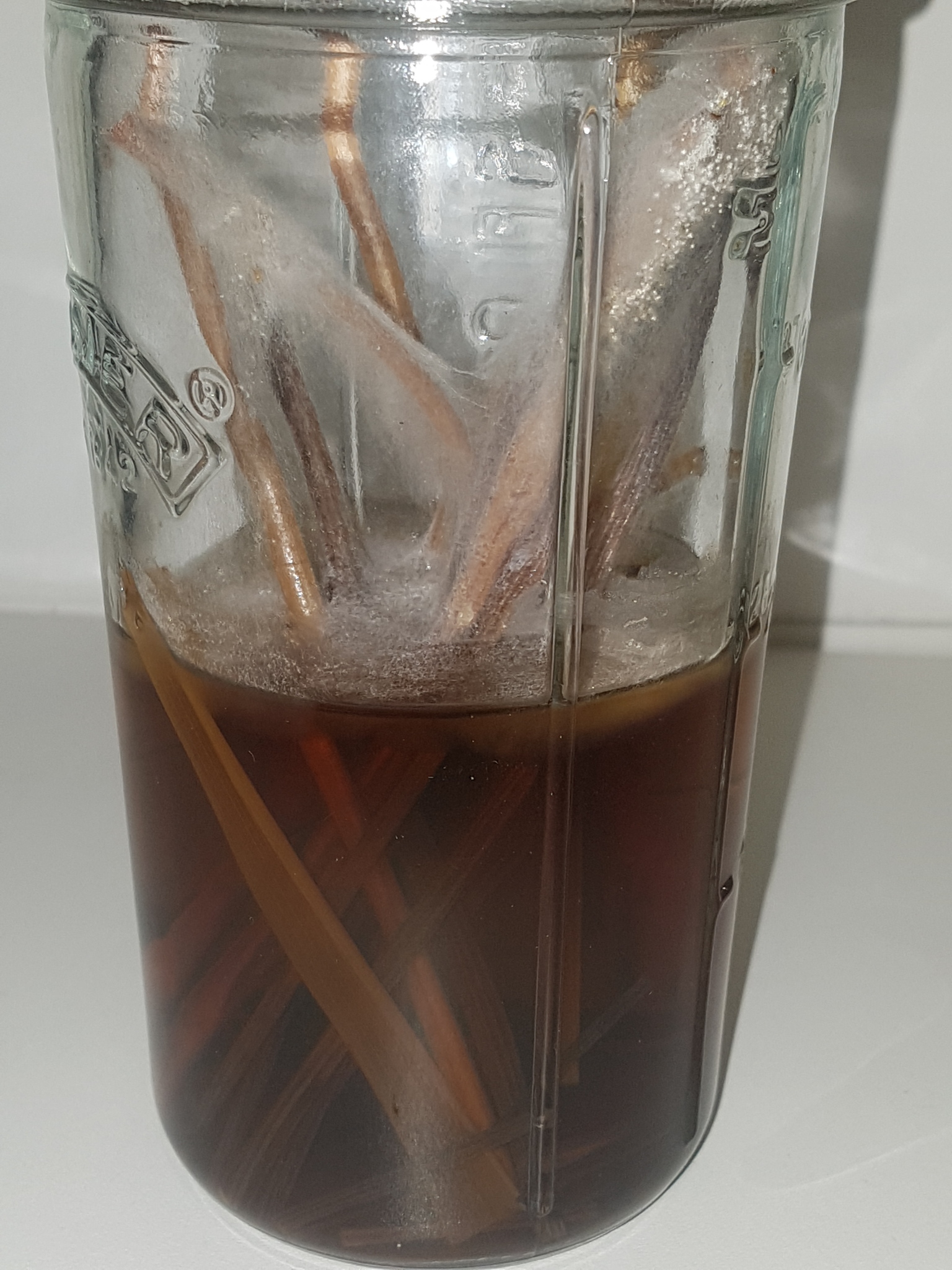
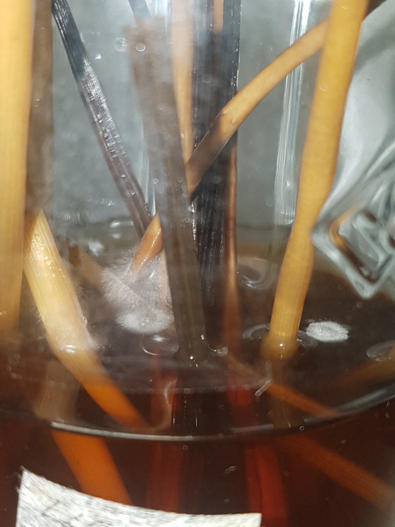
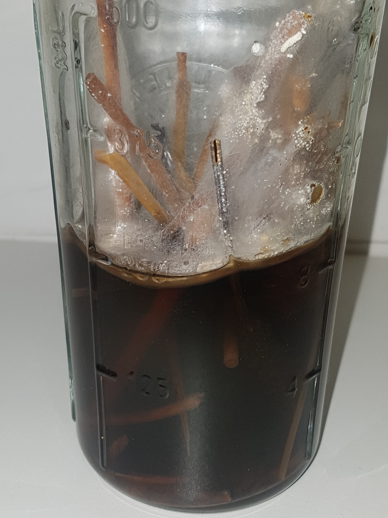
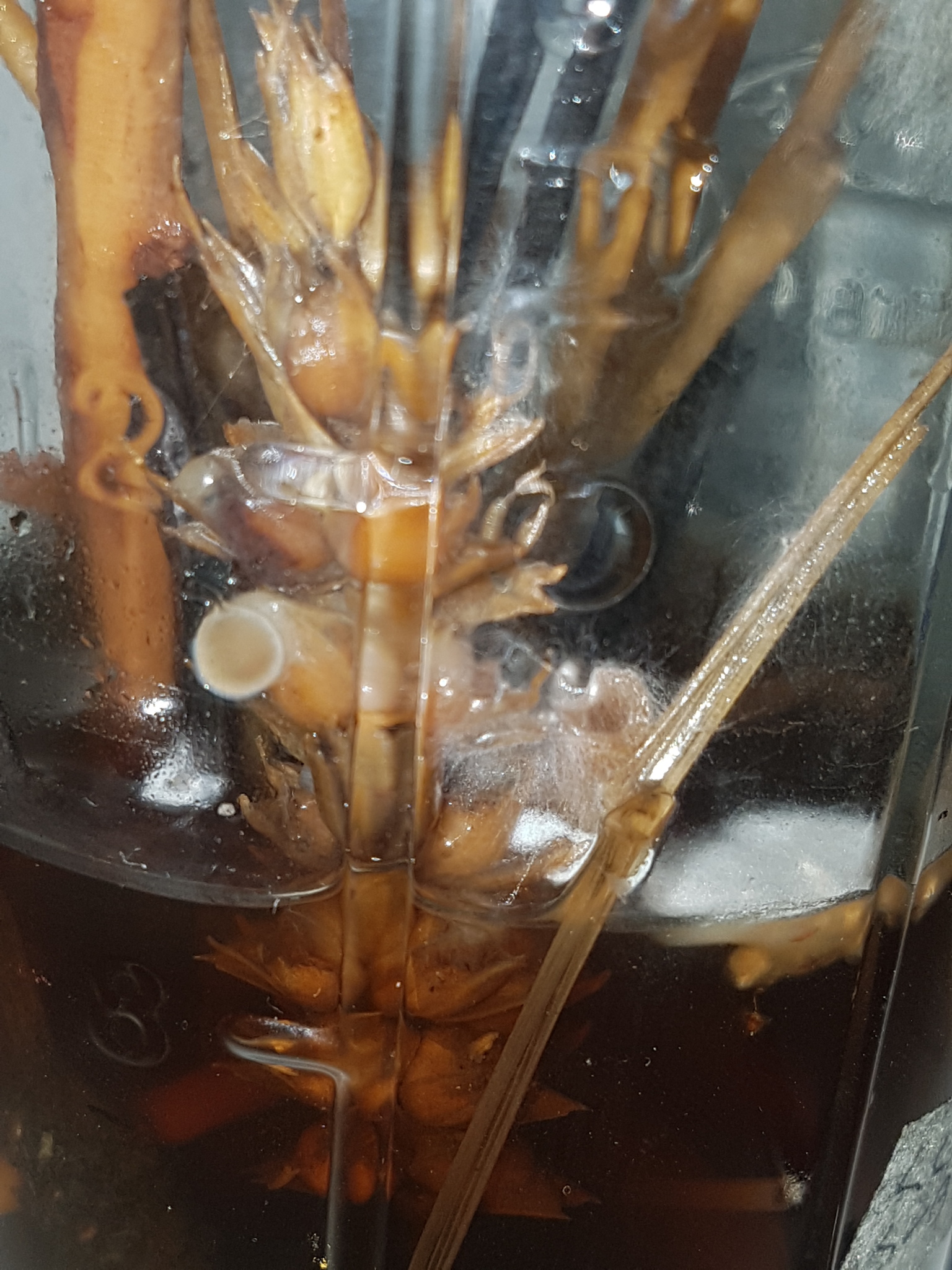
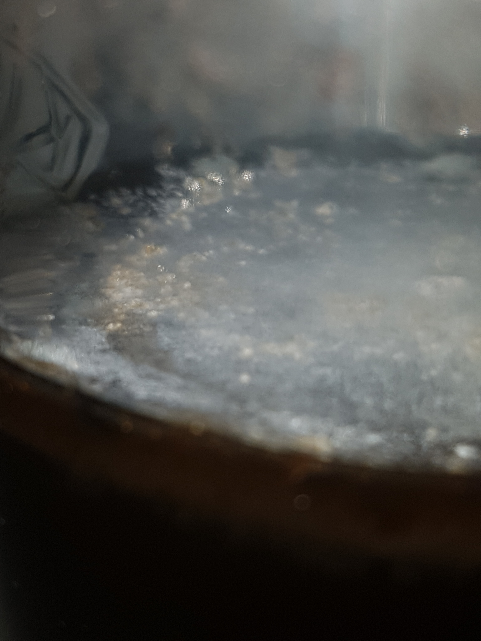
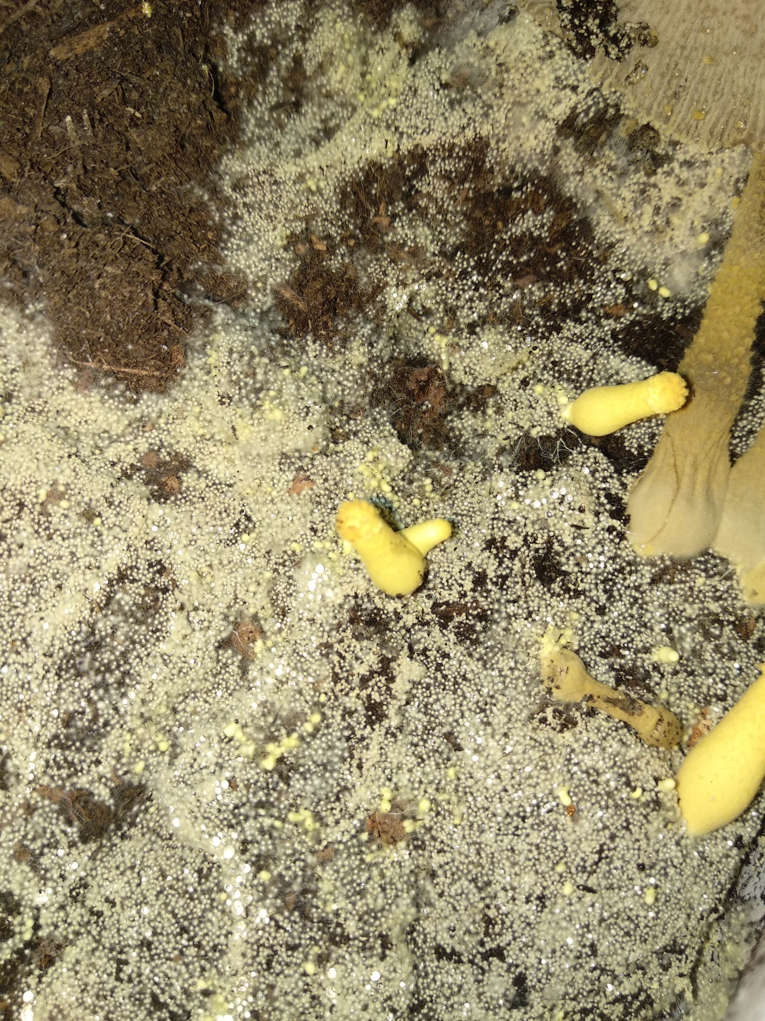
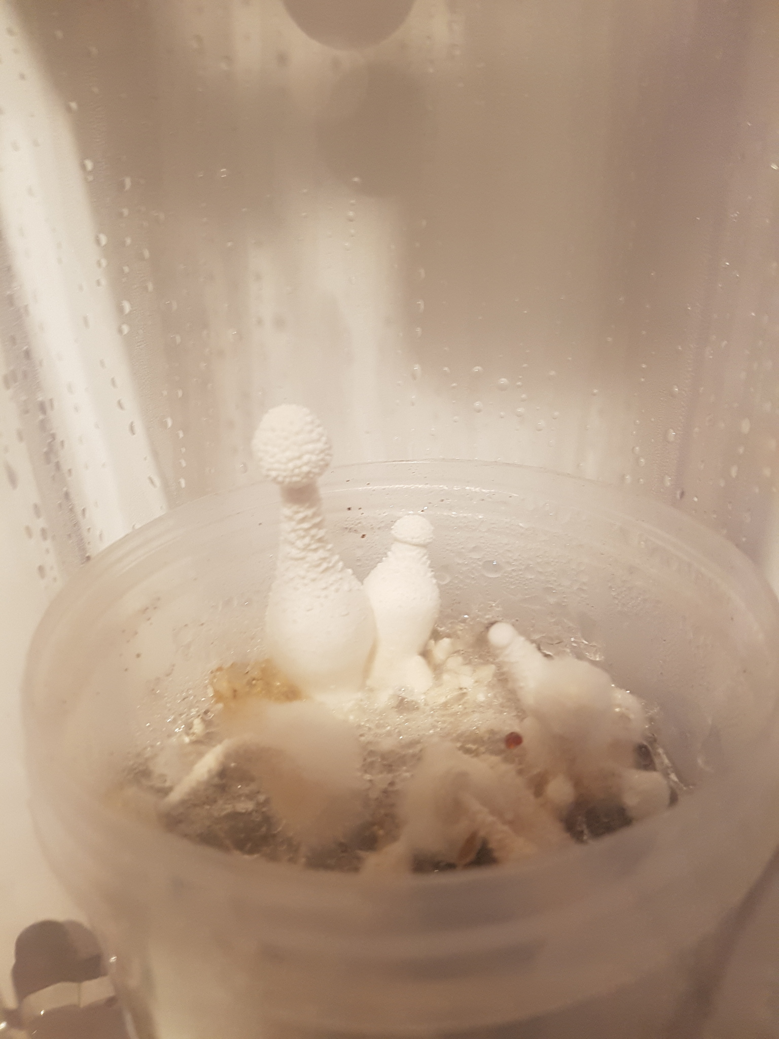
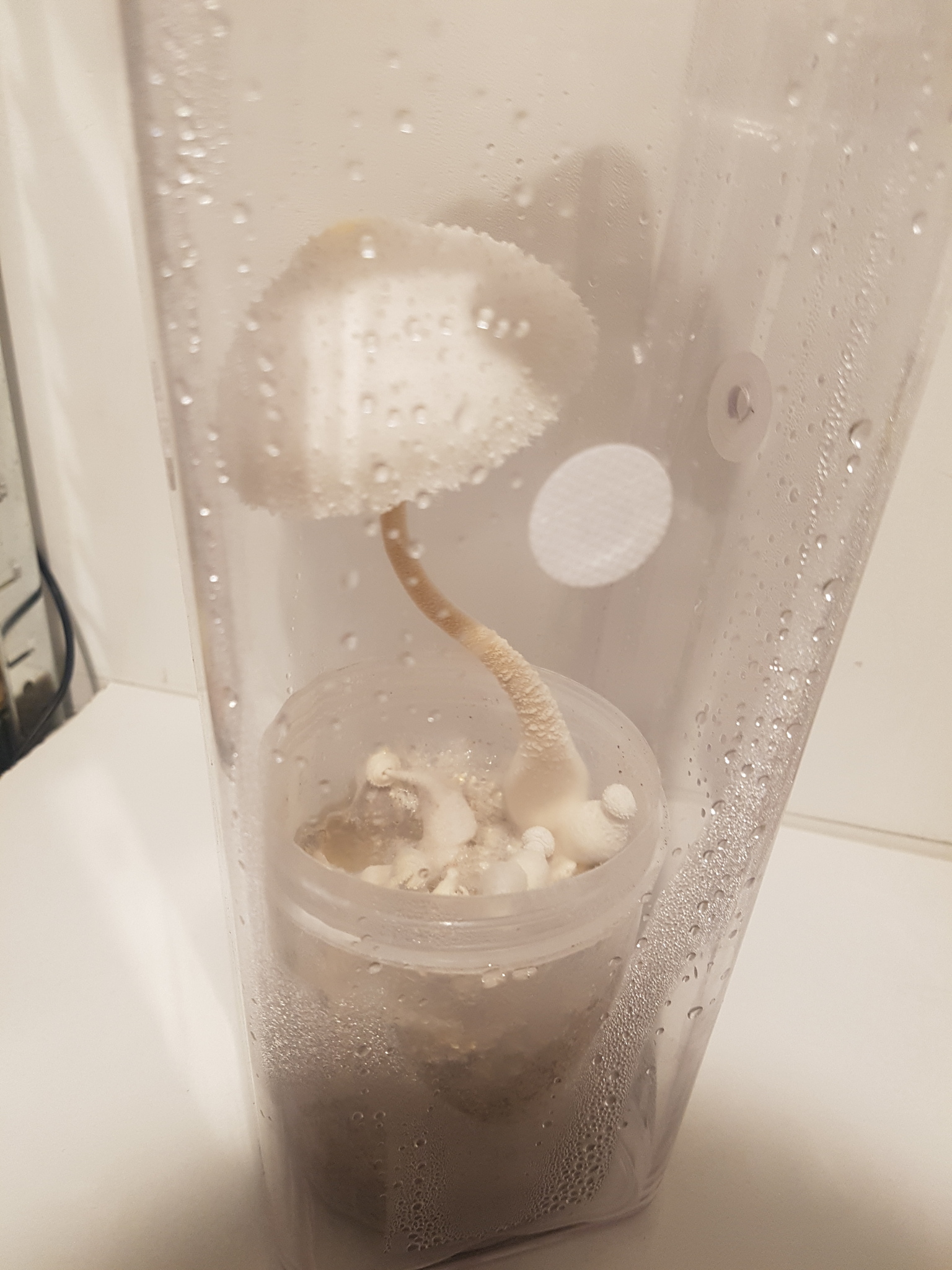
On 30/08/23 an over-mature mushroom was harvested from a 500ml jar with a substrate comprised of 50g potting soil, 15g coco coir, 10g wheat bran and 110ml water. The addition of wheat bran had resulted in faster colonisation in this jar as opposed to the ones with just soil and coir mixes however was not necessary to result in fruiting with mushrooms ultimately growing in all jars except for the one that was comprised of only coir.

The jar was opened and placed into a tall, clear plastic food storage container with four 0.3 µm filters covering 7mm holes to provide fresh air exchange. This did not allow much moisture to escape however and so condensation quickly built up resulting in water pooling on the surface of the substrate and in the bottom of the fruiting chamber. No contamination was evident but yellowish metabolites were visible in the liquid on the top of the substrate. When the jar was introduced into the fruiting chamber immature mushrooms were already present measuring a few cm in length. However upon dropping the jar down into the chamber the immature, unopened caps of some of these mushrooms simply fell off and even the undamaged ones aborted.
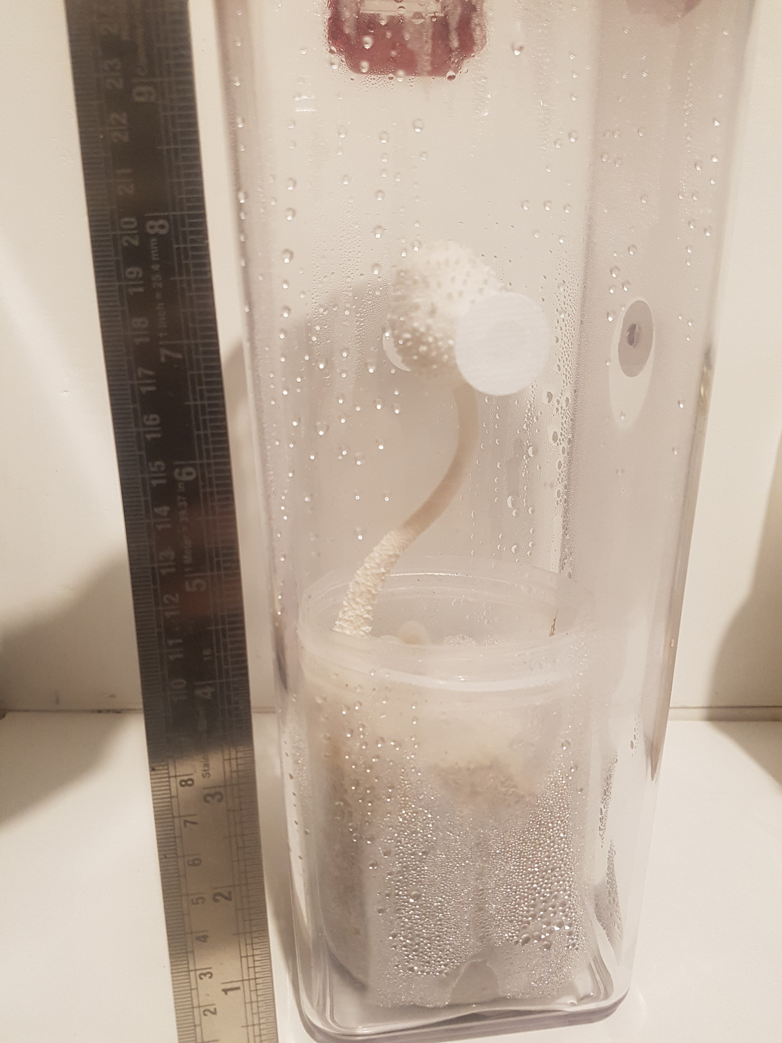
Fig. 17a - 26/08 22:30 Fig. 17b - 27/08 18:00 Fig. 17c - 27/08 22:30 Fig. 17d - 28/08 12:40 Fig. 17e - 29/08 14:00 Fig. 17f - 30/08 14:00

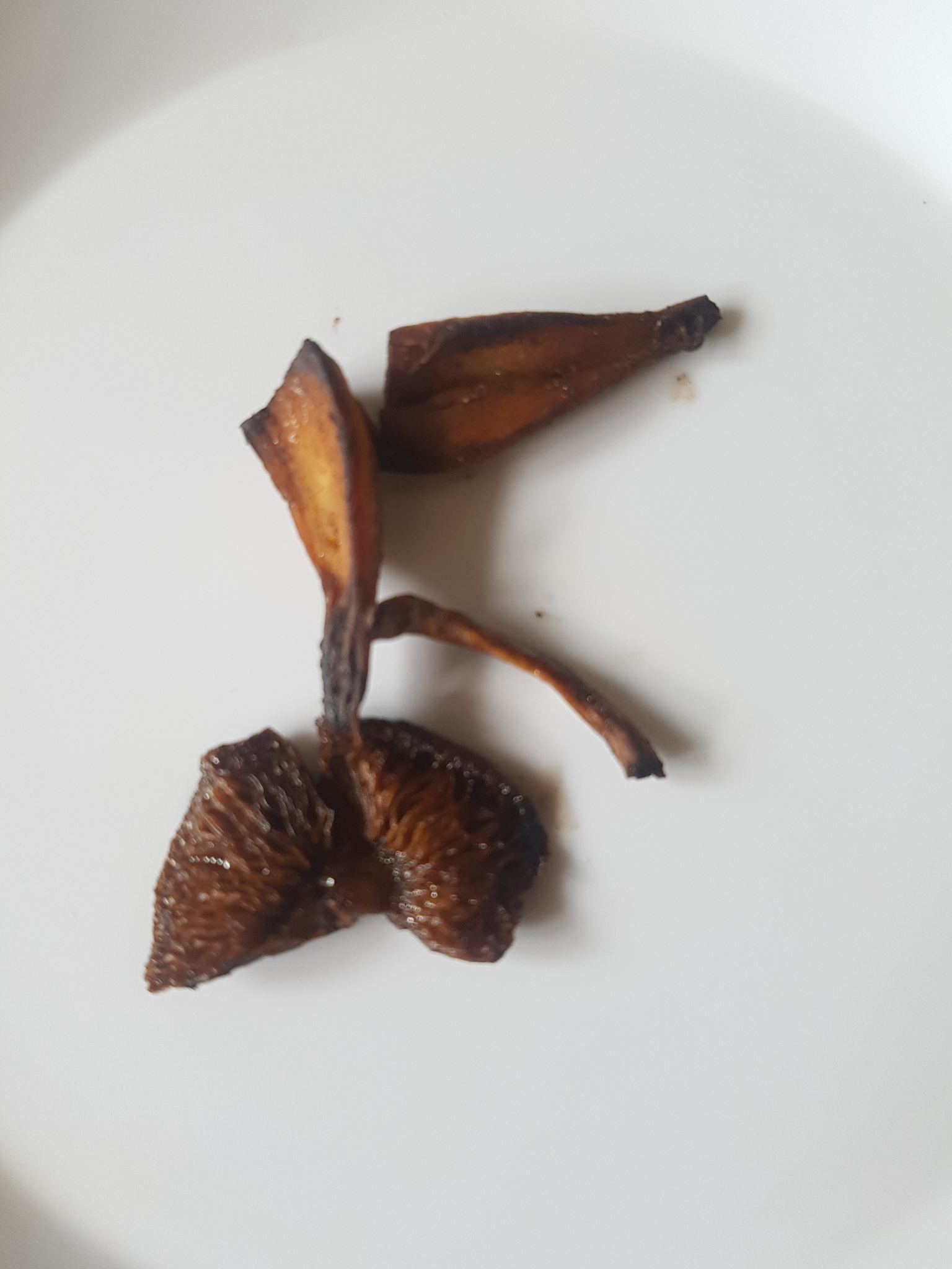
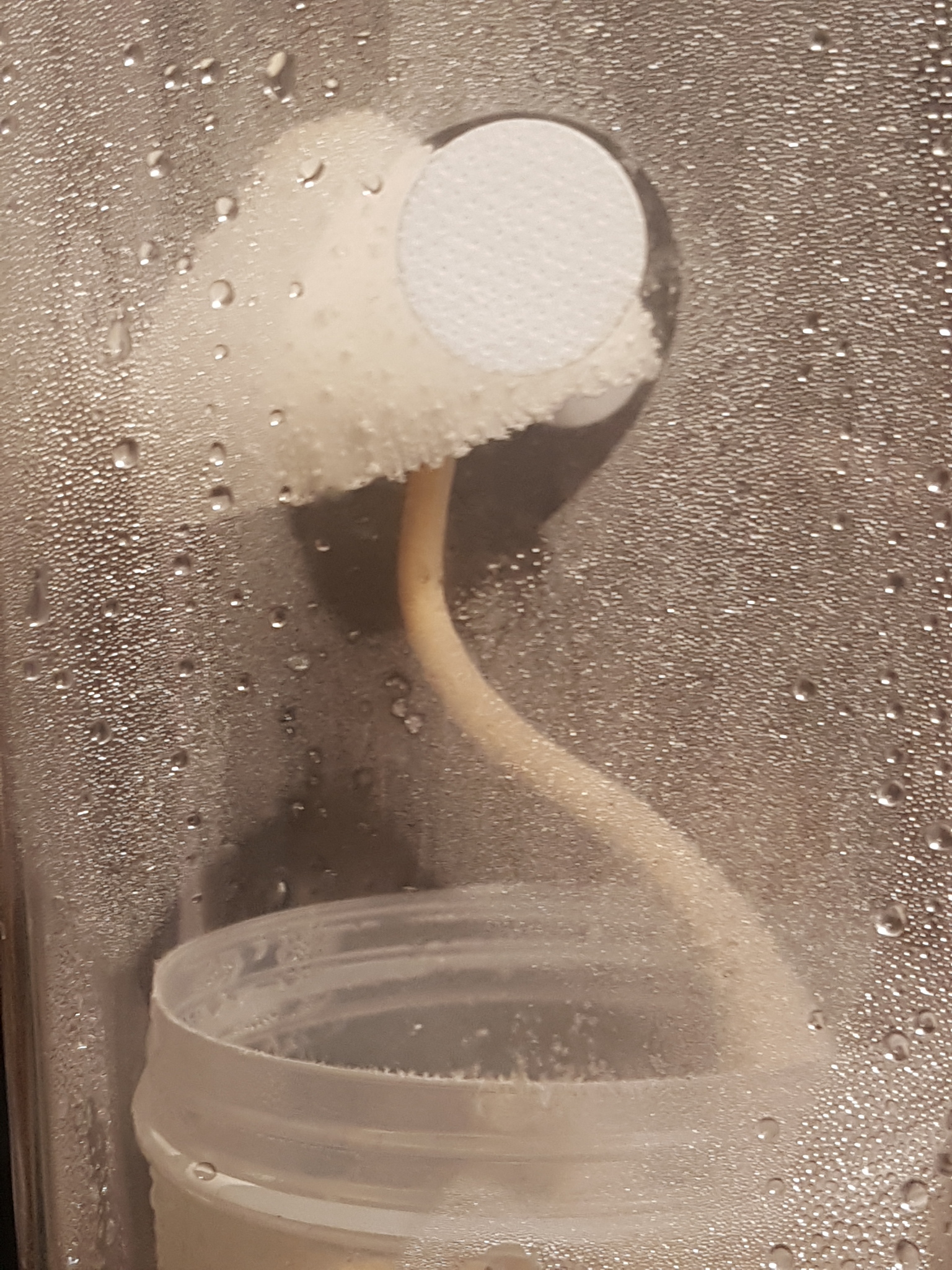
Figures 17a-17f shows the growth of this solitary mushroom over the course of four days, the second immature mushroom attached at the base to the larger one aborted and did not grow any more after the larger one began to develop. The cap did not expand and flatten as significantly as some other specimens but remained open for at least 51 hours before ultimately collapsing (additional photos were taken that have not been uploaded due to quality issues caused by the condensation). The window of time in which to harvest the mushrooms once the cap opens would appear to be around two days and this has likewise been observed in other jars where no mushrooms were harvested resulted in them deteriorating and ultimately smelling rather off putting.

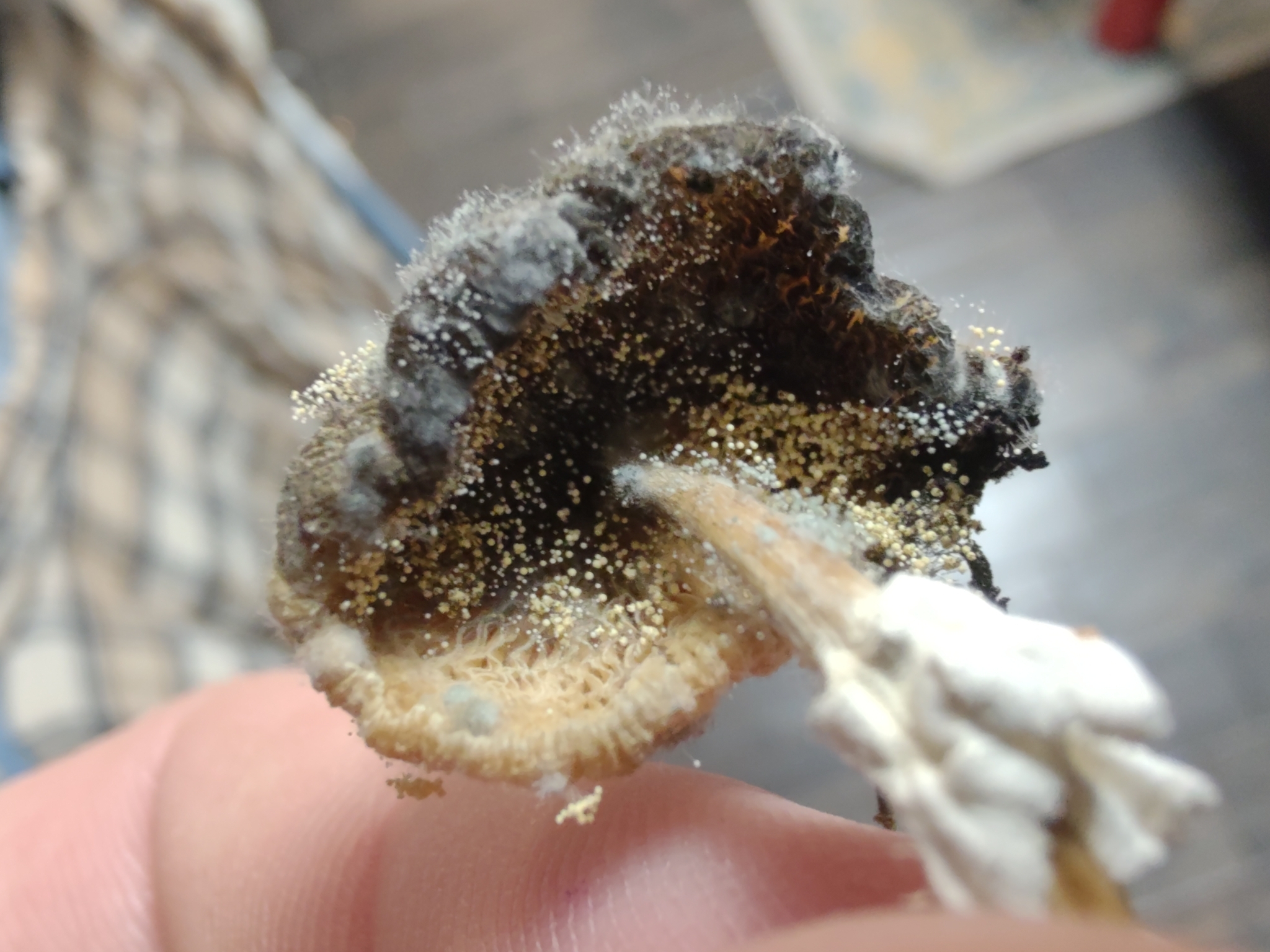
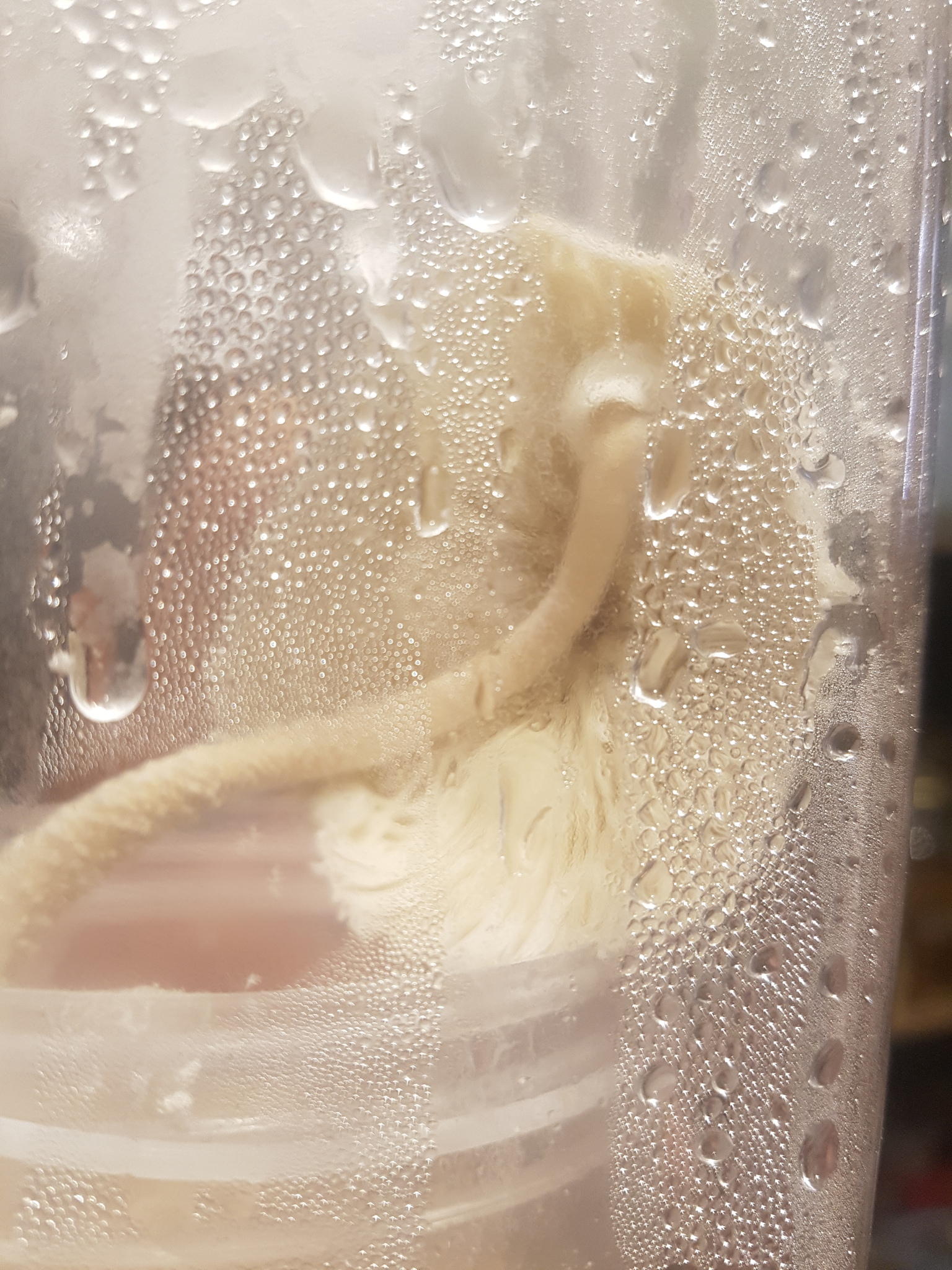
The last photo in this series shows the mushroom as it was harvested. It appeared as if the cap had detached entirely but in fact the stem had simply bent and it was still in one piece. This over-mature mushroom had a far more pronounced and unpleasant odour with the cap having a somewhat floral, but still unpleasant, scent and a much stronger and more unpleasant smell to the base of the stem and substrate which did indeed border on nauseating. The smell was off-putting enough to suggest that it would be no good to eat and so this mushroom was not cooked and consumed. After leaving it to sit in the open air for a couple of hours the floral smell was no longer present and all that remained was the unpleasant, chemical odour which if anything seemed stronger.

Despite the smell, the taste of the raw upper stem was not wholly unpleasant. It had an initial taste similar to the unpleasant smell with perhaps a very slight bitterness but it was not significant and it quickly gave way to a generally mushroomy taste that was reminiscent of the cooked taste of this mushroom. The cap however had a more unpleasant taste with more chemical overtones similar to the smell. The texture of the cap remained cottony and unappetising and whilst the stem had softened and become more water logged it still retained a reasonable texture, though squeezing it resulted in it yielding far easier than when fresh.

After tasting the mushroom and then smelling it again the smell appeared different. The cap smell seemed more pleasant and reminiscent of the taste but the base of the mushroom with the colonised substrate and sclerotia still attached retained the unpleasant smell. It is unclear if tasting the mushroom impacted the perception of the smell or if further maturation had occurred in the 20-30 minutes after tasting it.

The smell was compared against dried samples harvested on 11/08/23 which comprised of a small piece of cap and gills, a small section of the upper stem, the very bottom portion of the stem with some brown rice substrate and sclerotia attached and a few small primordia dissected on a slide. These are stored together in an air tight food grade plastic container and the smell was sampled by opening the container but not testing individual parts. The overall smell is very pleasant with a hint of the typical savoury smell found in many dried mushrooms but with a sweet, almost chocolate or toffee like smell on top of that.

This smell is not wholly dissimilar to the over-mature mushroom and when compared one after the other the same hints of fragrance can be noted. It appears that the more unpleasant smells come through as the mushroom ages and begins to deteriorate. This over-mature mushroom and another left to wither for even longer in another jar were stored in separate food grade plastic containers with the lids ajar to allow some drying. No noticeable decomposition occurred and they did slowly dry. They were sampled day by day by removing the lid and smelling the air trapped inside and the smell seemed more unpleasant each time, though not necessarily always the same perhaps due to the amount of time allowed between each sample. On some occasions the smell was strong, pungent and quite off putting but on others was simply unpleasant and a bit chemical in nature. It is different to the smell found in the substrate or immature mushrooms however which is not so unpleasant but more pungent.

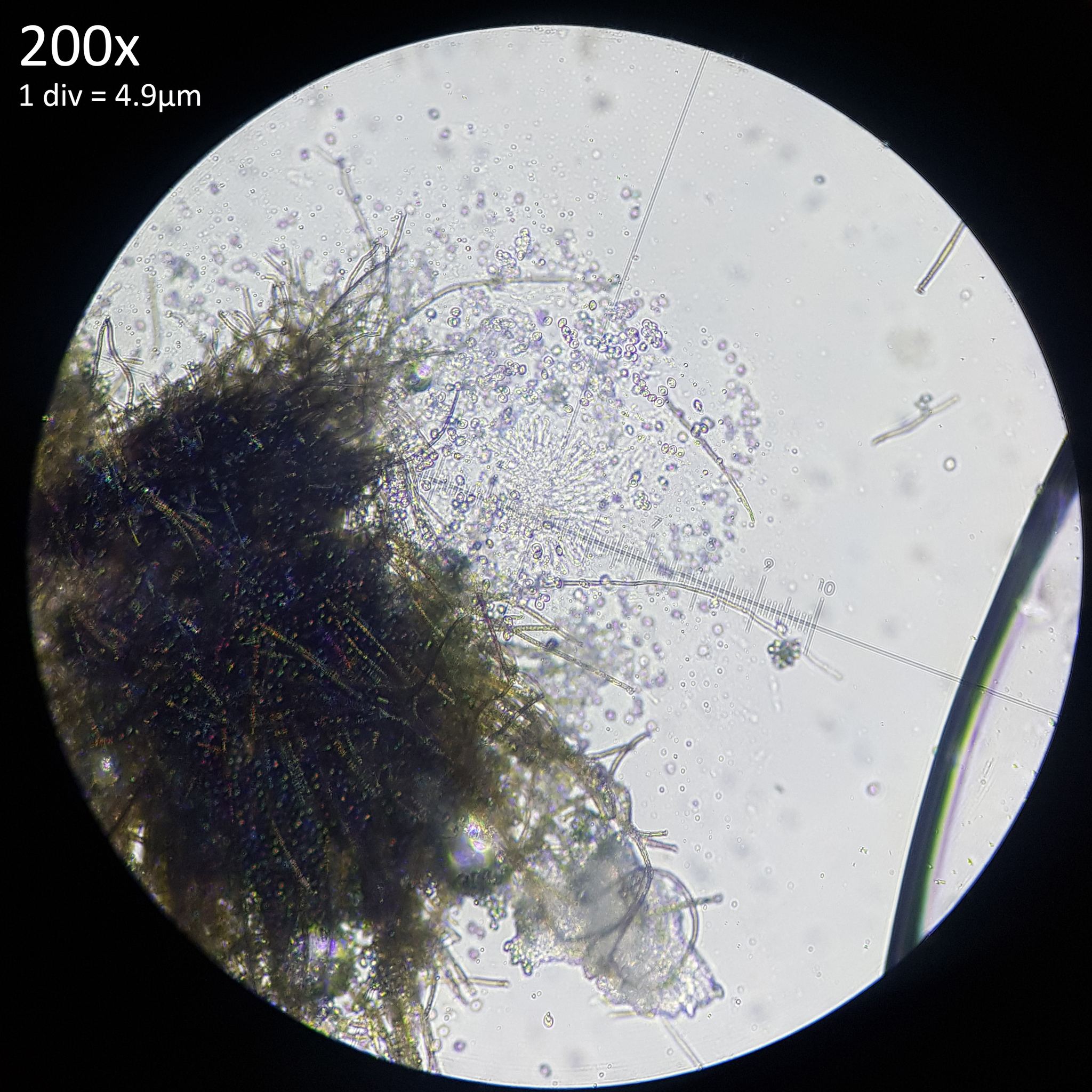
When a small piece of the soil substrate was examined under the microscope and cut in half to collect sclerotia for culturing, a strong smell was immediately noticeable, even whilst wearing a mask to avoid contamination. This smell isn't especially pleasant or unpleasant but simply comes across as chemical and a may be a little overpowering in larger samples. This piece of the substrate had been removed from the container an hour or two earlier and had sat in the open air but even so, upon exposing the interior the same pungent smell was noticeable as when the jar was opened initially. When a small piece of soil substrate with sclerotia, immature and aborted pins weighing 1g total was left out on the desk in a small room with little to no airflow or ventilation there was a noticeable smell upon entering the room.

This helps explain the variation noted in the descriptions of smell given by different authors. The mushrooms have a stronger, more pleasant smell when immature but this is masked by the far stronger, chemical smell of the myceliated substrate which clings to the base. When more mature and in their prime for picking the smell is not as significant and may not even be noticeable. It could easily vary from being described as generally mushroomy, pleasant but indistinct or simply having no smell. When over mature the smell becomes unpleasant and quite off putting with an initial floral smell that is lost to that unpleasant odour. Yet as they are left to dry the smell ultimately becomes more pleasant, appetising even.

The range of smells that are observable through cultivating this mushroom are really quite fascinating and whilst this description may go some way to explaining the variable descriptions given by others, it seems paltry compared to the experience as I cannot find the words to properly describe the smell at each stage of development.


6. Cultivation of tropical mushrooms as a matter of food security

This experiment in cultivating and consuming Leucocoprinus cretaceus was driven in part by my awareness of climate change and the concerns I have for the impact it poses to food supplies. It seemed worth exploring whether a mushroom that thrives in high temperatures and grows on readily available substrates could be grown for food.

Some issues that may potentially be faced with large scale cultivation are posed by the smell of the substrate, which is so surprisingly strong in just a small jar that it is hard to imagine what a warehouse sized grow would smell like. It could make for quite an overpowering environment to work in and it seems probable that the smell would lure in insects and pests. On the plus side however the spore production does appear to be far lower than with some commonly cultivated species and so health risks faced by the inhalation of large quantities of spores are probably reduced.

The presence of the sclerotia would also need to be addressed either by determining that they are safe to consume or formulating a removal method. With commercial cultivation of Agaricus bisporus it is common to simply cut the stem above the base to lose the bottom portion of the stem with the soil substrate that clings to it. This would be less than optimal for L. cretaceus as this is where the bulk of the flesh is found. Even if sclerotia do not pose a risk to consumption they would be less than ideal to cook as when heated on a metal surface they can be flung into the air and get lost. If they are not sufficiently damaged by the heat as to be rendered sterile then this could easily result in the mushrooms growing from floorboards or the base of skirting boards if water is spilled or otherwise leaks. Numerous observations on iNaturalist show the mushroom growing from beneath skirting boards or door frames and it seems possible that this may be due to sclerotia washing in with rainfall or floodwater. Since sclerotia production appears to be greatly reduced or entirely absent on substrates comprised of wood it may be viable to effectively eliminate sclerotia from the mushrooms with a top layer of wood on the substrate.

Some sclerotia would almost certainly survive in spent substrate so if this was sold as compost these mushrooms would become far more common and spread wherever this compost was used, similar to how L. birnbaumii spreads via potting soil and potted plants. It is probable that L. cretaceus already spreads in this manner given the numerous observations worldwide of it in potted plants and planters filled with commercial soil. However these observations are far, far fewer in number than those of L. birnbaumii and so for reasons that are not yet clear it would appear that L. cretaceus may be less proficient at spreading in this manner. Alternatively it could simply be that L. cretaceus has not been introduced into the potting soil/potted plant distribution network in such a widespread fashion.

Whilst it may be desirable to avoid sclerotia on the mushrooms destined for consumption their production could have other useful functionality. The carbon/nitrogen composition of the sclerotia is as yet unknown but it is probable that they are mostly comprised of carbon. As they are produced in enormous numbers such that they represent a sizeable weight of the spent substrate they could have a role in carbon sequestration. The sclerotia are very hard, durable and self-desiccate naturally and these traits appear to make them resistant to colonisation by mold or consumption by insects and mites. As such carbon locked away in sclerotia could be expected to remain there for a prolonged time, though mycelial growth may occur if they become wet. If sclerotia were treated as a sand-like aggregate and bonded together with a suitable resin they might make a viable construction material whilst also locking away carbon.

Leucocoprinus cretaceus does not appear to have been considered for cultivation before since the edibility of this species seems to have gone unnoticed. A 2021 review of the world's edible mushroom species lists Leucocoprinus birnbaumii as poisonous and Lc. cepistipes as conditionally edible and also includes Leucoagaricus americanus as conditionally edible, La. badhamii as poisonous and La. leucothites as edible.[30] Leucocoprinus cretaceus is not mentioned in this paper nor is it, or any Leucocoprinus species mentioned in a 2014 study on cultivating wild tropical mushrooms.[31]

However in Charles McIlvaine and Robert K. Macadam's 1900 'Toadstools, Mushrooms, Fungi, Edible and Poisonous: One Thousand American Fungi' L. cepaestipes is listed as edible and enjoyable when cooked in either its white or yellow form. Agaricus cretaceus is likewise listed as edible though again it is unclear which species this refers to.[32]

Nancy Smith Weber and Alexander H. Smith's 1985 'A Field Guide to Southern Mushrooms' also mentions an acquaintance of theirs consuming Leucocoprinus luteus several times without issue.[10] As recent sequences seem to suggest numerous yellow species which get identified as Leucocoprinus birnbaumii it is unclear which was consumed. It seems possible that reports on the toxicity of this species may be based on raw consumption with toxins that could be denatured when cooked. Alternatively it could be similar to other species in the Agaricaceae family which cause reactions for some people but not others. This too would also need to be ascertained for Leucocoprinus cretaceus by more people sampling them.

A 2019 review of mushroom cultivation in Bangladesh which focuses especially on Pleurotus species suggests that consumption of mushrooms may help address the malnutrition that is found to some degree in half of the population of the country. Based on yields produced, the optimal growing season for Pleurotus species was determined to be December-February with decreased growth from March onwards, moderate growth in April-July and minimum growth from August-October, increasing from November onwards. High summer temperatures are noted as one of the problems encountered with commercial cultivation.[33] A 2012 study on the cultivation of Agaricus bisporus in Bangladesh lists the problems faced by a sample of 30 producers with fly and cockroach issues being faced by 67% and contamination issues faced by 37%. Hot temperatures were faced by 40% of producers and amongst the technical issues that arose air conditioner problems were encountered by 50% and electricity by 60%.[34]

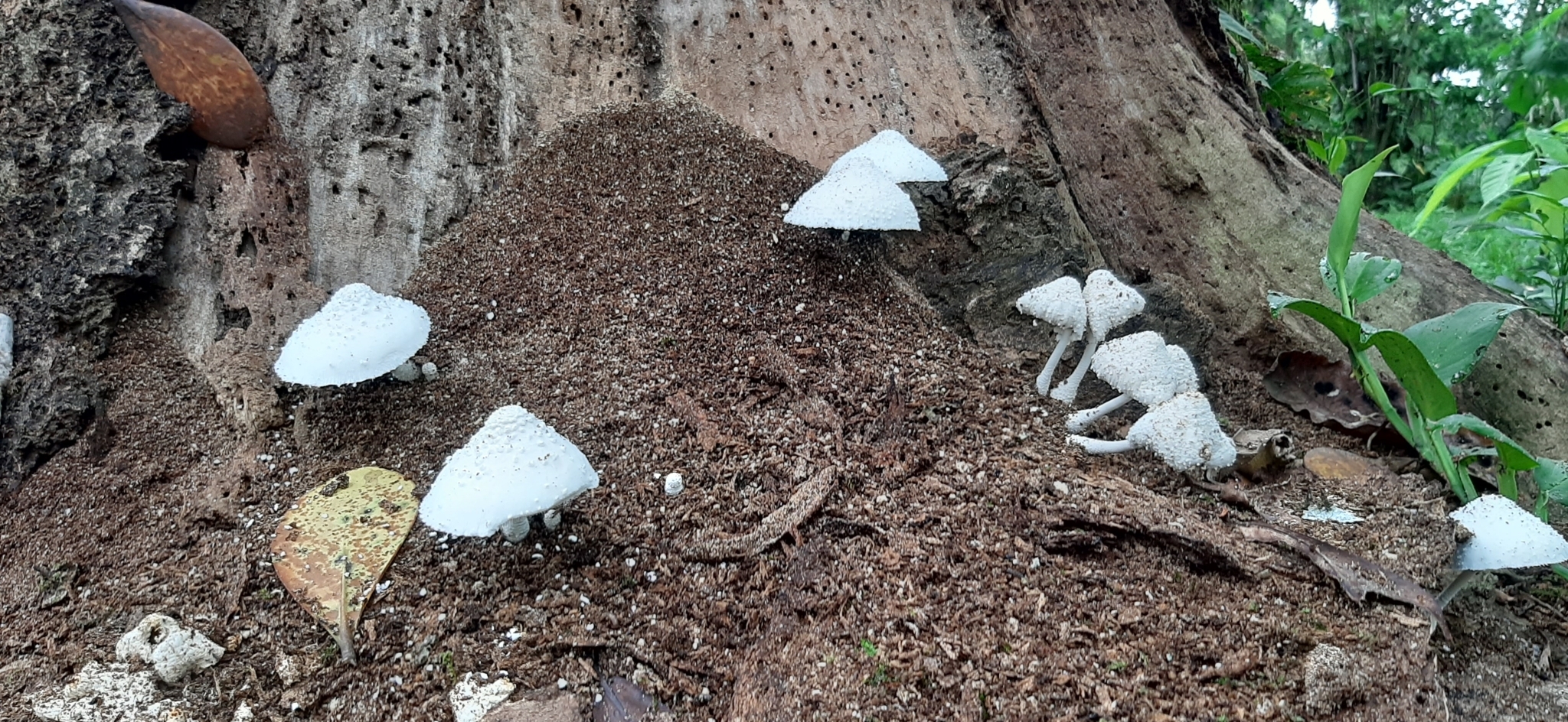
It seems probable that cultivation of L. cretaceus could address the issues faced in regards to heat and grow during the summer months in Bangladesh easily without air conditioning. iNaturalist observations for Leucocoprinus cretaceus in Bangladesh are few and temperature data from the region is not always available. Observation 105564290 by @biodiversityofbangladesh shows a nice flush of mushrooms in multiple stages of maturity spread across the floor of a forest near Khulna in Bangladesh on the 11th of June, 2021. However timeanddate.com is missing the weather data for a lot of Bangladesh during much of that year and so it isn't clear what the environmental conditions were and at best it can be approximated based on the 33°C recorded in Kolkata that day. Observations from India are greater in number and there are several from West Bengal for which temperature data is available.

Observation 172145361 by @wild_wild_nature was recorded on the 10/06/23 fruiting on wood near Kolkata. Time and date records a high temperature of 34°C and a low of 29°C on this day with similar highs and lows in the days before and after.

Observation 160716481 by @puspak_roy from 09/05/23 shows a moderately large flush fruiting on soil, compost or possibly manure in Bardhaman University. Some of the pins appear to be aborting and some larger mushrooms seem dry though several look like they are developing normally. Time and date records a high temperature of 42°C at midday and a low of 29°C this day with a high of 42°C on the previous day, 40°C before that and temperatures in the mid to high 30s in the previous week. Leucocoprinus cretaceus can grow to full maturity in just a few days so this suggests the mushrooms were able to develop despite these very high temperatures.

Observation 171760762 by @aniruddha_singhamahapatra from 08/07/23 shows a large flush of fully developed mushrooms growing on soil or woody debris beneath a forest canopy in the district of Bankura. Time and date records a high of 33°C and a low of 27°C this day with the same temperatures the day before, some rain in the preceding three days and temperatures in the mid to high 30s in the week prior.

Observation 95750750 by @banerjeed from 20/06/23 shows immature pins forming in a plant pot or planter in Asansol. Time and date records a high of 33°C and a low of 25°C this day with temperatures in the high 20s the week prior.

Observation 82690927 by @earthanki from 12/06/21 shows a single well developed specimen in a plant pot that was noted as growing in just one afternoon during the rainy season in Sonitpur, Assam, India. Time and date records a high of 33°C and a low of 27°C this day with a high of 29°C and rain the day before and rain and storms in the week prior with temperatures in the high 20s to low 30s.

A total of 200 observations of Leucocoprinus cretaceus worldwide from iNaturalist with the temperature data were collatedhere revealing many were growing at temperatures above 30°C with the highest temperature of 42°C. Whilst they will grow adequately at room temperatures in the mid 20s it would appear that their temperature tolerance extends far beyond this giving them potential for cultivation in hot climates.

7. Conclusion

Leucocoprinus cretaceus appears to be a perfectly edible mushroom with no side effects documented thus far however as the sample size for this experiment was only two people it would be prudent for others to sample this species and confirm that they have no reactions either.

Whilst these edibility tests cannot eliminate the possibility of longer term side effects these do not seem likely given that toxicity in members of the Agaricaceae family are gastrointestinal in nature with symptoms that emerge quickly rather than ones which bioaccumulate. The edibility of the sclerotia still needs to be explored as whilst it seems unlikely that they would be toxic or pose any health risk, the subject of sclerotia in Leucocoprinus species has not been sufficiently documented and so little is known about them. Given their significant hardness and resistance to chemicals it seems possible that they are not digestible and probable that they are best avoided. However the sclerotia are relatively easy to remove from the mushrooms with most dislodging readily when brushed off. One solution to reducing the amount of sclerotia present stuck to the base of the mushroom is adding a layer of wood chips to the top surface of the substrate. This seems to result in the production of fewer sclerotia compared to higher nutrient substrates like wheat bran or rice and is easier to clean from the mushrooms than soil.

The species is very easy to grow, performs well on numerous subtrates and will grow well simply on potting soil or soil and coir mixes that enable higher water content. Excellent growth is also observed on plant material like leaves and stems and so it may have potential for composting garden waste. It fruits so readily and quickly that most substrate tests resulted in mushrooms growing in the jar, often before fully colonising the substrate, meanwhile other Leucocoprinus species stored beside them in similar jars of substrate have yet to fruit at all. As yet however mushroom growth is sporadic and somewhat unpredictable with jars moved into fruiting chambers with heating sitting idle for prolonged periods meanwhile neglected jars left at room temperature may intermittently fruit when least expected. As such more experimentation will be required to assess the optimal fruiting conditions regarding temperature, humidity, air flow and lighting in order to improve yields and achieve predictable growth.


References

  • [1] Noordeloos, M.E.; Kuyper, TH.W.; Vellinga, E.C. (2001). "Flora Agaricina Neerlandica Critical monographs on families of agarics and boleti occurring in the Netherlands vol. 5".
  • [2] Beeli, M. (1927). "Contribution a L'étude de la Flore Mycologique du Congo: II". Bulletin de la Société Royale de Botanique de Belgique / Bulletin van de Koninklijke Belgische Botanische Vereniging. 59 (2): 101–112.
  • [3] Heinemann, P.; Goossens-Fontana, M. (1956). "Flore iconographique des Champignons du Congo".
  • [4] Bourniquet, Gilbert. (1943). "Champignons Nouveaux de Madagascar". Bulletin de l'Académie Malgache. 25, p. 16.
  • [5]Vellinga, E.C. (2009). "Nomenclatural Overview of Lepiotaceous Fungi (Agaricaceae) Version 4.7".
  • [6] Smith, Helen V.; Weber, Nancy S. (1982). "Selected Species of Leucocoprinus from the Southeastern United States". Contributions from the University of Michigan Herbarium. Vol. 15. pp. 300–301.
  • [7] Haller, R. (1950). "Beitrag zur Kenntnis der aargauischen Pilzflora". Mitteilungen der Aargauischen Naturforschenden Gesellschaft. Band 23.
  • [8] Locquin, Marcel. (1945). "Notes sur les Lépiotes II". Bulletin Menseul de Société Linnéenne de Lyon, 14ᵉ année, n°5, pp. 89-100.
  • [9] Josserand, Marcel. (1955). "Notes critiques sur quelques chapignons". Bulletin trimestriel de la Société mycologique de France, Vol. 71, pp. 68-74.
  • [10]Weber, Nancy Smith.; Smith, Alexander H.; Guravich, Dan. (1985) "A Field Guide to Southern Mushrooms". pp. 169-170.
  • [11] Bulliard, Pierre. (1788). "Herbier de la France, ou Collection complette des plantes indigenes de ce royaume". pl. 374.
  • [12] Elias, Fries. (1836). "Anteckningar öfver de i Sverige växande ätliga Svampar: 1-8". p. 34.
  • [13] Kummer, Paul. (1871). "Der Führer in die Pilzkunde". pp. 73-74.
  • [14] Gillet, Claude-Casimir. (1874). "Les Hyménomycètes, ou, Description de tous les champignons (fungi)". p. 563.
  • [15] Vittadini, Carlo. (1835). "Descrizione dei funghi mangerecci più comuni dell'Italia e de' velenosi che possono co' medesimi confondersi". p. 347.
  • [16] Jean-Jacques Paulet. (1793). "Traité des champignons". p. 359.
  • [17] Negrini, Valentina M.; Lonati, Davide; Pappalardo, Alfio; Schicchi, Azzurra; Petrolini, Valeria M.; Bernasconi, Lucia; Brolli, Benedetta; Grazioli, Cristina; Alonzo, Elena; Locatelli, Carlo A. (2022). "Case series of Chlorophyllum molybdites intoxication in Sicily: an "alien" mushroom species in Europe". Annali dell'Istituto Superiore Di Sanita. 58 (3), pp. 213–217.
  • [18] University of Veterinary Medicine Budapest. (2022). "Increasing number of intoxications by the mushroom Chlorophyllum molybdites in Italy". dfi.univet.hu
  • [19] Léveillé, Joseph Henri; Paulet, Jean-Jacques. (1855). "Iconographie des champignons de Paulet". Pl. CLXIII.
  • [20] Léveillé, Joseph Henri; Paulet, Jean-Jacques. (1855). "Iconographie des champignons de Paulet". p. 89.
  • [21] Mattirolo, Oreste. (1918). "Sul Ciclo di Sviluppo di due Specie Scleroziate del Gen. Lepiota Fr. e Sulle Loro Affini". Memorie della R. Accademia dei Lincei, Serie Quinta, Volume XII, Fascicolo XI. pp. 537-572.
  • [22] Elisha Hard, Miron. (1908). "The mushroom, edible and otherwise". figure 37, p. 54.
  • [23] Sowerby, James. (1797). "Coloured Figures of English Fungi Or Mushrooms". tab. 2.
  • [24] Hornemann, Jens Wilken. (1819–1823). "Floræ Danicæ Iconum". vol. 10, fasc. XXX, p. 13 & tab. MDCCXCVIII.
  • [25] Greville, Robert Kaye. (1828). "Scottish Cryptogamic Flora". Vol. VI, tab. 333.
  • [26] Barla, Jean-Baptiste. (1889). "Flore mycologique illustrée: les champignons des Alpes-Maritimes". fasc. 2, pl. 15
  • [27] Patouillard, N. (1889). "Tabulae analyticae fungorum". fasc. VII, fig. 612.
  • [28] Gillet, Claude-Casimir. (1874). "Les Hyménomycètes, ou, Description de tous les champignons (fungi)". vol.2, pl. 413.
  • [29] Cooke, Mordecai Cubitt. (1881). "Illustrations of British Fungi". vol.8, pl. 942.
  • [30] Li, Huili; Tian, yang; Menolii Jr., Nelson; Ye, Lei. (2021). "Reviewing the world's edible mushroom species: A new evidence-based classification system". Comprehensive Reviews in Food Science and Food Safety.
  • [31] Thawthong, Anan; Karunarathna, Samantha C.; Thongklang, Naritsada; Chukeatirote, Ekachai; (2014). "Discovering and Domesticating Wild Tropical Cultivatable Mushrooms". Chiang Mai Journal of Science, vol. 41, pp. 731-764.
  • [32] McIlvaine, Charles; Macadam, Robert K. (1900). "Toadstools, Mushrooms, Fungi, Edible and Poisonous: One Thousand American Fungi". pp. 46-47.
  • [33] Ferdousi, Jannatul; Riyadh, Zabid Al; Hossain (Md.), Iqbal; Saha, Satya Ranjan; Zakaria, Mohammad. (2019). "Mushroom Production Benefits, Status, Challenges and Opportunities in Bangladesh: A Review". Annual Research & Review in Biology, 34 (6), pp. 1-13.
  • [34] Barmon, Basanta Kumar; Abbasi, Parvez; Mamun, Al; Sharmin, Imrana; (2012). "Economics of Mushroom (Agaricus bisporus) Production in a Selected Upazila of Bangladesh". The Agriculturists 10 (2), pp. 77-89.

  • Publicado el marzo 19, 2024 10:47 MAÑANA por mycomutant mycomutant | 1 comentario | Deja un comentario

    22 de diciembre de 2023

    A preliminary investigation of sclerotia in Leucocoprinus cretaceus

    Abstract

    Sclerotia of Leucocoprinus cretaceus were observed growing in agar cultures and cultivated in various substrates with abundant sclerotia growth always preceding any mushroom development. The sclerotia are roughly spherical, ovoid or elliptical and range in size from (135) 245 - 735 (1150) µm with a mean average size of 475 x 413 µm. Immature sclerotia have pale yellow gelatinous flesh encased in a web like mycelial envelope similar to those of Leucocoprinus birnbaumii however at maturity they develop a hard, dark brown or black rind and become free from the mycelial envelope. The sclerotia are hard, durable structures that require force to cut or crush and new mycelial growth is easily cultured from loose sclerotia on agar, in solid substrates and in liquid. Sclerotia development can start within 1-2 weeks of inoculating a substrate and new sclerotia are produced constantly as the mycelium spreads with high nutrient substrates ultimately becoming filled with hundreds of thousands of sclerotia. The vast numbers of sclerotia appear to provide some resilience against mold and mycophagous pests but may also suggest that they serve not just as a means of surviving adverse conditions but also as an effective delivery mechanism for propagating the fungus. Sclerotia are readily separated from the substrate in clusters or individually and are prone to adhering to surfaces. Before losing the mycelial envelope the sclerotia are extremely hydrophobic resulting in them floating away in water and clinging to anything solid which could serve as a potential means of distribution in tropical rainforests via flood water. Considering the seed or insect egg like nature of the sclerotia as well as the very strong smell of the myceliated substrate possible interactions with animals including birds, termites and ants are also explored.


    Fig. 1 - Maturing sclerotia of Leucocoprinus cretaceus cultured on brown rice.

    1. Introduction

    Sclerotia are a commonly observed feature in Leucocoprinus birnbaumii and are routinely present in observations of this species from houseplants either with or without the associated mushrooms. It seems probable that the abundant sclerotia assist this species in spreading via potted plants and compost as they would be easily transferred between pots when plants are transplanted or repotted.

    Leucocoprinus cretaceus is also found in plant pots all around the world however to date fewer than a hundred observations of this species from plant pots have been collected on iNaturalist as opposed to the thousands from L. birnbaumii. Even taking into account the likelihood of multiple species being identified as L. birnbaumii it is still substantially more common to find in plant pots.

    Culturing Leucocoprinus cretaceus has demonstrated that it also produces very large quantities of sclerotia and is able to grow well in substrates with multipurpose compost and coir. This could therefore call into question the presumed role the sclerotia play in facilitating spread through plant pots. ie. If both species favour the warmth, produce sclerotia abundantly, grow well in potting soil and have global distribution then why is one species far more common in plant pots?

    It is possible that the answer lies in the observable differences between the sclerotia of these species and the differences in the growth behaviour. However without knowing what functionality the sclerotia provide these species in nature it would seem difficult to know how they function in plant pots. The more intriguing questions therefore would be what role the sclerotia play in the survival of these species in nature and what natural conditions is the artificial environment of a potted plant simulating?

    In captivity the sclerotia of Leucocoprinus cretaceus have proven to be an extremely effective means of propagation. The vast numbers that they are produced in and the properties they possess are suggestive of a role in nature not simply as a means of laying dormant and surviving but actively spreading through the environment and exploiting the unique ecological niches of their native habitat.

    The following study was initiated prior to carrying out a detailed review of the literature on sclerotia in Leucocoprinus species with the review conducted in order to see which species sclerotia had previously been documented in. The findings are presented in A review of the literature on sclerotia in Leucocoprinus species and other Agaricales. Conducting this review after having made so many observations of the sclerotia in Leucocoprinus cretaceus proved very advantageous with many 'aha' moments upon finding similar traits noted in other species. However no previous documentation of sclerotia in Leucocoprinus cretaceus was found and so this trait may not have been discussed previously with this species. The most comprehensive study on sclerotia in Leucocoprinus species appears to be Oreste Mattirolo's 1918 work however Mattirolo was unfortunately not successful in culturing these species due to contamination and he did not find sclerotia in L. cretaceus, stating that it did not produce them.[1]


    2. Sclerotia in Leucocoprinus cretaceus

    Sclerotia in L. cretaceus initially appear similar to those of L. birnbaumii, only without the yellow mycelium, but they soon darken and develop a brown/black colour as they mature. This darker colouration becomes apparent as they start to emerge from the mycelial envelope (fig. 1). At this stage of development they are small, hard structures with an approximate size range of 245 - 735 µm. The larger figures in this range tended to represent the length of non-spherical sclerotia with a narrower width. The average size was around 440 - 490 µm from samples measured growing on rice and agar which either still had the white mycelial envelope or were starting to lose it showing the black surface below.

    If they are isolated and have enough space to grow they form almost spherically or are ovoid or elliptical with relatively symmetrical proportions. However in the most densely clustered growth in which maybe 50 sclerotia were found on a single grain of the brown rice substrate they deform where they touch each other. Some are left with almost geometric looking shapes and flat edges. Subsequently measuring in situ is complicated by sometimes being unable to tell where one ends and another begins and in isolation by the non uniform shape with flattened surfaces.

    Fig. 2 - Mature sclerotia Fig. 3 - Dissected sclerotia in 70% isopropyl alcohol

    When sclerotia were again harvested from this brown rice substrate one month later as well as from a jar containing a soil substrate they were almost completely black having lost the mycelial envelope (fig. 2). The shapes appeared more irregular and shrivelled as if they had shrunk when drying. 70 sclerotia were isolated and measured with a range of 270 - 685 x 270 - 610 µm. The mean value was 475 x 413 µm, the mode was 490 x 367.5 µm and the median 465.5 x 416.5 µm. One larger specimen was found that was excluded from this range as it appeared to be two sclerotia joined in the middle with a narrower portion. This was 735 µm long with a width of 415 at one end and 345 µm at the other. Other specimens were found that appeared as if two sclerotia had formed together hemispherically with a distinct line where they touched which gave the appearance of a coffee bean. The larger measurements in the initial count of the less mature specimens may have been sclerotia that were likewise stuck together with this fact hidden by the mycelial envelope.

    Application of 70% isopropyl alcohol aided in the dissection of the mature sclerotia and revealed a yellow interior which is clearly distinct from the darker exterior surface (fig. 3). However a colour change was noted in the isopropyl alcohol with the black exterior becoming more reddish brown with gold flecks and such a deep yellow interior was not so noticeable with or without other reagents. This exterior dark layer measured ~5-10 µm thick and was not so readily distinguished and measured without the application of the isopropyl alcohol.

    In high nutrient substrates sclerotia are produced quickly and in great abundance. When a sterile substrate jar consisting of 60g brown rice and 90 ml rainwater was inoculated from liquid culture and left at room temperature sclerotia development was observed 9 days later and the substrate was deemed to be fully colonised by mycelium 23 days after inoculation, though new sclerotia growth continued even as mushrooms developed. The majority of the substrate was dissected 4 months after inoculation with some small amounts having been removed along the way as mushrooms were harvested or as donor material was taken to inoculate additional substrates.

    The sclerotia appeared most densely clustered and numerous on the exterior surface though were also found throughout the entirety of the 7cm diameter x ~6.5 cm height substrate. Breaking up the myceliated rice by hand and sieving it was sufficient to separate the majority of the sclerotia though many remained in amongst the rice and were not readily removed without accumulating more rice debris. Based on appearance, perhaps around 10% of the sclerotia remained in the rice and the recovered sclerotia also contained some fine debris. Additionally some dark brown to black irregularly shaped clumps of hard material that measured 0.5-1cm were found amongst the rice and were encrusted with numerous sclerotia that could not be easily removed. These clumps may have been pseudosclerotia and appeared to have their own brittle rind, though they were so densely packed with sclerotia that at this level of maturity it was difficult to distinguish any features. Sclerotia were not recovered from these structures though the brittle rind of the pseudosclerotia did contribute to the debris mixed in amongst the collected sclerotia.

    Before drying the collected material had a volume of approximately 35ml and weighed 19g, though some drying occurred during the time it took to collect the material. After one week of drying in the open air this material dried to approximately 10.5g. Some obvious debris like tiny immature primordia were removed manually and a minimal amount of larger material that did not pass through a 955-1,029µm mesh was discarded as debris as few sclerotia were present. The remaining material was then passed through a series of mesh screens and separated into size ranges. The meshes were measured under the microscope to find the range in hole sizes since they differed quite greatly to the size they were sold as.

    The 0.875g of material which passed through the 294-343µm mesh contained a lot of fine debris and powder which made it impractical to estimate a quantity of sclerotia. The smallest sclerotia found in this material were ~150 x 135 µm and appeared to have the colouration of mature sclerotia. The 1.235g of material which passed through the 955-1,029µm mesh but was caught by the 637-686µm mesh appeared to be approximately half debris so was likewise not included in the estimate.

    Microscopic examination of this material found some sclerotia that were much smaller than this range though clustered together or stuck to the debris. Some uniformly shaped sclerotia in the range of 800-900 µm were found in this material but many others were irregularly shaped conjoined pairs of sclerotia or clusters that appeared like several sclerotia that had formed together and merged. The largest sclerotia found ranged from 1,000-1150µm though many of these were highly irregular in shape and clearly the result of numerous sclerotia merging together so firmly as to become inseparable. A few were found with roughly uniform proportions which may have been single sclerotium though some appeared segmented and tightly stuck together such that it was uncertain.

    Debris was also present in the other size groups though was much less significant and represented only a small amount of the total material. The bulk of the material was larger than 294-343µm and smaller than 588-637µm. This was divided into three size groups (table. 1) with the 2.74g, 2.415g and 2.88g of each group containing mostly sclerotia with minimal debris. For each of these three groups five 0.1g (±0.005g) samples were weighed out, the material was then poured onto a white plastic tray in small portions at a time and manually counted with the naked eye. One difficulty with this approach was that some sclerotia appeared like clusters with irregular shapes and when rolled between the fingers would break up into numerous small sclerotia. Others that appeared similar however would not break up and may have been conjoined. As such the counts could not be expected to be exact.

    Minimum mesh size Maximum mesh size Weight collected (±0.005g) Sclerotia quantity in 0.1g (±0.005g) sample Estimated quantity range Estimated quantity average
    N/A 294-343µm 0.875g N/A N/A N/A
    294-343µm 441-490µm 2.74g 1,954 - 2,438 53,539 - 66,801 60,860
    441-490µm 490-539µm 2.415g 1,315 - 1,684 31,757 - 40,668 35,201
    490-539µm 588-637µm 2.88g 828 - 1,093 23,846 - 31,475 26,242
    588-637µm 637-686µm 0.185g 651 N/A 1,095
    637-686µm 955-1,029µm 1.235g N/A N/A N/A

    Table. 1 - Sclerotia size range and quantity estimate

    The quantity counted in the 0.1g samples was then scaled up to estimate the number of sclerotia in the total weight of material in each size group. Additionally for the 0.185g of material which was above this size range but below that of the debris filled larger group the entire amount was counted. The 0.875g of small material and 1.235g of large material was not included.

    Based on this the estimated quantity of sclerotia in the remaining 8.22g of material was 110,000 - 140,000 or 123,400 if the average amounts are used. This does not represent a full accounting of the sclerotia produced in this substrate jar due to those lost to the debris and the material previously removed from this jar. The substrate material that was discarded after sieving weighed approximately 17g when dry so it would appear that around a third of the dry weight of the substrate was sclerotia. However it is not yet known what the residual moisture content of the sclerotia is.

    Whilst these are only approximations the figures do help reinforce what is seen with casual observation of cultures - the sclerotia are produced in such high numbers that distribution via sclerotia would seem likely.


    2.1 Characteristics


    The sclerotia of Leucocoprinus cretaceus have a few traits which frustrate attempts to study them: they are very hard, grow in dense clusters, have a tendency to cling to surfaces and are extremely hydrophobic. Due to these traits freshly collected sclerotia will stick to the side of forceps or a needle when attempts are made to isolate them, or move them around on a slide. Dissecting them with a scalpel is not easy and they may just end up stuck to the side of the blade or being pushed aside when attempts are made to cut them. It feels like a grain of sand beneath the scalpel blade and they are prone to being forcefully flung aside when attempts are made to cut them much as a grain of sand would. It is interesting to note that Mattirolo made the exact same comparison to a grain of sand in the Leucocoprinus sclerotia he studied and also used the term 'mycelial envelope' or 'mycelial casing'.[1]

    Fig. 4 - Leucocoprinus cretaceus sclerotia crush mount Fig. 5 - Leucocoprinus birnbaumii sclerotia crush mount

    The greatest challenge in studying the freshly matured sclerotia is this hardness. When immature and still enveloped in web like mycelium the surface of the sclerotia in L. cretaceus is yellowish and does not pose significant resistance to cutting or crushing. However they harden as they mature such that mounting them on a slide requires deliberate force and the act of crush mounting sclerotia to observe them results in randomly splitting them open as they flatten out (fig. 4). Sectioning them into thin enough slivers to mount without crushing does not seem practical as whilst one cut is possible a second parallel cut near to the first would be exceptionally challenging without more delicate equipment. The sclerotia of L. birnbaumii (fig. 5) with their yellow colouration and mycelial coating look more like the immature sclerotia in L. cretaceus. They exhibit a similar hardness that requires force to cut or crush but appear more prone to squashing and flattening rather than splitting, as such it is possible that attempts to crush mount them can result in the cover glass cracking before they do. At present the sclerotia of L. birnbaumii have not been observed to develop a brown or black colouration with age though it is unclear how they change over a more lengthy period.

    The sclerotia of L. cretaceus are difficult to crush beneath the cover slip and require a steady pressure be applied in order to flatten the glass. Slowly pushing the tip of a blunt pair of forceps or the handle of a scalpel against the glass was sufficient to crush isolated sclerotium or small clusters, however they are quite resistant to the force and it feels as if the glass could break before it gives way - and did in a number of cases where pressure was applied too rapidly.

    Fig. 6 - Crush mount in distilled water Fig. 7 - Crush mount in Cresyl blue

    When viewed at 1000x magnification the only distinctive detail that is really observable with the sclerotia of Leucocoprinus cretaceus is the tight mass of hyphae giving the surface (fig. 6) and interior flesh (fig. 7) a highly cellular pattern. The terms pseudoparenchyma and prosenchyma have been used in previous documentation of sclerotia in other species to describe these traits, though this differentiation seems best conveyed simply by the photos as there is some variation in the appearance of the surface and interior between the sclerotia of different species.

    The interior flesh which spills out when crushed is hyaline or tinted yellow when amassed and the polygonal cellular texture becomes easier to visualise when stained. The exterior surface appears brown or orangy brown and differs in the texture with it appearing to have smoother, less uniform shapes as opposed to the rather geometric appearance of the internal flesh. Crushed sclerotia do not appear to split in any uniform fashion along seams or along the lines in this pattern.

    Before crush mounting, whole sclerotia occlude light quite effectively requiring top illumination to visualise them which sometimes results in a similar brown shade to the exterior surface being apparent, though they often appear much darker brown to black. The interior flesh colour can also be seen as distinct from the exterior surface when dissected, though this process is problematic as a result of the size, hardness and tendency to stick to surfaces. This does mean however that individual sclerotium or clusters can be easily picked up just by touching a needle or blade to it with them readily adhering to the surface and not easily dislodging (fig. 8). When mature sclerotia are left to dry they become slightly easier to cut and less prone to sticking to tools but more prone to rolling away instead.

    When harvesting mushrooms from jars loose sclerotia will often end up strewn around the work surface and must be collected after or else they will roll and blow around. On a smooth surface like paper they can be blown a significant distance with even a gentle breath but on the slightly dimpled surface of the melamine coated furniture board desk they are surprisingly hard to move. A short sharp breath will propel them a long way but persistent blowing, even relatively forcefully did little to move old, dry sclerotia. Harvesting mushrooms inside a still air box makes for an easy means of containment as the black specks are readily found on the plastic and can be collected just by touching a finger to them, to which they will stick readily.

    Fig. 8 - Sclerotium affixed to needle tip Fig. 9 - Dissection of fresh, mature sclerotia in 10% KOH

    When placed on a microscope slide with a drop of water, immature or freshly mature sclerotia will float and quickly move to the side of the droplet. They are extremely hydrophobic and any attempt to push them back to the centre will result in them moving to the edge of the drop virtually instantly or sticking to the instrument used to manipulate them. This effect is less noticeable with mature sclerotia that have dried out and it appears to be the mycelial coating of younger sclerotia which provides much of the buoyancy and hydrophobic reaction.

    Initially dissection of the sclerotia was achieved by suspending them in a drop of 10% KOH, which appeared to defeat the hydrophobic properties somewhat though it's unclear to what extent it may have saturated or softened the tissue as no perceptible change was noted to either colour or form (fig. 9). In the sclerotia of L. birnbaumii this application resulted in a yellow pigment, perhaps from the mycelium, diffusing into the medium but with L. cretaceus nothing was notable.

    Dissection was achieved several more times after the application of ethanol, methanol or 70% isopropyl alcohol although this was performed on older sclerotia which generally appear slightly easier to cut than the freshly matured ones. Isopropyl alcohol resulted in the most marked change in colour (fig. 3) and did appear to soften the structures such that they could be punctured easily with the tip of a needle. It seems probable that isopropyl alcohol is effective at sterilising the sclerotia though it is unclear if other chemicals had any real effect. The application of ethanol or methanol appeared to darken the surface immediately such that it looked dark black but similar was noticed simply by applying distilled water and so this apparent colour change could have just been due to immersion in liquid. After drying, the sclerotia treated with 99.85% methanol looked more matte and perhaps a little more brownish red but were not distinctly different to before. 36.2% hydrochloric acid resulted in a similar blackening to the surface though no obvious corrosion was noted with a brief exposure.

    Dissection was easiest to accomplish when sclerotia were viewed at 40X magnification with top illumination either on a slide in liquid to prevent them being flung aside and lost or in a small Petri dish to catch them. This task is more difficult when the sclerotia are freshly matured as they appear harder increasing the chance of them simply shooting out from under the blade. Additionally the mycelial envelope or the remnants of it increase the ability for it to cling to tools.

    The interior generally has a yellowish-orange colour but this can appear somewhat muted and grey in some specimens depending on age or with some reagents. Application of Melzer's reagent to the dissected sclerotia of L. cretaceus or L. birnbaumii results in a darkening of the exterior surface with the interior flesh similarly darkening though still retaining a slightly lighter orangy brown colour when excess Melzer's is rinsed away with distilled water. This may suggest that the exterior casing layer has a different composition to the interior, perhaps indicating that the brown or black surface observed in mature specimens of L. cretaceus sclerotia represents the development of a protective coating. Various studies found whilst reviewing the literature note that melanin is found in the brown exterior rind of sclerotia in some Coprinopsis species[2][3] so it seems probable that this also explains the colouration of the sclerotia in L. cretaceus.

    Fig. 10 - Dissection of dry sclerotium Fig. 11 - Dissection of burned sclerotium

    Dissecting mature, black sclerotia that had been left to dry for several days (fig. 10) was much easier and did not require the application of any liquid or reagent. Little resistance to the scalpel blade was encountered with the main difficulty being the tendency for them to be flung aside by the pressure.

    In order to crudely test their potential resilience to heat the mature sclerotia were exposed to the flame of a mini butane blowtorch resulting in them quickly glowing orange hot and being reduced to ash, after which they could simply be crushed between the fingers resulting in black soot. When placed on the flat side of a scalpel blade and heated from below with the torch they were reduced to white ash before the blade glowed orange hot. However with the lower temperature provided by burning wood they were better able to survive.

    Loose sclerotia and some clustered on small pieces of wood substrate (oak fuel pellets) were placed in an unlined steel bottle cap with small pieces of a wooden stirrer (probably Birch) piled on top which was ignited and allowed to burn for around a minute before burning out. The fire was not intense enough to completely destroy the wooden substrate although it was quite blackened and carbonised. Some of the sclerotia were carbonised and crushed easily between the fingers leaving black soot however some appeared to survive and remained hard when rolled between the fingers. Dissection of sclerotia from burned wooden substrate appeared easier with the structures being more brittle resulting in the blade snipping through them without much resistance. Some displayed a more orange interior possibly due to interaction with heat (fig. 11).

    Some surviving sclerotia were placed on the back of the scalpel blade which was then heated from below with a piece of burning wooden stirrer. The blade did not glow orange hot but was covered with black soot underneath and was too hot to touch. Some sclerotia shot into the air and were flung away as the blade was heated but others remained on the surface and still retained their form after. Less immediate heating with the blowtorch likewise resulted in sclerotia flinging into the air as the blade heated up. Whether sclerotia exposed to this heat remain viable to grow has not been tested at present as it would be preferable to heat them in a more controlled manner to determine survivability.


    2.2 Development of sclerotia and primordia

    Fig. 12 - Sclerotia forming from hyphal knots Fig. 13 - Sclerotia on agar

    The sclerotia form from hyphal knots as can be seen when cultured in agar (fig. 12) and further development results in the formation of a web like envelope of mycelium around the immature sclerotia (fig. 13). Both images here are from the same agar on the same day. At this early stage of the development the sclerotia lack a hard, dark exterior and can be easily dissected with a scalpel revealing pale yellow flesh that appears more gelatinous and lacking in the distinctive features which develop later. The sclerotia of L. cretaceus, L. cepistipes and L. birnbaumii all appear similar at this level of maturity. In sclerotia grown on agar abundant crystal formations are notable on the hyphal tips and crystals can be found inside developing sclerotia when they are dissected or crushed.

    These sclerotia were cultured on malt extract agar (1g agar powder, 1g light malt extract powder, 50ml water) which had been sterilised via the no pour agar method in a sealed polypropylene container with a PTFE filter disc. The agar was inoculated from spores on 26/05/23 and left at room temperature, which may have been suboptimal as the mycelium appeared slow growing and when it was harvested one month later on 27/06/23 the plate was only approximately half colonised.

    Fig. 14 - Sclerotia on agar with white mycelium envelope Fig. 15 - Maturing sclerotia with dark surfaces showing

    As the sclerotia develop further the white mycelial envelope (fig. 14) peels away to reveal the hard, dark surface of the sclerotia (fig. 15). Initially appearing yellow orange and remaining so on the interior, the exterior develops to brown or black with a roughly textured surface that shows a slightly iridescent effect when illuminated with a white LED. When left for longer the sclerotia ultimately become entirely free from the mycelium and to the naked eye appear as tiny black seeds scattered amongst the substrate. At this stage of development such structures could be almost imperceptible amongst soil or easily mistaken for insect frass or debris on wood. Without culturing them on a lighter coloured substrate such as rice or wheat bran in a sealed container they may not be so obvious.

    Sclerotia formation was not observed on this agar culture until after the container was opened to harvest colonised agar for propagation. After opening the container Trichoderma contamination developed in an otherwise uncolonised part of the agar and sclerotia growth appeared to begin all over the plate only after the L. cretaceus mycelium made contact with this patch of Trichoderma. This was first observed on 02/07/23, five days after harvesting the agar. At present it is unclear if the contamination prompted sclerotia growth, if the growth was prompted by damaging the mycelium during harvesting, the change in humidity, oxygen or CO2 concentration from opening the container or if it was simply coincidental. It may require a controlled experiment in which contamination is deliberately introduced in order to explore this however there is some reason to think that contamination may have been a factor.

    This has also been observed in L. birnbaumii when sections of stipe from a bag of potting soil were placed on agar. Sclerotia growth developed all over and around the stipe section in one plate in which Trichoderma grew from the soil. Whereas in another plate that showed no signs of Trichoderma contamination sclerotia were not observed to develop until much later when the L. birnbaumii mycelium had colonised the plate almost fully. Trichoderma is presumably still present in the non-sterile soil in which this sample was acquired from however a culture of L. birnbaumii continues to grow in this jar with abundant sclerotia and no noticeable mold.

    It appears as if the sclerotia may provide some protection against contaminants and if this is the case then contamination prompting earlier sclerotia growth could function as a survival mechanism. In a 500ml mason jar of sterilised wheat bran and wood inoculated with L. cretaceus from agar but contaminated with Trichoderma during inoculation the sclerotia appeared quite resilient to the mold. The substrate was 70g of willow pieces without bark, 10g wheat bran, 1g gypsum and 140ml water sterilised at 15 PSI for 1.5 hours with fresh air exchange faciliated via a 0.3µm PTFE filter disc covering a 7mm hole in the lid. The Trichoderma growth began on the opposite side of the jar to the L. cretaceus inoculation point but quickly swept through the substrate and overtook the leading edges of the L. cretaceus mycelium until more than half the jar was filled with the mold. As it did so however it left the sclerotia behind which remained as white dots scattered amongst the green sporulating contaminant. Even weeks after the Trichoderma had taken over most of the jar the sclerotia remained as white specks surrounded by green. The sclerotia that were surrounded by mold appeared to remain encased in the white mycelial envelope without maturing to black for longer than usual. Where sclerotia were densely clustered the growth of the Trichoderma slowed down or stopped as if unable to spread beyond the barrier created by the agglomerated sclerotia (fig. 16a). The mold appeared unable to colonise the sclerotia and growth of the L. cretaceus mycelium ultimately seemed to be resurgent and was able to recolonise some contaminated areas with new growth appearing around the sclerotia.

    Fig. 16a - Trichoderma contamination amongst L. cretaceus sclerotia Fig. 16b - Mature L. cretaceus sclerotia after surviving contamination

    This jar was left at room temperature and when checked around two months later (fig. 16b) almost no sign of the Trichoderma was found with only a couple green specks observable on the aborted, dried out mushrooms which had not formed fully likely due to low temperatures. The interior walls of the jar were covered with the mature L. cretaceus sclerotia which gave it a black/grey mold like appearance as is commonly seen in L. cretaceus substrate but the Trichoderma appeared to have lost the competition. After leaving this jar for a further three months no sign of the Trichoderma reappearing was seen. It seemed as if the sclerotia served to provide thousands of inoculation points for the L. cretaceus culture to recover from after the Trichoderma contamination. This has been observed multiple times with jars that were destined for the compost bin due to contamination appearing to only contain L. cretaceus when later checked. Fruiting bodies of L. cretaceus have also been observed to develop in jars that are fully colonised by green and black mold but where some small patches of sclerotia have managed to carve out space.

    More controlled experiments will be required to explore this further and determine which kinds of mold L. cretaceus is able to survive as casual observation would suggest that it is more vulnerable to Chaetomium. However if the sclerotia do serve to help the fungus survive competition from microorganisms like fungi and bacteria then it would also seem logical for their growth to be encouraged by their presence. If the sclerotia of Leucocoprinus species provide some resilience to Trichoderma then this could also be a factor in their survival in potting soil and compost.

    When Trichoderma contamination occurred in older cultures which had already fruited several times the mature black sclerotia likewise appeared to remain untouched by the mold with little black specks visible amongst the green. The fruiting bodies and old myceliated substrate easily fell prey to the mold however. The mature sclerotia also appeared to be able to survive infestation by mites (likely Tyrophagus putrescentiae) as whilst these pests ultimately bred abundantly, consumed the mycelium and mushrooms and destroyed the culture the same as they do for other species, the sclerotia appeared to remain untouched. It is possible that by virtue of exuding liquid during maturation the old sclerotia become too dry and hard to be consumed by mold or mycophagous pests.

    A study on sclerotia formation in Coprinellus congregatus found that the presence of low levels of bacteria in quantities difficult to detect appeared to induce sclerotia growth.[4] As such it would seem to be hard to categorically state that sclerotia growth has also been observed in uncontaminated cultures of Leucocoprinus cretaceus. At best I can say that it has also been observed abundantly in cultures that do not appear to be contaminated with every agar plate or substrate developing sclerotia. The number of sclerotia which develop and the time they take to grow appears linked to the nutritional content of the substrate.

    Cultures on water agar (1g agar, 50ml water) will also produce sclerotia though they are far fewer in number with only five or six specimens beginning to develop months after inoculating. By comparison cultures on malt extract agar were crowded with sclerotia within weeks which in some places were so numerous as to form piles of mature, black sclerotia several millimetres high.

    As the sclerotia mature on agar a crystal clear exudation forms on the surface of the sclerotia producing a glistening appearance. This persists for several days before discolouring yellow and ultimately evaporating or being reabsorbed into the agar, leaving the dry black sclerotia behind. This trait was noted by Mattirolo in his attempts to cultivate the Leucocoprinus sclerotia he studied however he noted that after the exudation discoloured, contamination from mold destroyed his cultures.[1] The exudation however doesn't appear to be related to contamination and is merely a part of the maturation process in which the sclerotia dry out.

    Exudation is reported in many of the studies on sclerotia in many species and is also covered in H. J. Willetts' comprehensive 1971 study of sclerotia which primarily focuses on plant pathogen species though does contain much information that appears universal to sclerotia. Specifically it is noted that sclerotia exude water after beginning to increase in size resulting in the sclerotia losing water content, which may increase their survivability. It is said that everyone who has studied the formation of sclerotia will have observed the moisture droplets on their outer surface.[5] Indeed this was very visible and hard to miss with the sclerotia of L. cretaceus or L. cepistipes when cultured on agar.

    Fig. 17 - L. cepistipes sclerotia with exudation Fig. 18 - L. cretaceus sclerotia on wheat bran

    The sclerotia of L. cepistipes are smaller than those of L. cretaceus but ultimately quite similar with their dark colouration at maturity. Observations of the exudation from this species (fig. 17) are interesting in that the droplets formed by the exudates can appear much larger than the sclerotia themselves. With either species these droplets are not readily apparent when sclerotia are grown in substrate. Even when a nutritionally rich substrate like wheat bran (fig. 18) produces abundant sclerotia growth exudation does not seem to be observable. It is not clear if this is the result of exudation being more quickly absorbed into the substrate, evaporating faster in the larger container or being formed slower due to the less saturated substrate.

    Even without a microscope the sclerotia are visible with the naked eye, starting as tiny, white, woolly looking specks on the substrate which were abundantly obvious in culture prior to any further investigation. In nature or in a plant pot however these could be easily missed or dismissed as mycelium unless the observer was actively looking for them. When the sclerotia were produced in jars of soil substrate they were readily seen against the side of the clear container whilst surrounded by the mycelial envelope but were not so apparent on the surface once mushroom production began. They were routinely collected along with harvested mushrooms but were not easily isolated from the substrate and would be hard to find when mature and black. Cultivation on brown rice proved optimal for study and collection.

    Fig. 19a - 29/07 - Sclerotia collected Fig. 19b - 02/08 - First primordia observed Fig. 19c - 04/08 - Mushrooms becoming distinctive Fig. 19d - 08/08 - Some aborted pins shrivelling

    Sclerotia growth was first observed most abundantly in a jar comprised mainly of brown rice with added nutrients where widespread growth was visible with the naked eye (fig. 19a). This was inoculated from the donor agar (fig. 14) on 27/06/23 and small numbers of sclerotia were first observed not long after finding them on the agar. The substrate for this jar was 75g brown rice, 0.75g yeast extract, 0.75g soluble starch (potato), 0.75g gypsum, 20g hardwood fuel pellets (oak), 105ml water in a tall, 500ml mason jar, filled about half full with a PTFE filter disc on the lid. This had been prepared and sterilised on 23/04/23 for another experiment but went unused and so the substrate appeared dry and likely was underhydrated to begin with. The wood on the surface was very dry and little growth was observed on it however the rice became covered in sclerotia, which grew continuously as the mycelium slowly spread down the jar. Note that none of the extra ingredients added are necessary and a substrate simply comprised of 40% brown rice to 60% water by weight produces abundant sclerotia and fruiting bodies.

    Sclerotia were harvested from this jar for study on 29/07/23 and within four days on 02/08/23 primordia began growing around the area of the substrate that was disturbed (fig. 19b). This growth was initially only observed at the site where the substrate had been dug into with forceps to collect myceliated rice and sclerotia but pinning spread gradually out from this area. It appeared as if disturbing the mycelium prompted fruiting however this site was where the most developed sclerotia were and likely was the site of inoculation from the agar. With the densely clustered growth of both the sclerotia and primordia it could appear as if the sclerotia were themselves growing into the mushrooms. However dissection of immature pins revealed solid white mushroom flesh with no traces of material from sclerotia. With larger mushrooms sclerotia were often stuck to the exterior surface and sometimes slightly embedded such that they had to be pried out rather than casually brushed off. However no sclerotia were found inside any fruiting bodies and it appeared as if sclerotia were mostly pushed out of the way by developing fruiting bodies rather than subsumed into them.

    As the mushrooms became larger and more distinctive in their form (fig. 19c) it became harder to observe the sclerotia amongst them. The largest mushroom that was initially produced filled the space where substrate was harvested from but it formed strangely and stopped growing. At this time the jar was just sat at room temperature and was only around 20C. This appeared less than optimal for growth and so the jar was placed on a heated plant tray on 06/08/23 which brought the base of the jar up to around 26-28C but the top still felt cool to the touch. The large mushroom however ultimately aborted, as did some smaller ones around it. These became shrivelled, looking almost deflated and gradually developed a yellow tinge (fig. 19d).

    The jar was elevated further from the heated tray to prevent overheating the substrate and an LED plant grow light was placed in front of the jar, putting out a reasonable amount of heat and warming the glass. With the combination of the heat pad and light placed on a shelf covered by a plastic sheet on the front and cardboard on the sides, the ambient temperature around the jar rose to 26-28C. Mushroom development appeared to speed up with the increase in temperature and many large specimens formed on the surface.

    Fig. 19e - 09/08 - Mycelium obscuring sclerotia Fig. 19f - 09/08 - Dense, caespitose growth Fig. 19g - 10/08 - Rapid growth in warmth Fig. 19h - 11/08 - Mature specimen harvested

    As the growth increased so did the spread of the mycelium with white, whispy mycelium beginning to obscure the sclerotia (fig. 19e) and the dense, caespitose growth of the mushrooms obscuring the substrate and the sclerotia upon it (fig. 19f). The heat appeared to result in numerous mushrooms developing well (fig. 19g) however after a mature specimen was harvested from this jar (fig. 19h) the remaining pins aborted and began to shrivel. Harvesting the mature mushroom resulted in the interior of the glass and the substrate becoming coated with powdery scales from the cap and stipe which further obscured the sclerotia.

    As a result it is not surprising that sclerotia go unnoticed in this species. The first point at which people are likely to notice this species growing in their plant pot, greenhouse or planter is when the mushrooms appear and by that point the sclerotia are already too hard to see. Scales from L. cretaceus readily detach in rain or when handled with white specks coating the area around them and so any immature sclerotia that are visible could easily be mistaken for scales or primordia. Whereas with Leucocoprinus birnbaumii the sclerotia are often observed before any mushrooms appear since they are distinctly noticeable with their persistent yellow colouration. The mature black sclerotia of L. cretaceus can often be found on the base of the mushrooms but if they are grown on soil these are not especially evident amongst the brown/black particulate matter of the soil itself. This may explain why Mattirolo stated that his Lepiota cretacea did not produce sclerotia as his observations were made on specimens collected from hothouses and he was never able to successfully culture Leucocoprinus species.[1]


    2.3 Propagation

    The sclerotia make for an exceptionally easy method of inoculating new substrates as when colonised rice grains are harvested and put to new substrates some sclerotia will invariably detach and end up strewn around the surface. This results in multiple inoculation points occurring from even small amounts of rice and in cases where single sclerotium have fallen into a gap between the substrate and the side of the jar growth can be observed quite rapidly.

    When jars of rice are sufficiently colonised they become filled with so many sclerotia in such dense clusters that it is not always apparent if taking a pinch of the substrate with forceps has resulted in harvesting any rice or if only sclerotia and mycelium are present. In practice it does not seem to matter as growth from clusters of sclerotia can be just as effective as from colonised grains of rice.

    For the purpose of these experiments it proved optimal to simply take a pinch out of a colonised rice jar and tap it on the side of the new jar to propagate. Numerous jars could be inoculated in this manner from a single donor jar using only a few grains per jar. After inoculation with a pea sized clump of mycelium and sclerotia if the jar is shaken gently side to side (without disturbing the substrate) these clumps can be broken up resulting in sclerotia being scattered all across the surface. The effect of this is reminiscent to using popcorn spawn to inoculate bulk substrate resulting in many inoculation points for rapid colonisation.

    This method of tapping the forceps on the side of the jar or shaking the jar afterwards also resulted in many isolated sclerotia being stuck to the inside walls of the polypropylene or glass jars. Even when they were not in contact with or close to the substrate these sclerotia were able to grow and mycelium was readily observed spreading from the little black dots on the glass. This growth was presumably the result of using the stored nutrients in the sclerotium to feed the mycelium and was likely triggered by the high humidity in the jars, which may have provided a water source via condensation. Growth on the glass or plastic did not appear to spread beyond a couple centimetres in diameter unless contact was made with a nutrient source, in which case new sclerotia formation occurred on the glass in a ring around the original and then clustered on the nutrient source.

    Even if attempts are made to inoculate a substrate in a controlled manner from a single grain placed in one location it is prone to multiple growth points from loose sclerotia as they are so easily scattered. The growth from single sclerotium often appears to be just as healthy and rapid as from a much larger clump of colonised rice. However it is not always clear if the growth is from the sclerotium itself or from residual mycelium stuck on the surface.

    Fig. 20 - Mycelium growing from sclerotium on agar Fig. 21 - Sclerotium dissected and crushed

    When a single sclerotium harvested from a soil substrate was placed on malt extract agar mycelial growth was first observed emerging in a neat circle around it six days later, though it was only being intermittently checked. This sclerotium appeared fully mature, black and without noticeable mycelium remaining on the surface so this growth appears to be from the sclerotium itself though more controlled experiments with surface sterilisation may be necessary. The agar container was opened 15 days after inoculation in order to harvest it for study (fig. 20). The exterior of the sclerotium was encrusted in mycelium and agar which was easily removed by manipulating it with a scalpel under 40x magnification. When removed the exterior surface appeared brownish black without discernible difference to the appearance when first collected. It remained hard and took force to dissect with the scalpel. After dissection the two halves were crush mounted in distilled water (fig. 21), which also took force. When compared against a mature black sclerotium that had been left to dry no discernible difference was noted, besides the presence of crystals, which may simply have come from the agar as examining an uncolonised section of the agar revealed similar crystalline structures.

    When a jar of brown rice (60g brown rice, 90ml water) was inoculated from sclerotia, mycelium spread out in a circle around isolated sclerotium that had fallen between the glass and the substrate. When the mycelium reached approximately 3-4cm in diameter new sclerotia began developing all around the edges forming a broad ring of scattered sclerotia surrounding the mycelial patch. The appearance was reminiscent of the mushroom distribution in a fairy ring forming species like Marasmius oreades though this effect was short lived as the entirety of the visible substrate soon became colonised and covered in large quantities of sclerotia. In jars containing a mix of wheat bran and wood or brown rice and wood the sclerotia formed primarily on the wheat bran/rice rather than the wood. Mushrooms harvested from wood had few to no sclerotia stuck to the base of the stipe as opposed to mushrooms harvested from rice or potting soil which were covered in sclerotia at the base. The addition of a layer of wood to the top of a rice or soil substrate was effective at producing mushrooms without sclerotia around the base.

    In all cases in which mushrooms were produced they only developed after sclerotia were widespread and mushroom production was so readily achieved as to occur in almost all substrates, even those using just small amounts of substrate intended only to study sclerotia growth. Whilst mushrooms would sometimes be seen to grow abnormally or abort and shrivel up if room temperature became lower sclerotia appeared to develop abundantly regardless.

    In cultures on wheat bran (10g wheat bran, 20ml water) the mycelium spread across the surface in rippling waves with sclerotia forming in the second wave behind the first, often elevated well above the substrate in the whispy masses of aerial mycelium. When sclerotia were harvested from a substrate of wheat bran and hops (20g wheat bran, 0.4g hops, 50ml water) which had produced mature mushrooms, the substrate appeared like black soil in places owing to the vast numbers of mature sclerotia that had amassed (fig. 18). Digging down into this substrate with forceps revealed that this black mass of sclerotia continued for at least a few centimetres below the surface. The substrate was not especially dense due to the vast numbers of sclerotia which seemed to make up a significant portion of the substrate creating a consistency somewhat like moist, coarse sand. Clumps consisting of dozens or hundreds of sclerotia were easily picked up with forceps to propagate to new substrates.

    Growth of sclerotia was also noticeable when jars of sterilised potting soil (verve multipurpose peat-free compost 50L from B&Q) were inoculated with glucose liquid culture, from agar or from a few grains of colonised rice with sclerotia. Greater numbers of sclerotia were produced in jars with some barley straw or wheat bran mixed in to the soil to provide additional nutrition and the addition of various leaves and plant debris to simulate leaf mulch likewise appeared beneficial. Sparse, slower growth of sclerotia was observed when pieces of wood (willow) were added to the soil and in substrates primarily comprised of wood sclerotia growth appeared much poorer than in those primarily comprised of soil. Whilst Leucocoprinus cretaceus is often observed growing from wood in nature it appears as if wood is less optimal for sclerotia growth.

    Development was quite abundant in jars containing only unamended potting soil with a ratio of 100g soil to 60ml water resulting in quick mycelial growth followed by widespread sclerotia development and ultimately fruiting bodies. The addition of coco coir to the substrate (Haxnicks growlite premium coir mix containing added nutrients, dried seaweed and hormones) proved even better with faster growth, greater numbers of sclerotia and more rapid development of fruiting bodies. A substrate comprised of 15g coir, 50g soil and 110ml water resulted in improved growth over the soil alone. In both of these jars sclerotia were observed 10 days after inoculating. It would seem probable that the increased growth is mainly the result of the coir enabling a higher volume of water without oversaturating the substrate though Leucocoprinus species may also be well suited to growing on coconut debris. Some Leucocoprinus species were described from waste material at coconut plantations and observations from terrariums or vivariums containing only coir are common.

    The added nutrient content of this coir is not known although it seems low as plants started in it grew very poorly or failed entirely and it has proven effectively useless for this purpose. It likewise proved unsuitable for bulk mushroom substrates as contaminating mold grew far better than the desired mushroom cultures, which may be the result of the added seaweed.

    In a jar containing 30g coir and 110ml water almost no growth from L. cretaceus was initially observed at all. Even whilst every other substrate test in this batch was well colonised this jar remained the only one without noticeable growth with at most a tiny patch of mycelium at the inoculation site. 46 days after inoculation maturing white sclerotia were observed in a reasonable quantity in some locations though were sparse in the substrate as a whole. They were primarily found towards the bottom of the jar on the side which had been facing the heat pad and light where condensation had collected. Growth may have been slow due to the lack of a nutritious substrate or inadequate hydration of the coir as in jars of underhydrated soil sclerotia are seen to develop much later.

    Mycelial growth from sclerotia can also occur in water even without nutrition being added. Sclerotia were dropped into non-sterile tap water, rainwater or distilled water with some individual mature, black sclerotium, some in small clusters, others in larger groups interwoven with mycelium or some tiny primordia and some still attached to pieces of rice or wood substrate. All initially floated besides those weighed down by rice. Stirring the water did not result in breaking up the clusters or sinking them, though some isolated, black sclerotium sank. The clusters that retained some mycelium were strongly attracted to the wooden stirrer or scalpel when it was placed in the water approximately 1cm away and quickly moved towards it, accelerating as they got closer. Upon touching the surface they stuck to it, perhaps as a result of surface tension and remained attached when it was lifted from the water. In some cases they stuck so firmly to the scalpel blade or wooden stirrer that they had to be scraped off as dipping them back in the water did not remove them. The mechanism of attraction and then adhesion was so reliable that it could be repeated multiple times without failing and when left undisturbed naturally happened with them sticking to the side of the polypropylene container.

    This effect however was short lived for most of the sclerotia. When the containers were examined again after five hours most of the sclerotia had sunk, including some patches with mycelium that had so easily stuck to the tools before but now lacked this trait. Isolated black sclerotium that lacked a coating of mycelium had all sunk and when more were added from material that had been left out to dry after harvesting they either sank immediately or floated but were easily sunk with a light touch. It therefore appears that the hydrophobic reaction and the buoyancy is not the innate product of the sclerotia but rather the mycelial envelope that coats them before they are fully mature. A cluster that contained two or three sclerotia attached to a very tiny and immature primordium remained floating and would not sink, though these did not so readily attract and cling to surfaces. Another cluster with a larger primordium became saturated and easily sank before the others. The sclerotia do have a tendency to become clustered around the base of the mushrooms, not swallowed up inside the flesh but stuck firmly enough to the exterior or tangled amongst the floccose coating so as not to be removed easily. When the mushrooms are very small this appeared to provide the sclerotia with something akin to a floatation device.

    4 days after introducing the sclerotia to distilled water most had sunk but a cluster of a few sclerotia that appeared to be attached to a tiny, very immature primordium were still floating in the water and white mycelium was visible spreading from the cluster and growing across the surface. One month later these were still afloat and the mycelium still appeared to be alive and growing, though had not spread substantially likely owing to the lack of nutrients in the water. Similar was observed in tap water where a cluster of approximately 50 sclerotia with mycelium remained floating and started growing new mycelium and in rain water where a cluster of 5 or 6 sclerotia as well as some isolated ones remained floating and growing. Besides these floating patches present in each container a month later, most sclerotia had sunk but some submerged mycelial growth was also observed. Some of the sunken sclerotia appeared to have a deep reddish purple colour when illuminated and there was a fuzzy white veil around them.

    This basic experiment as well as the observed traits of hardness, hydrophobia and sticking to anything that touches them may reveal some important clues as to the function of these structures in nature.


    3. Evolutionary advantages of sclerotia in Leucocoprinus species?

    Observation 182759740 - L. birnbaumii mycelium and sclerotia under bags of potting soil Credit: @rmacfie Observation 182863448 - L. cretaceus found in decomposing water hyacinth substrate Credit: @maricel-patino

    Observations of sclerotia in Leucocoprinus species are generally limited to L. birnbaumii in plant pots or from bags of compost with the sclerotia being much harder to see in observations of L. cretaceus or L. cepistipes such that there are fewer observations of them. Sclerotia are not commonly mentioned in the literature and their presence or absence does not appear to have been documented for most described species. Mattirolo's study appears to be the most comprehensive and he only documented sclerotia in a yellow species that he described as Lepiota flos-sulphuris[1] which appears to be Leucocoprinus birnbaumii and a pale species illustrated with a brown centre which he described as Lepiota incerta.[1] This may possibly be Leucocoprinus ianthinus or a similar species since L. lilacinogranulosus is considered a synonym though does differ in its description.

    Due to the lack of documentation it is difficult to assess what function the sclerotia may serve in nature since it is not yet known which other species produce sclerotia, what the properties of those sclerotia are and how these may relate to the habitat of the species. The properties of the sclerotia of Leucocoprinus cretaceus and the observations of its habitat however make for some intriguing possibilities with which to hypothesise on.

    Sclerotia being produced quickly and in vast numbers before any fruiting bodies appear, even when conditions are optimal for them, suggests that whatever role the sclerotia play it must be an important part in the fungal life cycle. The key question to answer is whether the sclerotia simply serve to survive and persist or actively spread.


    3.1 Surviving extremes?

    Willetts stated that 'It is generally accepted that sclerotia are able to survive conditions that are too severe for ordinary vegetative hyphae and spores' noting that sclerotia were a 'very formidable means of perpetuating a fungal species'. The paper also notes that the large size of sclerotia tends to confine them to the location where they develop rather than being spread via wind and air currents like spores, though the small sclerotia of Leucocoprinus species are not discussed. Sclerotia are said to provide some resistance to extremes of heat and cold but appear better adapted to surviving extremes of cold temperature. In the case of plant pathogen species this enables them to lay dormant in the soil during the cold months in order to survive until the next growing season.[5]

    However if Leucocoprinus cretaceus is assumed to be a tropical species that is native to South America, as is typically suggested, then it would seem unlikely for the evolutionary trait of sclerotia production to become so prolific and seemingly prioritised over mushroom production if the only purpose it served was surviving hardship like cold temperatures. The vast quantities of dense, hard sclerotia that are produced before any mushrooms surely requires some significant energy and nutrient usage that could instead go towards producing mushrooms or growing the mycelium out further, faster. Whilst it can be expected that the number of sclerotia produced in sterile, high nutrient cultures is likely greater than the amount produced in natural environments observations in which sclerotia are visible amongst the mycelium still suggest a great abundance.

    An assessment of the temperatures during 200 iNaturalist observations of wild fruiting bodies of L. cretaceus was carried out by looking up the temperatures provided by www.timeanddate.com and noting the highest and lowest temperature on the day based on the location given. The lowest temperature was -2°C although with this observation it was possible that the microclimate in the park in which it was found may have resulted in a higher temperature than was recorded elsewhere in the area. The next lowest was 4°C. The average minimum temperature was 21.64°C and the average high temperature was 29.23°C. The lowest high temperature was 14°C, the highest high temperature was 42°C and the highest low temperature was 30°C. The information has been collated in https://www.inaturalist.org/posts/84531-weather-table

    Reviewing the observations of L. cretaceus from South and Central America reveals year round growth with mushrooms growing in the wild during every month of the year. Most of these locations do not see significant cold periods with freezing temperatures being rare suggesting that it is unlikely for an evolutionary forcing to exist that requires the production of sclerotia to overwinter. The thick-walled spores of L. cretaceus may already be adequate at surviving the brief periods of cold sometimes seen in these South American countries and with fruiting bodies produced year round fresh spores are likely always present in the environment.

    In the temperate regions of Europe and North America observations of L. cretaceus are less common and often confined to plant pots or compost piles during the warmer Summer months. In Texas and Florida L. cretaceus can be observed in Autumn and Winter months however and is not uncommon to find growing outside. If the sclerotia do provide some resistance to the cold, which seems probable, that may be a useful trait in the environments this species has spread to however it would not seem to be of great importance to surviving in its native environment given the temperatures typically found there. Logically if a mutation occurred that resulted in fewer or no sclerotia being produced with mushrooms being prioritised instead then this genetic line could be expected to become dominant given the greater spread via spores this would result in. This may suggest an alternative purpose for the sclerotia that has resulted in the trait becoming dominant.

    A purpose which is often suggested for sclerotia in other species is surviving wildfire. This has been hypothesised as the purpose for the sclerotia in Conocybe cyanopus and some grassland Psilocybe species.[6] It is also well noted that Morels have an association with fire and the deep rooted, large sclerotia produced by some of these species do seem ideal for surviving fire and fruiting afterwards to take advantage of the freshly sterilised surface substrate. However the tiny sclerotia produced in vast numbers at or near the surface of the soil as can be seen in L. cretaceus do not seem like a logical adaptation to surviving fire.

    Studies of the sclerotia produced by the plant pathogen Sclerotinia sclerotiorum determined that stubble burning in fields was not an effective control method as sclerotia were capable of surviving the fires. The survival rate was dependent on the density of the stubble and the temperatures reached and fell in proportion to the higher temperatures created by denser stubble burns. Interestingly the size of the sclerotia was not determined as affecting the ability to survive stubble fires.[7] However the size range given for the sclerotia in this species is 800-1720 µm[8] and so they are larger than the typical size observed in L. cretaceus.

    Whilst the sclerotia of L. cretaceus do show some tolerance to heat it seems probable that this is going to be short lived owing to their diminutive size and lack of significant soil mass to shield them. Some sclerotia may get flung into the air by the heat as is seen when heating them on metal but in a wildfire scenario this does not seem like a reliable method of escape as most would surely just land in burning debris or hot ashes elsewhere. More importantly, rainforests generally are not prone to fire, or at least were not until people started burning them. It therefore seems unlikely that sclerotia production in L. cretaceus is an adaptation to surviving fire given this fact as well as the nature of the sclerotia produced.

    It seems probable that sclerotia do serve to aid this species in surviving extremes just as they do in other species - sclerotia appear to be naturally durable structures. However as the extremes of cold and fire are seldom found in this environment and as this species thrives in high temperatures with mushrooms produced year round these do not seem a satisfactory explanation for sclerotia being produced so abundantly. One simple explanation may be drought as these regions have in some cases experienced major droughts during El Niño years.[9] Sclerotia, with their tendency to exude water and dry out seem likely to be a good mechanism for laying dormant during periods of drought and then rapidly regrowing once the rain arrives. It seems highly likely that they would survive such conditions better than hyphae or spores but unlikely that they would all just remain where they formed once the rains return.

    Reviewing the observations of L. cretaceus from South and Central America has revealed an environment which seems ideally suited to inoculation via sclerotia if an appropriate distribution method is available. Splitting palm trunks, their exposed brush like roots or climbing vines against trees provide so many nooks and crannies for sclerotia to get stuck amongst and take hold as do the nutritionally rich termite mounds and ant nests.


    3.2 Distribution via sclerotia?

    Observation 179628228 - L. cretaceus on splitting palm trunk Credit: @primaberro Observation 178955584 - L. cretaceus on splitting palm trunk Credit: @pahill Observation 63310440 - L. cretaceus on palm roots Credit: @alexa_mg

    Whilst the rainforests are not prone to natural disasters like fire they are prone to something else which could prove equally catastrophic without adaptation: flood. Seasonal flooding is a characteristic of many of the locations in South America in which L. cretaceus is naturally found.[9] The tiny, numerous, highly hydrophobic (when immature) sclerotia found on the surface of the soil could readily be dislodged by flooding or even heavy rainfall. Some amount of these sclerotia, where attached to enough mycelium or immature primordia could remain floating for quite some time and would be prone to clinging on to any debris that floated past like sticks, leaves and clumps of soil. Isolated mature sclerotium would sink, be swept along by the current and may end up buried in the freshly deposited soil or falling into crevices on the bottom of the flood channel. Sclerotia could be readily distributed through the flooded environment with the brush like texture of palm roots catching them and every flooded termite nest providing countless holes for sclerotia to end up in. The properties possessed by the sclerotia seem virtually ideal for distribution in this manner and they may be able to survive and regrow after such upheaval better than the spores.

    This principle was tested by introducing clumps of sclerotia to a variety of sterile, submerged substrates including some with various plant material (straw, sticks, stems) placed vertically in a jar half filled with rain water. A few hours after introducing the sclerotia some were found clinging to the sodden stems well above the water whilst others floated on the surface or clustered around stems at the water level. Tilting the jar to raise or lower the water level on one side resulted in floating sclerotia sticking to the plant material and then remaining there, above the water line when the water level fell. Sclerotia remained floating days later with mycelial growth visible on the surface but when this tilting was repeated some clusters remained affixed to the plant material and were not relocated whilst others remained floating but with mycelial strands anchoring them to the debris.

    Fig. 22a - Jar 1 - 07/11/23 Fig. 22b - Jar 1 - 27/09/23 Fig. 22c - Jar 2 - 07/11/23 Fig. 22d - Jar 2 - 27/09/23

    Jar 1 (fig. 22a) had a substrate comprised of 1.5g plantain (Plantago) stem, 3g willow wood, without bark and 1g wheat straw cut into lengths to stand upright in the jar above the 260ml of rain water. These jars were inoculated on 18/09/23 from pieces of a wheat bran substrate (fig. 18) that was well colonised and full of sclerotia. Subsequently there were numerous inoculation points as a result of sclerotia coming loose from the substrate as it was placed into the jar. Immature sclerotia and clusters of mycelium floated whilst mature sclerotia sank. 9 days after inoculation mycelial growth was present both on the plant material and floating on the surface of the water (fig. 22b).

    Jar 2 (fig. 22c) had a more nutritious substrate comprised of 1.6g plantain (Plantago) seed heads and stem, 1g plantain stem, 1.5g wheat seed heads, 1g wheat straw, 5g willow wood, without bark with the pieces likewise cut to stand upright in the jar of 260ml rain water. This was inoculated at the same time in the same manner. Much greater mycelial growth occurred with more sclerotia produced on the plant material and immature primordia forming however Trichoderma contamination was also present in this jar and so whether the more numerous sclerotia growth was due to the contamination or the more nutritious substrate is not clear. The jars were stored at room temperature which was adequate though probably suboptimal for growth as the control jar of rice substrate developed malformed or aborted primordia consistent with lower temperatures. 9 days after inoculation many floating sclerotia had become stuck to the wheat grain and mycelium was quickly spreading from the base up the seed head (fig. 22d). 80 days after inoculation the mycelium from L. cretaceus appeared healthy on both the water's surface and the plant material with sclerotia at all stages of development still present and some aborted primordia. The floating patches of Trichoderma were not able to spread as significantly as the L. cretaceus.

    Fig. 22e - Glucose solution - 07/11/23 Fig. 22f - Yeast extract solution - 07/11/23 Fig. 22g - Soil solution - 07/11/23 Fig. 22h - Sclerotia on surface - 07/11/23

    Jars containing liquid substrates without plant material were inoculated at the same time and in the same manner. Mycelial growth was also observed from floating clusters of sclerotia placed in 14g glucose in 350ml rain water (fig. 22e), 7.5g yeast extract and 12g glucose in 300ml rain water (fig. 22f) and 60g potting soil in 300ml rain water such that the soil was entirely submerged (fig. 22g). It was expected that the yeast extract and glucose solution would perform best as this mix is capable of resulting in significant submerged cultures in Cordyceps militaris and other species probably due to the high nitrogen content however surprisingly the best mycelial growth was noted in the submerged soil jar, despite the only nutrition added to the water being suspended soil sediment.

    Growth in the suspended soil jar rapidly outpaced that of the glucose or yeast extract solutions and new sclerotia were formed floating on the surface which were readily seen when exudation began (fig. 22h). The exudation was first observed in the soil jar 15 days after inoculation when the floating mycelial patch was only 2-3 centimetres wide, so production of sclerotia will have started a few days to a week prior to this but they were not clearly apparent until exudation occurred. This is consistent with the control jar of rice in which the top centimetre of the substrate had become crowded with maturing white sclerotia 15 days after inoculation.

    No new sclerotia were observed to grow in the glucose or yeast extract. In the glucose jar mycelial growth was also present from the sclerotia which had sunk to the bottom of the jar, each of which became surrounded in a fuzzy orb of mycelium. Similar growth also appeared to be present from the sunken sclerotia in the yeast extract when the jar was illuminated to view them but was not observed in the other jars.

    Floating sclerotia growth was also seen when a jar of sterilised submerged soil was inoculated from sclerotia but then left open to the environment via uncovered holes in the lid in a warm grow tent with chilli plants which were heavily infested with numerous fungus gnat species. In this non-sterile environment Trichoderma quickly appeared floating on the surface of the jar and ultimately the liquid became filled with bacteria with some microbial mat production and a smell like a bog. Within the first weeks however Leucocoprinus cretaceus was able to grow a floating patch of mycelium a few centimetres wide which produced many new sclerotia before the culture succumbed and diminished.

    Preliminary tests with non-sterile environments have also been conducted by placing soil that has already been colonised in a large, sealed container without air holes and filling it with rain water that had been sat outside in an open barrel. The bulk of the soil substrate was submerged so as to crudely simulate a flooded environment with some pieces floating and forming a layer on the surface. This initially resulted in very good surface growth with floating patches of mycelium and sclerotia being seen to rapidly produce new growth across the surface of the debris filled water. This growth appeared to be able to persist for some months but ultimately fell prey to bacteria with slimy microbial mats and foam replacing the previously healthy looking growth. When the flooding test was repeated with the addition of a freshly unearthed sweet potato root crown or sweet potato stems sitting in and above the water in a jar with filtered air holes this material quickly became colonised but as it became oversaturated and rotted growth on the plant material subsided, though the floating mat remained thick.

    No new growth is seen below the water line on submerged plant material or soil in any of these experiments and in oversaturated, but not submerged, soil jars growth is limited to the top layer. In a naturally flooded environment then it could be expected that the mycelium of Leucocoprinus cretaceus which remains in the soil beneath the water may suffer, cease growing and fall prey to microorganisms better suited to anaerobic or submerged conditions. The sclerotia that remain in the soil might help serve as a survival mechanism to recover after the water recedes whilst some clusters of mycelium and sclerotia from the surface could be expected to float away. The mycelium is quite fragmentary due to the high numbers of sclerotia so flooding may result in many separate pieces being carried away. It would seem unlikely for sufficient floating growth to occur and persist for long outside of a sterile environment though potentially new sclerotia could form in floating patches if they are able to survive for a couple weeks. In such a flooded environment it seems more likely that these clusters would become stuck to plants, trees and debris that sit above the water line. This may result in rapid growth which anchors the clusters to the debris and quickly colonises the material to escape the flood water. The rapidity with which new sclerotia can be formed and their tendency to form in aerial mycelium on saturated plant material would help ensure survival if further flooding occurs and waters rise.

    It will be necessary to further test this idea in non-sterile substrates with various plant material and compare results against similar saprotrophic species which do not produce sclerotia. Comparing growth in this environment against inoculation from spores may also help determine whether sclerotia provide a sufficient advantage at surviving and spreading through a flooded environment.

    In a region that is as prone to flooding as South America the ability to effectively spread via flood waters and then quickly colonise the disturbed ground and amassed debris could be highly beneficial. In a flooded environment with gradual drainage this mechanism could result in sclerotia being distributed all through piles of debris as the water level falls resulting in inoculation from multiple points. This may provide an explanation for many of the observations in which the mushrooms are observed growing from naturally occurring piles of sodden debris and perhaps could also explain why this species seems well at home in compost piles in temperate regions.

    Possibly it could also provide an explanation for incidents where Leucocoprinus cretaceus mushrooms are observed after flooding in human environments.

    Observation 140409142 - L. cretaceus on flood damaged carpet Credit: @katra Observation 16695204 - L. cretaceus growing from door frame Credit: @darren23 Observation 180854333 - L. cretaceus under stone planter Credit: @stephaniagl99

    Observation 140409142 shows Leucocoprinus cretaceus mushrooms growing from the carpet in a house in Florida that was flooded after a hurricane. The owner of the property describes having 8 inches of level 3 contaminated water in the living room with black mold also being an issue. They did not return to the house until a month after the hurricane, which is when the photos were taken. It is of course possible that spores just washed in with the flood water, blew in after and settled on the damp carpet or that myceliated debris was brought in spreading the growth. Alternatively it seems possible that this growth could have been started by sclerotia washing in.

    There are many observations of Leucocoprinus species growing from the walls and floors of properties on iNaturalist and it is not uncommon for ID requests to be posted on forums after people find the mushrooms growing from their buildings or furniture in tropical regions. Sometimes this appears to happen in temperate locations such as observation 16695204 made at some point during September of 2018 in Illinois, US. It is unknown if flooding was involved but it cannot be ruled out as a cause given the flooding which occurred in that area during May of that year as a result of subtropical storm Alberto[10] and during September as a result of thunderstorms.[11]

    Observations 91736012, 69295193 and 90986172 also seem like cases where sclerotia could have found their way into these cracks to start growing, possibly via water inundation but where not enough information is provided to support this as spores could likewise be responsible. Observation 180854333 was made in Mexico at the end of August and shows a solitary mushroom growing beneath a stone slab used as planter with debris having washed into the corner from the rain. As such it is not clear if the fungus has been introduced with the rain via sclerotia or if it is just growing out from the planter. The prolific mycelial growth on the surface of the debris however may be indicative of the type of habitat it prefers.

    This tendency to grow from cracks beneath skirting boards and doors is not unique to Leucocoprinus cretaceus with many observations of L. birnbaumii, L. cepistipes and other Leucocoprinus species growing in the same human environments. Likewise many species in the Psathyrellaceae family, Gymnopilus species and occasionally some Pluteus species amongst others may be found in this habitat. Such cracks and gaps may form ideal areas for humidity to build up to allow the formation of fruiting bodies and spores could readily find their way in there. As such it cannot be guaranteed that such growth is the result of sclerotia, merely it seems possible that they could help facilitate this behaviour.

    If sclerotia serve as a means of asexual propagation then distribution via water would seem the most probable mechanism though further testing and observation would be necessary to determine whether this does happen in nature. Distribution of sclerotia via wind does not seem especially feasible or at least not as an explanation for why they have evolved in the manner they have. The sclerotia are small and light enough that they could potentially blow around like grains of sand in a desert or on a beach but in the dense canopy of a rainforest with the debris and plant cluttered undergrowth it seems doubtful that they would spread very far via this mechanism. Leucocoprinus cretaceus is also observed in North America, Africa and Australia but there do not appear to be any observations on iNaturalist of it growing in deserts, dunes or overtly sandy environments.

    There are also many observations of L. cretaceus from Asia in locations with a similar tropical climate to South America and observations in the wild in especially warm regions in Europe also occur. Sequences suggest that this species has a global distribution and the consistency of the results would not appear to indicate any similar species. iNaturalist observation 135186560 from Florida by @lester34 shows a 99.70% match with observation 102740682 from Arizona by @jonaleef, a 99.54% match with observation 33746808 from Indiana by @dcchris and a 99.68% match with observation 91485340 from the Cayman islands by @jhaakonsson. 99-100% ITS matches to this Florida specimen are also found in collections from: Dominican republic, Phillipines, Malaysia, Laos, Thailand, China, India, Australia, Nigeria, Egypt. The lowest matches submitted under the name Leucocoprinus cretaceus are three sequences from Pakistan with two at 97.90% from the same study and 97.73% from another.

    Another noteworthy submission is a sample submitted from Greece which is a 98.53% match to the Florida specimen but this is based on a query cover of only 81%. The sample was isolated from soil in an experiment on using the wastewater from olive mills as a fertiliser for pepper plants. The soil used in the experiment was loamy sand or sandy loam collected from olive groves which was first passed through a 3mm sieve. One finding of the experiment was that the Leucocoprinus cretaceus fungus appeared to be inhibited by the olive mill wastewater application.[12] It is also interesting to note that if sclerotia were present in this soil then a 3mm sieve would not be sufficient to remove them but as this sample is titled as 'uncultured' it is probable that the authors were not aware of sclerotia production in this species.

    It is interesting to consider whether this apparent global distribution is due to anthropogenic factors or whether this spread occurred well before international aircraft and ship travel. As observations of L. cretaceus in plant pots are far scarcer than L. birnbaumii it would seem unlikely that distribution via potted plants and potting soil is the primary means in which it has spread. The sclerotia seem like a mechanism that is worth exploring to explain this distribution as air travel perhaps makes it inevitable that the species would spread globally via sclerotia. Tiny sclerotia produced abundantly on the surface of the soil seem ideal for getting stuck in the tread on shoes much as plant seeds do and so international tourism would make such a distribution method possible. However if sclerotia serve as an effective means of spreading in this manner then it can be expected that such distribution would have occurred prior to human travel.

    It has been documented that spores can be distributed via birds on their feathers and as such it is already probable that migratory birds are responsible for spreading some fungi species globally. Although documentation of this appears to have been primarily with molds and some plant pathogen species[13] it would seem feasible that mushroom forming species could spread in the same manner. Whilst this could provide a plausible explanation for the spread of L. cretaceus globally it would also seem worth exploring whether the seed like appearance of the sclerotia results in any interactions with birds.

    The peridioles of species in the Nidulariaceae family have been hypothesised to engage in seed mimicry resulting in birds eating them and distributing the spores further than could be achieved only by the peridioles being splashed out of the cup by rain water.[14] This has not been demonstrated in nature however but in captivity canaries were observed to consume the peridioles only if mixed in with other food and spores were capable of passing through the digestive system without damage. Digestion resulted in the peridioles breaking up and the spores being released.[15] Whilst the sclerotia of Leucocoprinus species are not spore bearing structures they are similar to the peridioles in their form, colour and hardness and so it seems plausible that a similar interaction with birds could occur. If mature, dried sclerotia were present in the top soil amongst scattered seeds it would seem possible for birds to incidentally ingest the sclerotia in the course of foraging.

    Sclerotia are found in many plant pathogen species[5] and the size and appearance of these structures is often similar to seeds. Subsequently sclerotia may often be found on the ground in fields at the base of contaminated crops where fallen grain may also be present. Despite this it would appear that possible interactions between birds and sclerotia have not been well explored and as such it is not clear if such interactions even occur. A 2020 study notes that interactions between birds and fungi in general is not well documented with only 54 bird species documented as eating fungi. This study does suggest that birds serve as a distribution mechanism for fungi and also discusses the many brightly coloured, truffle-like fungi of New Zealand which are hypothesised to attract ground nesting birds with some species potentially mimicking fruit. Interactions between birds and subterranean mycorrhizal truffles in Africa and the Middle East have also been documented with animals, including birds hypothesised as distribution methods as a result of mycophagy.[16]

    Compared to these species, Leucocoprinus cretaceus would not appear to have any specialisations that suggest an evolutionary forcing by bird distribution. Even if birds are not intentionally consuming sclerotia it seems possible that some sclerotia will incidentally be consumed by some birds in the process of foraging for insects, worms or slugs which are attracted to the mushrooms or living in the top soil amongst the mycelium and sclerotia. Depositing red wiggler worms from a wormery into a jar with spent substrate of Leucocoprinus cretaceus has demonstrated that sclerotia will stick to the worms' bodies in the same way as particles of soil or sand can. As the worms move, clumps of sclerotia stuck to them or solitary mature sclerotium free from the mycelial envelope are transported with them. This mechanism in itself could provide some distribution via sclerotia and could also be expected to occur with slugs and snails. It would seem unlikely however that such creatures by themselves would travel further than wind-borne spores could. It is not yet clear if the worms will eat the sclerotia or if they survive this process but sclerotia stuck to the exterior of the worms seems like a viable means in which birds may ingest them.

    It is also not clear if or how well the sclerotia would survive digestion by birds and other animals although the structures have shown some resilience to chemical degradation such that it seems potentially possible. In this scenario however it could also be expected that spores from the mushrooms would be coating whatever nearby animal or plant material birds would be feeding on. The thick-walled spores of Leucocoprinus cretaceus may already be well suited to surviving digestion resulting in some amount of spread via birds though this may not be ideal. The addition of chicken manure to substrates appeared detrimental for the growth of L. cretaceus and resulted in very poor growth or no colonisation of the manure at all so this would not appear to be a preferred habitat.

    The sclerotia are also likely to become stuck to the feet and fur of animals that walk over the colonised soil or forage amongst it. It would seem unlikely however that the sole or primary evolutionary purpose for the sclerotia is distribution via birds and other animals in this manner. If distribution via animals or interaction with them is a factor in the ecology of this species then the most likely candidates would appear to be ants or termites.

    After the strongly aligned sequences listed above the next nearest matches are in the range of 91.69-92.21% and all are undescribed Leucocoprinus species isolated from the nests of fungi-farming Mycocepurus, Mycetosoritis and Myrmicocrypta species ants. Unlike the commonly known leafcutter ants in the higher attine genera of Atta and Acromyrmex these lower attine species do not farm Leucoagaricus gongylophorus and there is little information about the Leucocoprinus species they do farm. The studies for which these sequences were performed[17][18][19][20] do not mention sclerotia and so it is unclear if the Leucocoprinus species farmed by these ants produce them. A 2023 study on fungus growing ants in Brazil described two new species of Leucocoprinus with L. attinorum cultured from the nests of Mycocepurus goeldii and L. dunensis from the nests of Mycetophylax morschi. Unlike Leucoagaricus gongylophorus neither of these species have been documented as producing gongylidia. These new species are closely related to L. cretaceus and are now placed in the same clade.[21]

    This photo and this one of Mycetosoritis hartmanni taken by Alex Wild does appear to show objects which look similar to sclerotia in amongst the mycelium. These may just be frass or debris though their similarity to the appearance of sclerotia in Leucocoprinus cretaceus is intriguing. Likewise the photo of Mycocepurus smithii carrying frass to dispose of looks oddly like the occasional conjoined sclerotia found in cultures of L. cretaceus.

    Considering that the nearest known relatives of Leucocoprinus cretaceus appear to be species associated with ants and that the sclerotia are of a size, shape and texture that is similar to other objects the ants interact with and may likewise be appropriate for manipulation by ants it would seem prudent to explore possible interactions between them.


    3.3 Association with ants or termites?

    Observation 145474868- L. cretaceus with ant damage Credit: @indianara Observation 64534407- L. cretaceus harvested by ants Credit: @kkrockytop Observation 145529097- L. cretaceus with possible sclerotia Credit: @j_blockeydaintree

    Reviewing observations of Leucocoprinus cretaceus from the wild in South and Central America turns up a few observations of ants eating the mushrooms with the pieces carried off by each ant being of a similar size to the sclerotia. Where the damage is extensive enough to litter the ground below with pieces of the mushroom it would not seem inconceivable for ants to collect sclerotia along with this debris. Interestingly observation 145529097 from Australia appears to show a mushroom with sclerotia on top of the cap, or at least structures that look very much like them. It is not clear how this came to happen but if heavy rainfall is sufficient to displace sclerotia from the soil with force enough to coat the cap of the mushroom or nearby leaves this could increase the opportunities for them to be collected when the mushrooms are harvested by ants.

    As of yet it has not been explored whether ants are in any way interested in the sclerotia however it is worth noting that the smell upon opening a jar of cultivated Leucocoprinus cretaceus is very strong and that this smell appears to be from the myceliated substrate itself. In general this species has some very strange and powerful odours associated with it which raise the question of whether these are to attract animals like insects or mimic ant pheromones in some manner. Fresh mushrooms with caps that have either just opened or are about to open do not smell particularly of anything or are mildly pleasant, after opening they persist for a couple days before deteriorating at which point the smell becomes increasingly off putting with chemical or unpleasant floral notes. These smells change over time as the mushroom is left to dry naturally with it initially becoming more unpleasant day by day until the dried material develops a reasonably pleasant, savoury smell. The strongest smell however is in the myceliated substrate that is full of sclerotia and in the tiny, immature primordia. This is immediately noticeable upon opening a jar, even for a second inside a still air box whilst wearing a mask. It is neither especially pleasant or unpleasant but simply pungent and unusually like chemical aromas and this is likewise noticeable when sampling a small piece of the substrate which can be more pungent than a whole mushroom.

    It therefore seems worth exploring whether this smell attracts the ants and affects their behaviour in any way. It seems unlikely that the ants would attempt to eat the mature sclerotia or collect them for food as the hardness is such that they may not be able to pierce the exterior layer. However if the sclerotia mimicked eggs or frass via their size, texture and smell this could result in the ants depositing the sclerotia in an environment optimal for growth such as a nest or waste pile.

    Observation 138624470 - L. cretaceus on possible ant debris Credit: @annie_tremblay

    Whilst this review was carried out in order to look for signs of possible association with ants it found few observations that showed any sign of interaction. However it did turn up a significant number of observations from termite mounds, pieces of arboreal termite nest that have fallen to the ground and wood with signs of termite damage or termite mud tubes. Many observations in which the mushrooms are growing from holes in the wood or from wood with holes in it might be explained by beetles with the spores either being caried in by the insects or drifting into the holes. Observations in which termite mud are present however are interesting as L. cretaceus would likely grow faster on this substrate than on the wood itself and then be able to spread into the wood from the mud. Observations in which some signs of termite or ant association may be present have been collected in a project to compare with a broad approach applied so as not to exclude observations where it is unclear but possible. Observations on termite mounds have also been found in Africa and Australia and are likewise included in the project.

    Observation 169876009 - L. cretaceus on arboreal termite nest Credit: @azalia_criollo Observation 152435121 - L. cretaceus on fallen piece of arboreal termite nest Credit: @sam_afm Observation 148538734 - L. cretaceus on insect damaged wood Credit: @abigail8182

    Mushrooms growing on termite mounds is of course far from unique, the substrate is evidently highly nutritious and many species can be observed growing on old mounds. Termitomyces and Podaxis are known to be associated with termites but all manner of saprotrophs can be observed growing on termite mounds when browsing fungi observations in regions with a lot of termites. Additionally standing or fallen wood that is peppered with termite holes is going to be naturally prone to colonisation by fungi either from spores spread via the wind or brought in by insects. A review of observations for a wood loving genus like Gymnopilus will likewise turn up many observations growing on termite damaged wood.

    It could therefore be easy to ignore any possible association between termites and Leucocoprinus cretaceus and just dismiss it as another opportunistic saprotroph whose spores happened to end up there. The sclerotia however may provide an alternative explanation which should be seriously considered as there is a species of fungus associated with termites which produces sclerotia of a similar size and shape to those of L. cretaceus in order to engage in egg-mimicry.

    Observation 149068086 - Athelia termitophila 'termite balls' fungus Credit: @kim_fleming

    Athelia termitophila is a fungus that produces sclerotia, commonly known as 'termite balls', which have been extensively documented from the nests of Reticulitermes species termites where they mimic eggs resulting in the termites tending the sclerotia. They were initially described by Kenji Matsuura as the sclerotia of a Fibularhizoctonia species considered to be an anamorph of an Athelia species.[22] The sclerotia are globose, starting white before maturing to pale brown, orangy-brown or brown with a size of 240 - 410 μm.[23] Rarely oval shaped sclerotia are formed due to the fusion of two sclerotium,[22] a trait that is also seen rarely in Leucocoprinus cretaceus sclerotia.

    The termite ball sclerotia use a chemical signature to mimic the eggs and also mimic their size and shape with sclerotia that are of a similar diameter to the eggs being tended by the workers whilst smaller ones are not. The globose shape of the sclerotia does not match the longer, oval shape of the eggs and so they appear very different to the human eye but as workers carry eggs via the shorter side the sclerotia apparently do not need to match the shape in order to trick the termites.[24] Likewise the colour would not appear to be of any importance since termite workers are blind[22] but the texture may be a factor as both the sclerotia and eggs are similarly smooth. There may therefore be a selective pressure for the fungus to produce sclerotia that are similar in size, shape, smoothness and chemical signature to the eggs but no pressure for colour.

    This mimicry prompts the termite workers to pile up the sclerotia as if they were eggs and tend to them by keeping them moist with saliva. The behaviour appears to inhibit germination of the sclerotia with fewer germinating in the presence of the termites than those left untended.[24] Whilst experiments without nest material initially suggested that the sclerotia improved the survival rate of the eggs, possibly due to them inhibiting fungi or bacteria[24] it was later concluded that this seemed unlikely to occur in the natural environment with nest material already providing some antibiotic effect.[22] Rather than having a mutually beneficial symbiosis with the termites the fungus appears to be parasitic. Whilst the fungus only rarely kills and colonises the eggs, the number of sclerotia is sometimes greater than the number of eggs in a nest subsequently resulting in time and energy being wasted by the colony for no reward. The termites do not consume the sclerotia or obtain any nutritional benefit from the presence of the fungus.[22]

    Basidiomes of the fungus appear to favour cooler temperatures with observations of them in nature taking place between 3.9 - 17.5°C whereas the sclerotia are produced, in culture, at 15-30°C. The sclerotia therefore may serve as a survival mechanism for warmer temperatures when fruiting bodies cannot be formed.[23] During colder months most termites burrow underground with egg production ceasing resulting in the sclerotia being ungroomed by the workers, thus removing the inhibition to germination from the saliva. The fungus shows a high tolerance to cold and may then be able to colonise the decaying wood in the absence of the termites.[25]

    Temite balls have been found in the nests of Coptotermes formosanus, Reticulitermes amamianus, R. kanmonensis, R. miyatakei and R. speratus in Japan, R. labralis in China and R. flavipes and R. virginicus in the United States.[23]

    iNaturalist does have a couple observations of R. flavipes in South America but otherwise these species and others in the Reticulitermes genus are not found there and are not native to this location. With Athelia termitophila appearing better adapted to cooler temperatures it would seem ill suited to this environment. South and Central America do have many species of termites distributed across many genera with a wide variety of nesting behaviours however. Given the number of observations of Leucocoprinus species on nests and the likelihood of more Leucocoprinus species producing sclerotia which are as yet undocumented it would seem prudent to consider whether a similar relationship may exist which fills the same ecological niche. Alternatively the presence of L. cretaceus mushrooms fruiting from nests may be explained by the properties of the termite mud material.

    Observation 128186445 - L. cretaceus on old termite tube Credit: @mularo1 Observation 29182887 - L. cretaceus on wood with mud tube remnants Credit: @jmeerman Observation 148876887 - L. cretaceus on termite tubes Credit: @vpeechatt

    Observations in which Leucocoprinus cretaceus is fruiting from termite mud tubes or the remnants of them on trees appear to be quite common. In culture L. cretaceus performs well on potting soil and benefits from the addition of plant material mixed into the soil whilst growth directly on wood is slower. Termite mud tubes are made of soil and can contain fecal matter and organic material mixed with termite saliva whilst Nasute termites build tubes out of 'carton' material.[26] The carton material that arboreal termite nests are built from has a consistency like papier-mâché with an interior made of chewed wood and an outer wall of hard soil material. Termite fecal material and saliva are used like cement to bind material together.[27] This may form optimal conditions for L. cretaceus to grow with the termite holes in the wood then providing an easy ingress for the fungus to begin to colonise the wood.

    A study on termite nests in Indonesia that sampled three sites found a carbon content in the nests of 2.82, 3.05 and 3.46% showing an increase compared to the 2.08, 2.21 and 3% in the surrounding soil. Whereas the nitrogen content was 0.23, 0.24 and 0.25% in the nests compared to 0.27, 0.15 and 0.20% in the soil. The additional carbon was derived from the feces and saliva of the termites as well as organic matter which was higher in the nest material than in the surrounding soil. The nest material also contained a higher moisture content than the surrounding soil owing to the higher quantities of organic matter and cellulose mixed with the clay from soil.[28]

    It's probable that spores from L. cretaceus would germinate and grow well in this environment and so colonisation of this termite mud may simply be explained by this mechanism. Whilst L. cretaceus does have many observations growing on nests it is not unique in this behaviour and other species such as Leucocoprinus parvipileus and L. tephrolepis were documented growing on old nests[29] and other species are observed in this environment with casual browsing on iNaturalist.

    However as the sclerotia are of a size that could be carried by termites it would seem conceivable that they could be picked up and placed in the mud during nest or tube building behaviour. This would likely be difficult to observe in nature as the mature sclerotia have a colour and texture that would make them difficult to find amongst the termite mud. Additionally if the material has been colonised by spores of L. cretaceus then some sclerotia will almost certainly grow before any mushrooms do. When old termite mud tubes and carton material break apart and fall to the ground sclerotia and myceliated substrate could be distributed into the soil and leaf litter below. Whereas sclerotia produced in hollows in wood would be less likely to be distributed. This may provide an explanation for why L. cretaceus appears to produce few to no sclerotia directly on wood but many on soil material or more nutritious substrates whereas the fruiting bodies are produced adequately on wood, soil and high nutrient substrates alike.

    As sclerotia will grow in the light and appear to favour the surface of substrates it could be expected that sclerotia would be produced on the exterior surface of termite mud tubes or carton material and then fall to the ground below as they mature and become free of the mycelial envelope. In this manner some limited wind distribution might be expected before they hit the ground, especially with the smallest sclerotia. Since sclerotia are produced continually as the mycelium spreads resulting in sclerotia of all levels of maturity being present simultaneously the colonisation of elevated termite nests may result in a steady supply of sclerotia raining down on the surrounding area.

    In order to assess whether any such relationship with ants or termites exists it would be necessary to see if/how workers interact with sclerotia during different stages of maturity and whether the strong smell of the myceliated substrate alters their behaviour in any manner.


    4. Conclusion

    The sclerotia of Leucocoprinus cretaceus may confer a number of advantages rather than performing only one specialised function. As the sclerotia naturally dry out during maturation it would seem probable that they are able to survive periods of drought or cold and remain viable for extended periods of time in the soil. Further experimentation should reveal how long they can persist in this state and what extremes of temperature they are able to survive.

    It is unclear how abundantly sclerotia are produced in nature compared to in culture or to what extent distribution via water may be a factor. However the basic physical properties of the sclerotia would suggest that some sclerotia will invariably be relocated via water flow. Further experimentation with simulated flooding and inoculation of non-sterile material via sclerotia will be necessary to determine how viable this distribution mechanism is.

    The experiments detailed here with mold were unplanned and the result of accidental contamination. Some controls were present by virtue of the other jars that remained uncontaminated but it would be preferable to assess the ability of sclerotia to survive contaminants via more detailed experimentation as well as the inoculation of substrates with multiple competing species. It would appear that the sclerotia confer some ability to survive mold which could further aid survival during dormancy in soil or improve their ability to survive competition on fresh substrates after flooding.

    Sclerotia production appears to be increased in higher nutrient substrates though sclerotia size, whilst quite variable, appears to be in a consistent range regardless of substrate. In nature sclerotia could be expected to be produced more abundantly in soil, termite mud or on nutritious plant material than on wood. The impact of temperature and light on sclerotia production still needs to be explored and doing so in conjunction with another species which forms similar sclerotia such as Leucocoprinus cepistipes may be prudent so as to compare the ecology of these species.

    Relationships with ants or termites are entirely speculative at this time. Ideally the experimental methodology outlined in the studies on Athelia termitophila in which termite workers are exposed to sclerotia and similarly sized synthetic objects could be repeated for Leucocoprinus sclerotia. Accumulating more observations in the wild would also be preferable and it would seem probable that sclerotia will be found if colonised termite mud material is dried, crumbled and sieved.

    As the sclerotia are produced so readily and in such vast numbers on a variety of substrates it may be worth exploring possible applications for them such as carbon sequestration. It would first be necessary to determine the carbon content of the sclerotia in order to assess viability for this purpose. If they do contain a significant enough carbon content it would seem quite viable to lock away carbon from decomposing organic material in the readily cultured sclerotia. The irregular shape, size and texture of the sclerotia could make for a sand like aggregate that would be easily bonded by resin to form construction material.


    References

  • [1] Mattirolo, Oreste (1918). "Sul Ciclo di Sviluppo di due Specie Scleroziate del Gen. Lepiota Fr. e Sulle Loro Affini". Memorie della R. Accademia dei Lincei, Serie Quinta, Volume XII, Fascicolo XI, pp. 537-572.
  • [2] Badalyan, Susanna M.; Navarro-González, Mónica; Kües, Ursula. (2011). "Taxo­nomic significance of anamorphic characteristics in the life cycle of coprinoid mushrooms". Conference: ICMBMP7, Vol. 1, pp. 140-152.
  • [3] Traquair, James A.; Gaudet, Denis A.; Kokko, Eric G. (1987). "Ultrastructure and influences of temperature on the in vitro production of Coprinus psychromorbidus sclerotia". Canadian Journal of Botany, Vol.65, No. 1, pp. 124-130.
  • [4] Choi, Hyoung T. (1987). "Formation of Sclerotia in Liquid Cultures of Coprinus congregatus and Their Phenoloxidase Isozymes". Mycologia, Vol. 79, Issue 2, pp. 166-172.
  • [5] Willetts, H. J.; (1971). "The survival of fungal sclerotia under adverse environmental conditions".Biological Reviews, Vol. 46, pp. 387-407.
  • [6] Stamets, Paul. (1996). "Psilocybin Mushrooms of the World: An Identification Guide" p. 24.
  • [7] Hind-Lanoiselet, T. L.; Lanoiselet, V. M.; Lewington, F. K.; Ash, G. J.; Murray, G. M. (2005). "Survival of Sclerotinia sclerotia under fire". Australasian Plant Pathology, Vol. 34, pp. 311-317.
  • [8] Nicot, Philippe C.; Roy, Christine; Duffaud, Magali; Villeneuve, François; Bardin, Marc. (2018). "Can sclerotium size of Sclerotinia sclerotiorum be used
    as a predictor of susceptibility to Coniothyrium minitans?".
    Biological and integrated control of plant pathogens, Vol. 133, pp. 181-186.
  • [9] Cavalcanti, Iracema Fonseca Albuquerque. (2012). "Large scale and synoptic features associated with extreme precipitation over South America: A review and case studies for the first decade of the 21st century". Atmospheric Research, Vol. 118, pp. 27-40.
  • [10] Malagon, Elvia; Weinberg, Tessa. (2018). "Remnants of Subtropical Storm Alberto hit Chicago, trigger rains breaking record for wettest May". Chicago Tribune.
  • [11] National Weather Service. (2018). "September 3, 2018: Storms Bring Flooding and Isolated Damage". National Weather Service.
  • [12] Karpouzas, Dimitrios G.; Rousidou, Constantina; Papadopoulou, Kalliope K.; Bekris, Fotios; Zervakis, Georgios I.; Singh, Brajesh K.; Ehaliotis, Constantinos. (2009). "Effect of continuous olive mill wastewater applications, in the presence and absence of nitrogen fertilization, on the structure of rhizosphere-soil fungal communities". FEMS Microbiology Ecology, Vol. 70, Iss. 3, pp. 388-401.
  • [13] Warner, Gloria M.; French, D. W. (1970). "Dissemination of fungi by migratory birds: survival and recovery of fungi from birds". Canadian Journal of Botany, Vol. 48, pp.907-910.
  • [14] Reichholf, Josef H.; Lohmeyer, Till R. (2012). "Regentropfen oder Samen-Mimikry? Evolutionsbiologische Gedanken über Verbreitungsstrategien der Teuerlinge". Mycologia Bavarica, Band 13, pp. 1-7.
  • [15] Sarasini M, Pina G. (1995). Nidulariaceae; prima parte. Ciclo vitale e caratteri generali: il genere Crucibulum. Rivista di Micologia, Vol. 38, pp. 237–252.
  • [16] Elliott, Todd F.; Jusino, Michelle A.; Trappe, James M.; Lepp, Heino; Ballard, Guy-Anthony; Bruhl, Jeremy J.; Vernes, Karl. (2019). "A global review of the ecological significance of symbiotic associations between birds and fungi". Fungal Diversity, Vol. 98, pp. 161-194.
  • [17] Júnior, Rodolfo Bizarria; Rodrigues, Andre; Nagamoto, Nilson S. (2020). "Lack of fungal cultivar fidelity and low virulence of Escovopsis trichodermoides". Fungal Ecology, 45:100944.
  • [18] Sosa-Calvo, Jeffrey; Ješovnik, Ana; Vasconcelos, Heraldo L.; Bacci Jr, Mauricio; Schultz, Ted R. (2017). "Rediscovery of the enigmatic fungus-farming ant "Mycetosoritis" asper Mayr (Hymenoptera: Formicidae): Implications for taxonomy, phylogeny, and the evolution of agriculture in ants". PLOS ONE, 12(5):e0176498.
  • [19] Solomon, Scott E.; Rabeling, Christian; Sosa-Calvo, Jeffrey; Lopes, Cauê T.; Rodrigues, André; Vasconcelos, Heraldo L.; Bacci Jr, Maurício; Mueller, Ulrich G.; Schultz, Ted R. (2019). "The molecular phylogenetics of Trachymyrmex Forel ants and their fungal cultivars provide insights into the origin and coevolutionary history of ‘higher-attine’ ant agriculture". Systematic Entomology, Vol. 44, Iss. 4, pp. 939-956.
  • [20] Sosa-Calvo, Jeffrey; Schultz, Ted; Fernández, Fernando. (2018). "Phylogeny and evolution of the cryptic fungus-farming ant genus Myrmicocrypta F. Smith (Hymenoptera: Formicidae) inferred from multilocus data". Systematic Entomology, Vol. 44, Iss. 1, pp. 1-24.
  • [21] Urrea-Valencia, Salomé; Bizarria, Rodolfo Júnior; Kooij, Pepijn W.; Montoya, Quimi Vidaurre; Rodrigues, Andre. (2023). "Unraveling fungal species cultivated by lower attine ants". Mycological Progress, 22:66.
  • [22] Matsuura, Kenji. (2006). "Termite-egg mimicry by a sclerotium-forming fungus". Proceedings: Biological Sciences, Vol. 273, No. 1591, pp. 1203-1209.
  • [23] Maekawa, Nitaro; Yokoi, Hiroaki; Sotome, Kozue; Matsuura, Kenji; Tanaka, Chihiro; Endo, Naoki; Nakagiri, Akira; Ushijima, Shuji. (2020). "Athelia termitophila sp. nov. is the teleomorph of the termite ball fungus Fibularhizoctonia sp." Mycoscience, Vol. 61 (6), pp. 323-330.
  • [24] Matsuura, Kenji; Tanaka, C.; Nishida T. (2000). "Symbiosis of a termite and a sclerotium-forming fungus: Sclerotia mimic termite eggs". Ecological Research, Vol. 15, pp. 405-414.
  • [25] Komagata, Yasuyuki; Fukasawa, Yu; Matsuura, Kenji. (2022). "Low temperature enhances the ability of the termite-egg-mimicking fungus Athelia termitophila to compete against wood-decaying fungi". Fungal Ecology, Vol. 60, 101178.
  • [26] Termite Web. "Termite mud tubes".
  • [27] Krishna, Kumar. "Termite - Nests, Mounds, Colonies". Encyclopedia Britannica.
  • [28] Prasetya, Angga; Utami, Sri Nuryani Hidayah. (2023). "Identification of Termite House Cementation as an Effort to Make Synthetic Cementation for Soil Conservation". Jurnal Teknik Pertanian Lampung (Journal of Agricultural Engineering), Vol. 12, No. 3, pp. 745-755.
  • [29] Justo, Alfredo; Angelini, Claudio; Bizzi, Alberto; Tatti, Alessia; Vizzini, Alfredo. (2020). "Three new cryptic Caribbean species in the Leucocoprinus heinemannii complex (Agaricaceae, Agaricales)". Mycological Progress, Vol. 19, pp. 1445-1457.
  • Publicado el diciembre 22, 2023 05:15 TARDE por mycomutant mycomutant | 16 observaciones | 2 comentarios | Deja un comentario

    26 de octubre de 2023

    A review of the literature on sclerotia in Leucocoprinus species and other Agaricales

    Abstract

    Whilst there are many fungi known to produce sclerotia, documentation of them in Leucocoprinus species is lacking with only a few species recorded as producing them and little information on each. As L. birnbaumii is one of the most common species to find in plant pots and has sclerotia which are readily visible owing to their size, colouration and vast numbers spreading across the top of the soil this may be suggestive of sclerotia facilitating transmission via potting soil. It seems probable that other Leucocoprinus species found in plant pots can also spread via sclerotia, but that they simply produce less visible sclerotia which go unreported. Culturing Leucocoprinus cretaceus in a sterile substrate has produced abundant sclerotia followed by mushrooms and sclerotia have also been found in cultures of L. cepistipes and L. cepistipes cf. var. rorulentus both from plant pots. This review was initially intended to research which Leucocoprinus species sclerotia had previously been documented in. However besides from Mattirolo's 1918 study there appears to be little documentation of sclerotia in Leucocoprinus species. Sclerotia are not documented in many Agaricales in general but studies of sclerotia in some Coprinopsis species reveal some universal traits that are applicable to other species. Minute sclerotia have subsequently been found stuck to the stipe of Conocybe cf. macrospora from a plant pot demonstrating how easily these structures could be overlooked if not specifically looking for them. This literature review is therefore presented in order to summarise information such that it may assist in future research into sclerotia.


    1. Introduction

    The presence of sclerotia in Leucocoprinus species appears to be an under-studied subject with little to no documentation of sclerotia in anything other than Leucocoprinus birnbaumii and only a small number of Agaricales documented as producing sclerotia.[1] The large number of observations of L. birnbaumii provides good evidence for how abundantly sclerotia are produced by this species as they are readily seen but it may not be so apparent with other species as they do not get documented and are not easily seen in observation photos. The yellow to whitish-beige sclerotia of L. birnbaumii are frequently visible on the top of soil in plant pots, in bags of compost or against the glass of terrariums and vivariums growing amongst the coir. At a little under 1mm in size (500-800µm),[2] these small, hard structures are easily seen with the naked eye and indeed frequently become noticed.

    As these sclerotia are produced in such vast numbers and are of such a discreet size they could be easily distributed through potting soil when plants are repotted or propagated from root suckers. This may provide an explanation for how this species was introduced into greenhouses and plant pots from tropical plants brought back by early explorers and why it continues to be a common find in plant pots. Such structures could function similarly to resting spores allowing the species to lay dormant in soil until conditions are right to grow.

    Houseplant blogs and forums are a good source for observations of these sclerotia because when people find them in their pots they tend not to know what they are looking at and so automatically worry that it may harm their plants and hence are inclined to seek advice online. Posts asking if they are mold, spider mite or insect eggs are common - as are people responding and diagnosing it as one of these things. The fear of the unknown is such that people will often panic and replace all their soil or dowse it in vinegar, hydrogen peroxide or even odder home remedies which seem unlikely to work and more likely to harm the plant than taking no action would.

    Search engine image results are therefore full of observations of Leucocoprinus sclerotia misdiagnosed as something else entirely. Many people do however recognise them as a common fungus that is harmless for the plant and so reassurance usually comes, occasionally with an identification of them as L. birnbaumii but very rarely with the explanation that they are sclerotia. When they do get identified as L. birnbaumii, they're usually either dismissed as primordia, mycelium or no further description is provided. Despite sclerotia in L. birnbaumii being documented a long time ago and being commonly observed ever since it appears that information about them just has not been widely disseminated. Given the lack of awareness and scarcity of information on the sclerotia which are so readily visible in L. birnbaumii it is perhaps no surprise that there appears to be little information on sclerotia in other Leucocoprinus species. It may be that it is only via culturing these species that their sclerotia production can properly be documented and explored.

    Observation 68805306 - Leucocoprinus birnbaumii sclerotia Credit: @zorille Observation 175638202- Leucocoprinus cretaceus sclerotia in culture Credit: @mycomutant Observation 134794364- Leucocoprinus sp. sclerotia Credit: @pogona_vitticeps

    2. Taxonomic history of Leucocoprinus sclerotia and previous studies

    Sclerotia of Leucocoprinus species appear to have been observed and described many times since the 1800s with some early classifications of them as fungi imperfecti belonging to an unknown species before observations of them were made with the associated mushrooms. The taxonomy on them however is confusing and filled with many erroneous entries and mistaken information, much of which is addressed by Oreste Mattirolo in his incredibly comprehensive 1918 text (in Italian).[3] This work appears to the be the most comprehensive study of Leucocoprinus sclerotia to date with much of the taxonomy addressed by Mattirolo and so the key points will be summarised here along with collating the other sources.

    Paul Christoph Hennings may have been the first to describe the sclerotia in a Leucocoprinus species along with the associated mushrooms in 1898 when they were noted growing on soil in the hothouses of Heinrich Hildmann's cacti gardens in Birkenwerder, Germany. The early taxonomy of Leucocoprinus species however is just as confusing and conflated as that of the sclerotia and so Hennings attributed them to L. cepistipes, though described the mushrooms as golden yellow suggesting he was actually describing L. birnbaumii. This confusion is common owing to a number of early illustrations and descriptions which conflated several species or considered them all the same, only varying in colour. The sclerotia are described as small, barely 1mm in size, light yellow and felt-like which does match the appearance of sclerotia in L. birnbaumii. Hennings compares them to Sclerotium mycetospora Nees ex Fr.[4]


    2.1. Sclerotium mycetospora and S. sinapispermum

    Nees covered Sclerotium species extensively in his own work[5] but the description of Sclerotium mycetospora comes via Fries in 1822. They are said to be like mustard seeds in habit, gregarious, starting pubescent and white. He notes they favour the light, grow amongst the bark in hothouses and grow into Agaricus volvaceus and so they are compared with Sclerotium pubescens which he says also grow into Agarics:[6]

    S.? mycetospora, liberum, subtectum, globosum, lævo, stramineo-album, basi tenui byssinae insidens.
    S. mycetospora. Fr. Nees in litt.
    Habitus seminis Sinap. albæ, gregarium; primo obsolete pubescens, album; mox glabriusculum, stramineum. Observante Cel. inventore astate, favente luce! (h. e. non obtectum in Agaricum volvaceum excrescit. Inter corticem in Caldariis. (v. s.)
    Cum status modo elementaris & e vegetatione, luce priva, personatus sit, strictissime omittendus; tamen ad indolem reliquorum, quibus simillimum, illustrandam maxime confert, quare inserere non dubitavi, Cf. Scl. pubescens, fungorum etc, quæ etiam Agaricos progignunt.

    In 1863 Gérard Daniel Westendorp described Sclerotium sinapispermum as growing from tan bark in a hothouse in Menen, Belgium. They were noted as being spherical, smooth, 0.5-1mm in diameter and starting yellowish before maturing to orange and then red-brown with a distressed surface when dry and free from the mycelium:[7]

    Scler. sinapispermum n. sp.
    Péridium sphérique, d'un demi à un mill. de diamètre; à l'état frais d'abord jaunâtre puis orangé, lisse et adhérent par un point; à l'état sec libre, d'un rouge brun et légèrement chagriné à la surface. Chair cornée blanche.
    Sur la tannée, dans une serre chaude à Menin, chez l'horticulteur Vander Plancken.

    This description seems to strongly align with that of Leucocoprinus sclerotia in regards to the dimensions, colour and habitat. The note of them adhering to a point is also of great relevance as this trait becomes very apparent and at times quite frustrating when trying to study the sclerotia in various Leucocoprinus species. They will readily stick to a needle tip, scalpel or any instrument used to manipulate them. As such attempts to locate them on a slide or dissect them can prove frustrating although in practice it does mean that isolated sclerotium or small clusters can be purposefully collected for study simply with the tip of a needle.

    In 1867 Jean Kickx suggested that S. sinapispermum was synonymous with S. mycetospora.[8] In 1889 Victor Fayod noted that Sclerotium mycetospora belonged to Volvaria volvacea (now considered a synonym of Volvariella volvacea).[9] This association however seems to only be based on the previous one with Agaricus volvaceus and Fayod does not provide any more information. Sclerotia were also noted by Adalbert Ricken in 1915 who cited Hennings description and attributed them to Lepiota cepaestipes, describing the cap of the mushroom as whitish, yellowish or even sulphur yellow.[10]

    In 1918 Sclerotium mycetospora was categorised by Mattirolo as having white sclerotia and classified as Lepiota incerta.[3] In 1948 Karel Cejp reclassified this as Leucocoprinus flos-sulphuris var. incerta.[11]

    The association with Volvariella volvacea makes the identity of the species Fries was describing unclear and the mention of Sclerotium pubescens also growing into an Agaric species is confusing. Christiaan Hendrik Persoon described Sclerotium pubescens in 1801 as gregarious, globose and pale with a hairy base and said they were found in Autumn on the gills of a decaying Agaric mushroom, the type of which is not further specified. This does not sound like a species which grows into an Agaric mushroom but rather a fungus that decomposes them. Little other detail is provided besides them being 1 line wide.[12] This would correspond to ~2mm or ~2.25mm so seems too large for the sclerotia of a Leucocoprinus species unless it is greatly rounded up. Collybia tuberosa and similar species grow on decomposing mushrooms and produce sclerotia which are around this size or larger so may provide an explanation.

    Additional sources of confusion for Leucocoprinus sclerotia may be some Aspergillus and other mold species growing on decomposing mushrooms. It is not uncommon to see observations in which sclerotia of L. birnbaumii are found alongside Aspergillus or other molds which are either decomposing the mushrooms or consuming material in the plant pot. If both were found together it could easily cause confusion with the assumption that they were related due to their similar appearance. The sclerotia and associated mycelium do have an appearance similar to many molds.

    Observation 142511321- Aspergillus growing on a mushroom Credit: @pipsissewa Observation 142511321- Aspergillus close up Credit: @pipsissewa
    Observation 150294254- Hypomyces species on polypore Credit: @komille277 Observation 3922092- Moldy Leucocoprinus Credit: @williamgarner

    2.2. Sclerotium hirsuta and Periola hirsuta

    Periola hirsuta was Elias Magnus Fries' 1822 reclassification[13] of Sclerotium hirsuta as described by Heinrich Christian Friedrich Schumacher in 1803.[14] As Mattirolo points out however there are numerous issues with the reclassification and description as Periola. This description is provided by Fries:[13]

    P. hirsuta, obconica, hirsuta, ochroleuca.
    Scl. hirsutum. Sehum. Saell. 2. p. 187. Fl.Dan. t.1320,
    Sparsa, simplex, superficialis, 2 lin. fere longa, subturbinata, intus pallida, versus basin umbrina. Substantia carnoso-gelatinosa. Ad Rhizomorpham subcorticalem supra truncos vetustos fagineos. Sept. (v. ic.)

    The habit described here misses some critical features of the original description and seems to suggest growth on old Beech trunks. Fries also cites Florae Danicae Iconum tab 1320 (MCCCXX).[15] This illustration appears to be something unrelated. Based on the illustration and the description provided by Fries it would seem doubtful that this is a description of Leucocoprinus sclerotia at all. However the original description by Schumacher[14] provides more detail:

    S. hirsutum, obconicum subturbinatum, hirsutum, ochroleucum, intus pallidum basin versus umbrinum; substantia carnoso-gelatinosa.
    In vasis exsiccatis subcorneis trunci cæfi Fagi sylvaticæ. Septembr.

    It specifically notes that they were growing in vessels containing the dried Beech bark - a common habitat in which to find Leucocoprinus species with many of the early descriptions being from bark beds in hothouses. Given this context the more appropriate translation of vasis may be vases or plant pots but no further information is provided.

    Mattirolo addresses these issues and states that the illustration represents greatly enlarged structures and that it did not correspond to the truth, the specimens having been communicated to the artist by Schumacher rather than observed and drawn by the same person. He notes that the size given by Fries of 2 lines long corresponds to 4.5mm, stating that this is about five times larger than the actual size and that Fries arrived upon this size by measuring the fungi depicted in the illustration rather than observing specimens personally.

    Mattirolo notes the importance of the colour description given and provides a definition[3] (translated from Italian via Google):

    Ochroleucus, means exactly one color, albo-flavidus, corresponding to that of candle wax; but not to that of beeswax. This name derives from ochros == yellow earth, and leucos == white. Since it is important for our study to fix exactly the value of the Chromotaxia colors of P. A. Saccardo. Editio alter. Patavii, 1894.

    Kew's website notes that confusion can arise over colour citations given in Chromotaxia due to the alteration of the colours in the book with age.[16] So this definition, which is just buried amongst the references in the footnotes on the page is more useful than it may appear as the surface of the sclerotia can indeed appear wax like with some variation in colour between species.

    Mattirolo goes on to cover, in great detail the string of errors surrounding P. hirsuta that resulted in another seemingly erroneous illustration being created by August Karl Joseph Corda in 1837-38 in Icones Fungorum. A citation of volume II, tab XIII, fig. 160 is given however this appears to be a transposition error as the illustration is actually figure 106.[17]

    Corda describes the entire outer surface as being covered in flakes and polygonal spores arranged one after the other like beads on a thread and says that these spores were able to germinate and produce new fruiting structures.[17] In 1910 Teodoro Ferraris also illustrated and described P. hirsuta with conidia and stated that it was affiliated with Volutella.[18]

    Mattirolo concludes, based on his own contamination prone attempts at cultivation of the sclerotia, that the spore bearing conidia described by these authors as being present on the surface of P. hirsuta are either the result of mold such as Penicillium growing on the sclerotia or the internal tissue of the sclerotia spilling out when crushed. Based on my own observations however I would posit another theory - that these authors may not have even been observing the sclerotia.

    Fig. 1 - Chaetomium contaminant found in L. cretaceus culture. Fig. 2 - Corda's illustrations including Periola hirsuta Fig. 3 - L. cretaceus sclerotia from agar erupting with crystals.

    The Chaetomium species (fig. 1) that has contaminated many of my own attempts to culture Leucocoprinus species would appear to resemble Corda's illustration of Periola hirsuta (bottom left corner of fig. 2). In some cultures this was observed to be growing side by side with the sclerotia and the immature forms of them were initially indistinguishable to the naked eye, appearing only as tiny white specks scattered gregariously across the substrate. As the Chaetomium developed further it became greenish yellow and ultimately dominated in most of these cultures eventually colonising the entire jar and appearing like a greenish black or grey mold throughout. This appearance is not wholly dissimilar to a jar colonised by L. cretaceus in which the vast number of mature, black sclerotia amongst the white mycelium results in an overall grey, mold like look.

    In others where Leucocoprinus sclerotia were noted and aborted primordia were present, spores resembling those of Chaetomium were also found amongst the crushed sclerotia when examined under the microscope. This initially caused confusion in identifying the sclerotia as whilst the number of spores was comparatively few it could appear as if they were spilling out of the crushed sclerotia. Chaetomium spores are somewhat similar in appearance to those of L. cretaceus so it was not clear if the spores were left over from inoculation or if they belonged to the Chaetomium species. Attempts to culture these sclerotia however instead produced jars filled with Chaetomium. They are immediately distinguishable from sclerotia when examined microscopically due to the vast numbers of spores that spill across the slide when crushed - a feat that was easy with the Chaetomium, which offered little resistance compared to the sclerotia that took force to crush.

    Additional confusion was caused by the crystals that spilled out of some crushed sclerotia that were cultured on agar (fig. 3), at low levels of magnification these looked as if they could have been spores and were quite polygonal in form, as many crystals are. At higher levels of magnification it became abundantly clear that they were crystals and they were also noted in agar around the tips of the hyphae that were forming into sclerotia. This potentially suggests that this aggregation of crystals represented the crystals collected from many hyphae as a store of nutrients in the centre of the sclerotia however whether this phenomenon is just the product of growing in an agar mix with a high sugar content has not been evaluated yet. Distilled water was not used for this agar mix and the water has a high mineral content so Calcium oxalate crystals are also a possibility. Some crystals appeared colourless whilst others had a brownish yellow colour, similar to that of the malt extract agar itself so there may have been multiple different crystalline structures present. Some crystals have also been noted in the sclerotia of L. cretaceus and L. cepistipes cultured on water agar but such crystalline structures have so far not been observed in sclerotia collected from other substrates so may simply be from the agar.

    With or without crystals, the sclerotia and the Chaetomium could reasonably be conflated and confused and it may be that they are only easily distinguished with a modern microscope and camera to compare images. This common soil fungus which appears somewhat similar to the sclerotia of Leucocoprinus species may explain the confusion in this illustration and Corda's description. When the outer casing of the sclerotia turn black with age they appear quite similar to the black centre of the Chaetomium fruiting bodies and the yellowish green hairs on them could appear like some other mold. Additionally, as neither Corda or Ferraris remark on the hardness of the structures or note any difficulty in crushing them beneath a cover slip it does not seem likely that they were actually examining Leucocoprinus sclerotia. The hardness is such that mounting them for examination is not trivial and so it seems like it would be impossible not to make note of this trait.

    Interestingly it was Corda who gave L. birnbaumii the specific epithet that it has today in the third volume of Icones Fungorum, it famously being named for a Mr Birnbaum, a gardener in Prague who found the species growing amongst pineapples in hothouses in the Salmovský gardens. Yet in his description of L. birnbaumii Corda does not note sclerotia.[19]

    Other mycologists of the time seem to provide vastly different descriptions of P. hirsuta that more closely fit those of the sclerotia of a Leucocoprinus species. In 1899 Domenico Saccardo provided a description of P. hirsuta from sphagnum moss in plant pots in a greenhouse in the Treves garden in Padua, Italy:[20]

    Sporodochi superficiali, rotondi, internamente duretti o carnoso-gelatinosi. Conidi globoso-ovoidei, tipicamente disposti a catenella, fra le setole, continui, ialini.

    — Sporodochi sparsi, quasi rotondi, un po' pelosetti, 0,5-0,7 mm. diam., esternamente bianchi ed all'interno duretti e di color nocciuola pallido; ife del contesto irregolarissime, intrecciate, ramose, 6-10 μ. di grossezza, le periferiche ialine, filiformi, un po' ramose, grosse 3 μ., spesso granulose. Conidi globoso-cubici, ialini. Sulle foglie degli Sphagni sparsi sui vasi, in una serra del giardino Treves a Padova.

    Whilst this description still mentions conidia the description otherwise sounds like a good match for the sclerotia of a Leucocoprinus species. The size of 0.5-0.7mm, almost round and hairy structure and the external white colour with a pale hazelnut colour internally are consistent if examining sclerotia that are still encased in their mycelial envelope. As is the description of them as hard or fleshy and gelatinous since this can change with maturity. Dried material from this collection is stored in a herbarium so could be further evaluated.

    Mattirolo states that he communicated details on this species in 1900 to P. A. Saccardo having made a collection from the sphagnum moss used as a substrate in orchid greenhouses in Florence and Turin. This was followed by a further collection by D. Saccardo in 1904 from orchid greenhouses in Rome where it was described as growing easily even on the bare backs of vases and not being harmful to the orchids or other moss covered plants. This is describing a typical trait of L. birnbaumii sclerotia which is commonly seen in observations on iNaturalist and houseplant forums. They have a tendency to grow on the underside and around the base of terracotta pots and in the drip trays, often in vast numbers. The association with orchids has also been frequently noted with professionals in the modern Orchid industry regarding them as problematic due to their appearance reducing the decorative value of the plants.[21]

    In 1918 Sclerotium hirsuta was categorised by Mattirolo as being the species with the ochroleucus sclerotia and was classified as Lepiota flos sulphuris,[3] a reclassification of Schnizlein's Agaricus flos sulphuris.[22] In 1948 Karel Cejp reclassified it as Leucocoprinus flos-sulphuris.[11]

    Observation 20014723 - L. birnbaumii sclerotia in sphagnum moss Credit: @captaxon Observation 35644569 - Leucocoprinus sclerotia in drip tray Credit: @carlesbeebe Observation 18189317 - Leucocoprinus sclerotia Credit: @mira_l_b Observation 160794482 - Leucocoprinus sclerotia in drip tray Credit: @bmack

    2.3. Lepiota flos-sulphuris and Lepiota lutea (Mattir.)

    In 1851 Adalbert Schnizlein described Agaricus flos sulphuris growing from moss used to top the beds in a hot house in the botanical garden. He says it often appeared after the moss had been in use for one and a half years and whilst he does not explicitly describe sclerotia, he does note that the mushrooms arise in many places simultaneously in great numbers in masses agglomerated by mold, which he described as a very pretty sight. This is accompanied by an illustration of Agaricus (Lepiota) cepaestipes Sowerby, but it is completely yellow, again suggesting that it was L. birnbaumii which was observed.[22] Schnizlein cites Ludwig Rabenhorst's description of Agaricus cepaestipes that is similar to his observation but white[23] and so the name Agaricus flos sulphuris meaning flower of sulphur was used instead because of the yellow colouration.[22]

    Mattirolo acknowledged that William Withering's Agaricus luteus, Corda's 'strangely named' Agaricus birnbaumii and others may have been analogous to the yellow species he was observing however he settled upon Lepiota flos-sulphuris to distinguish it from Lepiota lutea which he also described. Lepiota flos-sulphuris was described as scaly, flocculose and bright sulphur yellow with concolourous gills and orange flecks in the centre of the cap. A spore size of 8-10 x 4-6 μm is given. Mattirolo only noted this species growing in greenhouses with tropical plants so concluded it was a species of tropical origin that was introduced to Europe with these plants, though noted it could spread to others and was not dependent on them.[3]

    Mattirolo observed ochroleucus sclerotia associated with this species which had a creamy coloured mycelial envelope that was 10 µm thick on average whilst the overall sclerotia reached just 1mm diameter on average. The mycelial envelope hardly disappeared when rolled between the fingers and a repeated force was required to remove it, resulting in yellowish brown colouration showing through and the sclerotia becoming smaller. When fresh they resist crushing and 'escape the razor cut' and when dried they have the hardness of grains of sand. They retain their colour in alcohol with the mycelial coating not changing in character or colour. The sclerotia grow very close to each other with a yellowish bysoid mycelium holding them together.[3]

    Lepiota lutea was described as very close in its general appearance but with the sulphurous yellow colour of the mushroom being lighter and lacking the orange colouration. It has a slightly swollen but not bulbous stipe which is scaly or hairy but lacks the flocculose coating. More importantly the spore size is far smaller at 4-5 x 3-4 μm and so it is clearly distinct. Mattirolo noted this species as growing on terra di castagno and on old trunks of various species. It was not exclusive to the greenhouse environment with it being found outside in the regions of Piedmont and Lombardy, Italy always on terra di castagno. From this he concluded that this species was not of tropical origin and was native to Europe. Sclerotia were not observed in this species.[3]

    Terra di castagno is a substrate of decomposing chestnut wood which literally translates as 'chestnut earth' or 'chestnut soil', though does not yield results in English for these search terms. Italian results describe it as very acidic, nutritious and excellent for almost all plants.

    Recent ITS sequences posted to iNaturalist with accompanying photos do suggest multiple species going under the name of L. birnbaumii and genbank results are fairly inconsistent with many sequences submitted under this name that are only 90-95% matches with others. It appears that there are in fact a number of similar yellow Leucocoprinus species found in plant pots which routinely get confused. Observation 135980665 by @sarahmycena and 26909898 by @pycnoporus with sequences provided by @stevilkinevil appear to show paler specimens than is seen with 126103447 by @kev317. The sequences for these are distinct as is observation 19645434 by @alan_rockefeller from a pile of wood chips in California which likewise differs genetically and appears to have a slightly different colouration. Appearances can vary however due to rain or watering washing out the yellow pigment in some specimens resulting in a pale looking cap with brighter yellow centre and so distinguishing specimens via macroscopic characteristics is complicated by environmental conditions.

    Observation 57857449- Immature mushroom Credit: @roalvagcoral Observation 57857449- Washed out mushrooms Credit: @roalvagcoral Observation 107519035- Immature mushrooms Credit: @kwhelan Observation 107519035- Washed out mushrooms Credit: @kwhelan

    This does not appear to be the case with Mattirolo's Lepiota lutea however as it has a spore size more consistent with that of Leucocoprinus straminellus or L. medioflavus. The photos Mattirolo includes are in black and white but do look similar to the few confirmed photos that are available online or in papers for these species and his illustration (fig. 4) also confers with them, showing pale mushrooms with a brighter yellow centre. By contrast Mattirolo's illustration of Lepiota flos-sulphuris (fig. 5) looks like L. birnbaumii.

    Differentiating Leucocoprinus straminellus, L. flavescens and L. medioflavus in photos is as yet unclear to me especially given the virtually identical white variants Leucocoprinus straminellus var. albus and L. medioflavus var. niveus.[24] Observations for these species are not as common as for L. birnbaumii but are by no means rare either so in time it should be possible to acquire enough samples to try and distinguish them. For the time being I have just been collecting observations of them here in order to better compare them. Many of these observations appear to match what Mattirolo was observing with Lepiota lutea. I have so far only examined the spores of one such specimen with a similar macroscopic appearance and a small spore size that fell within this range. Attempts to culture it however resulted in very slow growth that ultimately succumbed to contamination with efforts to save it proving futile vs the contaminants that grew far quicker. A culture was left to grow on wheat bran for some months however and in this period no sclerotia were observed though further study would be needed.

    iNaturalist observation 134794364 by @pogona_vitticeps seems to show a pale species with a brighter yellow centre developing abundant sclerotia in a bag of potting soil however the mushrooms are under developed or possibly deformed and so it is not clear what they are. A similar appearance is found in observation 173147298 and 174912287 by @jarenjaren and this one has recently been sequenced with the results showing only a 92-93% match with L. birnbaumii and a 96% match with L. straminellus. So L. medioflavus might be the correct identity for this observation but microscopy will be required to confirm. Observation 183108232 by @hikasukepon also appears similar and has microscopy which also suggests L. medioflavus as a probable ID.


    Fig. 4 - Lepiota lutea Fig. 5 - Lepiota flos-sulphuris Fig. 6- Lepiota incerta with sclerotia

    2.4. Lepiota incerta

    Mattirolo described Lepiota incerta as a new species so no synonyms are listed. The cap and gills are described as straw-white (stramineo-albo) with a stem that is flocculose with a bulbous white base and Isabelline (pale cream brown) around the annulus. The spore size is given as 8-10 x 4-6 μm, the same as for Lepiota flos-sulphuris but also similar to many species of Leucocoprinus that have been described by others. He describes minute scales in the centre of the cap but when he says 'ad centrum tantum carnosulo' 'only fleshy in the centre' it is unclear if this is in reference to the thickness of the flesh or a fleshy colour, since his illustration (fig. 6) does show a brownish or creamy brown colour around the centre that is not otherwise noted in the description.[3]

    With this species, Mattirolo observed white sclerotia with a diameter greater than 1mm with a tomentose, milky white mycelial envelope that was 3-4 µm thick. It disappeared easily when handled between the fingers resulting in the sclerotia becoming much smaller and a hazelnut colour showing through. They had a more gelatinous consistency making them more easily crushed and dissected and when dried they were not as hard as the ochroleucus ones. In alcohol they immediately lost the white colour with the mycelial envelope becoming transparent and a distinct brown colour showing through. The sclerotia grew spaced apart and linked together by white mycelial cords but lacked the bysoid mycelium.[3]

    Mattirolo compared the mushrooms with Lepiota cristata, Lepiota tenella (=Leucocoprinus tenellus), Lepiota serena (=Leucoagaricus serenus), Lepiota morieri (=Cystolepiota seminuda) and Lepiota brebissoni (=Leucocoprinus brebissonii). The greatest affinity was noted as being with Agaricus (Lepiota) straminellus (=Leucocoprinus straminellus), the dried specimens of which are said to be so similar as to appear identical.[3]

    It is unclear to me which species Mattirolo was observing but based on the illustration I believe the best contender could be Leucocoprinus ianthinus given that it is also commonly observed in plant pots. However based on some differences apparent in the macroscopic features and different microscopic features noted by many authors it seems likely that there may be two similar species so some of these observations could be L. lilacinogranulosus. As this species is currently considered a synonym by Species Fungorum and Mycobank observations of L. ianthinus may represent a collection of both species.

    iNaturalist observations would suggest that Leucocoprinus brebissonii is commonly found in the wild worldwide with many observations especially on the West coast of the United States but it does not seem to be a common find in plant pots. A review of the over 700 observations of this species on iNaturalist (research grade, casual and 'needs ID') has not turned up any definite observations of this species in plant pots.

    By contrast the over 300 observations of L. ianthinus on iNaturalist suggest they are almost exclusively found in plant pots. There are some observations from the wild in Florida, Georgia, Alabama and Louisiana which commonly get suggested as L. ianthinus by the iNaturalist algorithm and indeed do look quite similar, though it is currently unclear which species they are. There are also similar observations from Australia, though these generally have some slight differences in appearance to the American ones and it is possible that L. austrofragilis may match some of the Australian ones.

    It is not clear where L. ianthinus is actually native to or how it became introduced into Europe and the rest of the world. Exploring the taxonomy on some of the common houseplants of tropical origin like Pothos (Epipremnum aureum), Montserra and Dieffenbachia with which these mushrooms are commonly found could suggest possible origins, especially in cases where the plants were introduced to hothouses in Europe shortly before the mushrooms were described (Pothos and Agaricus ianthinus were both described in the 1880s). However as the description of L. ianthinus does not give any information on the nearby plants in the hothouse at Kew in which it was found and the text on Pothos does not explicitly mention when or if specimens were sent to Kew this is at most speculative and inconclusive.

    Flora Agaricina Neerlandica gives a spore size for Leucocoprinus brebissonii of 8.5-13 x 5-8 μm or 9.3-11.5 x 5.6-7 μm on average and for L. ianthinus it notes 8-12 x 5.5-7.5 μm or 9.4-10.4 x 6.5-6.7 μm on average.[25] Mattirolo's Lepiota incerta is close at 8-10 x 4-6 μm but would not seem to be a strong match to the range given here. Derek A. Reid gives a smaller spore size for L. ianthinus of 6.5-10 x 5.75-6.5 μm in contrast to a size for L. lilacinogranulosus of 7.5-9.75 x 5-7 μm or 6-7 x 4.75-6 μm. L. lilacinogranulosus var. subglobisporus is noted as having globose or subglobose spores at 4.5-6 x 4.2-5 μm.[26] Jean Louis Émile Boudier produced illustrations of Leucocoprinus tenellus which appear similar to Mattirolo's illustration of Lepiota incerta[27] however a spore size of 12-14 x 7-8 μm is given.[28] Mattirolo noted this species as being related but differing in colour, type of annulus and spore size.

    Other similar looking species have been described such as Leucocoprinus otsuensis from Japan in 1953 by Tsuguo Hongo which was said to be closely related to L. brebissonii but being easily distinguished by the scales on the cap, which are dark brown. The spore size given is 9-11 x 6.5-7.5 μm or 10-13 x 6-8 μm.[29] Species Fungorum lists the current name as Lepiota otsuensis Hongo whilst Mycobank has it as a synonym of L. brebissonii.

    The uncertainty of which species Mattirolo was dealing with is reflected in the name incerta which is Latin for uncertain, a name which Mattirolo said he gave the species as it signified his thoughts. It is easy to see why Karel Cejp considered this species to be a variant of Leucocoprinus flos-sulphuris given the identical spore size, the presence of similar sclerotia and Mattirolo's uncertainty as to its identity.

    It could even be tempting, were Mattirolo's work less detailed and thorough, to dismiss them as just the same species. The illustration of Lepiota incerta has a colouration quite similar to that of many Leucocoprinus species when they are old or dried where the yellow colours become more grey brown or beige. The larger, white sclerotia that are gelatinous and less hard could represent a more immature form which appears larger before the softer tissue hardens and loses water content. Collecting hundreds of observations of Leucocoprinus sclerotia in an iNaturalist project has made it possible to easily compare them and look for differences and similarities. Colours appear quite variable from a distinctly yellow to white or more of a beige colour but bright yellow mushrooms that appear to be L. birnbaumii are present in enough of these observations to suggest that these sclerotia belong to the same species regardless of colour, hence distinguishing the sclerotia by colour may not be reliable. Sclerotia in L. cretaceus and L. cepistipes also appear white when immature and it seems likely that if other Leucocoprinus species produce sclerotia they will be similarly white like the mycelium.

    Additionally the yellow pigment present in the sclerotia or mycelium of L. birnbaumii leaches out and diffuses into a solution of 10% Potassium hydroxide (fig. 7). This may be Birnbaumin A or B, the same 1-Hydroxyindole pigments that have been isolated from the fruiting bodies via methanol extraction.[30] Considering the solubility of this pigment it may be possible that when tap water is used to water potted plants the Sodium hydroxide that it can contain results in sclerotia that lose the yellow colouration and appear white. Possibly reactions could also occur with naturally occurring hydroxide ions produced in water as a result of an ionisation reaction or with other substances either added to drinking water, such as chloramine or chlorine or found naturally in it such as various dissolved minerals.

    Mattirolo notes that the sclerotia of Lepiota flos-sulphuris and of Lepiota incerta were confused by previous authors due to both living on the same substrate in greenhouses. However his own study was also conducted in this environment with attempts to culture either the sclerotia or mushrooms being unreliable and failing due to contamination. Therefore the bulk of his observations were made on specimens growing in the greenhouse and the associated sclerotia were identified by their proximity or attachment to the mushrooms rather than through observation in isolated cultures.

    Fig. 7 - Yellow pigment of L. birnbaumii sclerotia diffusing in KOH Observation 176917949- L. birnbaumii and L. ianthinus Credit: @tx_tea

    Since Mattirolo's work is comprehensive and was conducted over many years with sclerotia illustrated at the base of the mushrooms and as the appearance of dried Lepiota incerta is compared to other herbarium specimens, it seems safe to rule out these species simply being the same.

    It is however worth noting that he was never able to successfully culture the sclerotia to produce mushrooms and every attempt to grow them, by his own admission, succumbed to contamination and failed. He was able to demonstrate that no conidia were present on the sclerotia, as other authors had claimed, by observing their growth for years in the greenhouses of Turin and Florence. He was regrettably not able to further study their growth in isolation however due to cultures failing. He describes his attempts at cultivation as fruitless despite trying many times over the years.

    The main issue that plagued Mattirolo's efforts to culture these species appears to be a combination of contamination and perhaps less than ideal substrates. He tried sphagnum moss, grapes, plums, terra di castagno and 'various jellies' or gelatines though does not mention what processes were used to sterilise these substrates or to provide a sterile growing environment. He describes sclerotia and cultures readily falling prey to mold, often after the appearance of colourless 'brilliant' water droplets/exudation which turned yellow and opaque followed by mold appearing.

    In a culture of Leucocoprinus cretaceus grown on malt extract agar I observed similar exudation appearing shortly after the development of the sclerotia, which were produced in abundance compared to the water agar formula that had little growth. The droplets were at first crystal clear and gave the top of the substrate a brilliant, glistening appearance like early morning dew on grass. They persisted for several days and grew to a similar size as the sclerotia themselves before some yellow orange discolouration occurred within the liquid. These droplets ultimately disappeared, either through evaporation or reabsorption leaving the sclerotia looking dry, however no contamination occurred within this culture. The exudation appears to be related to the development of the sclerotia and the result of losing water as they mature, harden and dry. This has only been noted on agar however and not observed in jars of rice, wheat bran or wood so it is unclear if it is affected by the saturated substrate providing excessive water. Such exudation is commonly remarked upon in studies of sclerotia in many different species however and appears to be a common trait.

    Mattriolo's experimentation appears limited by the tools, materials and knowledge of the era. He notes that attempts to cultivate cultures from isolated sclerotia only resulted in contamination whilst cultures from myceliated substrate were able to produce new mycelium and hyphal knots that formed into small sclerotia, before they too fell prey to contamination. The sclerotia production never reached that which happened naturally in pots in greenhouses even when attempts were made to control the temperature.

    It may be for this reason that he stated that Lepiota cretacea (now known as Leucocoprinus cretaceus) did not produce sclerotia since they are not easily observed in specimens growing naturally in soil but are readily apparent in cultures, especially in high nutrient substrates.

    In his observations of the species that he described as Lepiota flos-sulphuris, Mattriolo notes that the sclerotia and mushrooms are only observed on the Sphagnum moss lining the tops of pots after they have been in the pot for at least one year. He states that if the moss is removed and replaced with new moss it takes another year before sclerotia or mushrooms reappear. He also notes similar with the growth in terra di castagno when used to cultivate tropical plants.

    These observations match those we see today with people regularly noting mushrooms or sclerotia only appearing in their plant pots after they've already had the plant for several years, which adds to their surprise and confusion when they find them. It is also important to note that Mattriolo was writing from a time before the modern potting soil mixes we use today were formulated. Commercially available potting mixes comprised of peat moss wouldn't arrive until the 1960s and it is only more recently that coco coir has come to replace this out of environmental concerns. Earlier observations of Leucocoprinus species almost always note bark beds as these were commonly used in hothouses however Sphagnum, terra di castagno and modern potting soil mixes may provide a more ample habitat that encourages greater production of sclerotia. These substrates may provide a similar environment to the light, airy and moist topsoil of rainforest floors and are likewise rich in undigested organic material. This could explain why the sclerotia of Leucocoprinus birnbaumii are observed so readily today yet were not so commonly remarked upon in earlier observations.


    2.5. Cenococcum xylophilum & Leucoagaricus meleagris

    There are some additional descriptions which resemble the sclerotia of L. birnbaumii which were not covered by Mattirolo. In 1829 Fries described the Cenococcum genus as containing two species with C. geophilum citing Sowerby's Lycoperdon graniforme as a synonym and C. xylophilum as a new species. Cenococcum are described as small black balls about the size of vetch seeds which are not rare but are easily overlooked amongst black soil. At first the interior is similar to the exterior but then becomes filled with powdery spores and leaves a hollow in the centre with brown spores noted in C. geophilum and white spores in C. xylophilum. However no spore measurements or descriptions are otherwise given.[31]

    Lycoperdon graniforme was found in woodland in England where it appeared like shot pellets lying on top of the ground. They were brittle and cracked easily revealing a black powder and are illustrated as little more than rough black balls containing this powder.[32] This can surely be disregarded as Leucocoprinus sclerotia however as the description of Cenococcum xylophilum is heavily based on direct comparison with C. geophilum it was necessary to explore this.

    C. geophilum was found in England amongst beech woodland or dried up basins in Autumn and a variant is also noted from France. C. xylophilum is described as having a pale purplish floccose thallus with mature peridia that are only loosely attached. The outside is said to be similar to sclerotium but the inside is floury whitish. It was found in woodland in Petropoli, Russia.[31]

    The sclerotia of C. geophilum are similar in appearance to the sclerotia of Leucocoprinus cretaceus appearing as they do as small black balls. However as it is an Ascomycete fungus that is ectomycorrhizal this is clearly unrelated.[33]

    In 1900 Jean Louis Émile Boudier and Narcisse Théophile Patouillard reclassified Cenococcum xylophilum as Coccobotrys xylophilus. It was noted as being a true sclerotium and described as having minute 1-2mm wide, hard irregularly round or pear shaped structures with an ochreous-yellow exterior with a black surface beneath followed by a reddish layer under that and a paler centre. This centre area is more friable with a pale ochreous colour that often turns red or whitish when it dries out. They were found during April in bark beds in a hothouse growing palms. These numerous growths arise from rhizomorphic mycelium that is ochreous-fawn in colour. The outer layer of the sclerotium is said to be exfoliated by boiling lactic acid and not coloured by the addition of a hot solution of cotton blue added to the acid. The interior of the cells is filled with granular protoplasm which starts white before becoming ochraceous. The protoplasm is stained with iodine in the same manner as glycogen/starch. The granular structure simulates the appearance of spores though Boudier and Patouillard do not describe finding any spores and since no mushrooms are described it does not appear that fruiting bodies were observed and associated with the sclerotia.[34]

    The colour, hardness and habitat described here as well as the black and ochreous-yellow layers does sound like the sclerotia of L. birnbaumii. However the size given is much larger and it is unclear what the repeated mentions of a red colour refer to. The French text only says 'rouge', several times and the Latin uses 'rufis' so it is not clear what particular shade is being described. Reddish brown could be an adequate description of the orangy brown colour seen prior to the black colour developing in the sclerotia of L. cretaceus and when L. cepistipes is cultured on agar a reddish brown colour is sometimes present in the old mycelium surrounding the sclerotium, with similar noted in descriptions of some Coprinopsis species however this colour is more brown than red.

    Coccobotrys xylophilus is described as being very reminiscent to the Emericella genus with a citation that points to Patouillard's Emericella variecolor (now known as Aspergillus stellatus). The similarity that is referenced appears to be the presence of the hairy, yellowish, globose or pear shaped tubers which are 1-3mm high and scattered or grouped on rotten wood.[35] However it is noted that C. xylophilum is very different from this species by virtue of the sclerotia.

    In 1900 Charles van Bambeke described the 'very rare and hardly known spherical grained mycelium' of Coccobotrys xylophilus as an asexual morph belonging to Lepiota meleagris (now known as Leucoagaricus meleagris). The specimens studied by Bambeke were found growing on the tan bark in a hothouse in Belgium. Little further detail is added to describe them.[36]

    However it is noted by Else Vellinga that the material examined by Boudier and Patoulliard and Bambeke was not the same as the Cenococcum xylophilum examined by Fries.[37] This is indeed very evident by the differences in the descriptions given and as such it is unclear which species were being described by these authors, suffice to say that some of them do resemble the sclerotia of a Leucocoprinus species.


    2.6. Additional studies

    In 1948 Karel Cejp reclassified Mattirolo's Lepiota flos sulphuris as Leucocoprinus flos-sulphuris and Lepiota incerta as Leucocoprinus flos-sulphuris var. incerta. Cejp personally observed L. flos-sulphuris growing in greenhouses as well as L. cretaceus, L. cepistipes and L. meleagris. Little further information is provided on the sclerotia of L. flos-sulphuris however and sclerotia were not observed in any of the other species.

    Cejp states that L. flos-sulphuris can be easily confused with the similar yellow species described from greenhouses Leucocoprinus luteus (With.) Pat. and Lepiota lutea (With.) Godfrin. He notes that the authors of them were probably not familiar with the sclerotia always created by L. flos-sulphuris but goes on to state that Agaricus cepaestipes and Agaricus cretaceus did not produce sclerotia.[11]

    This speaks to the distinctive nature of sclerotia in Leucocoprinus birnbaumii such that they are far more easily observed than those of L. cepistipes or L. cretaceus which would be easily missed without culturing these species.

    Cejp states that Corda's Agaricus birnbaumii is probably identical to L. flos-sulphuris. His reclassification of Lepiota incerta does not appear to have been based on personal observation however and no further information was provided on this species with Cejp stating that it differs only in the colour of the mushrooms. Cejp also says that Leucocoprinus flos sulphuris var. nigrescens-minor is a variety which is just a coloured form with mainly black coloured scales on the cap. The citation given points to Agaricus cepaestipes var. nigrescens Baglietto[38] but there does not appear to be any information in this description to assume an association with L. flos-sulphuris. There does appear to be a variant or species similar to L. cepistipes but with a darker centre to the cap which is seen occasionally in observations so this is likely what Baglietto was describing.

    A study in 1962 was successful in culturing sclerotia of Leucocoprinus luteus (now considered a synonym of L. birnbaumii) on wheat chaff until they fruited. Warcup and Talbot state that they were unable to determine whether sclerotia had previously been noted in this species. However they cite L. cepaestipes as a similar species that possesses pale or light yellow sclerotia so this may just be another example of the taxonomy being confused with these species.[39]

    In 2011 a study on sclerotia of L. birnbaumii found in a plant pot in Japan confirmed via genetic sequencing that the sclerotia were the same species as the mushrooms.[2] This study does not cite any of the sources used here that reference sclerotia or any of the early descriptions of the Sclerotium species so it seems that the authors were unaware of them. This would not be surprising as this study appears to be the only modern one which discusses sclerotia in a Leucocoprinus species in any detail.


    3. Sclerotia in Agaricales

    The 2014 study 'How many fungi make sclerotia?' provides an excellent place to start researching sclerotia with many sources collated. When it comes to Agaricales the list of species is short and incomplete but it does provide some genera to research further. The study lists the following saprotrophic Agaricales as producing sclerotia: Leucocoprinus luteus, Pleurotus tuber-regium, Coprinus lagopus, Coprinopsis sclerotiorum, Agrocybe arvalis, Psilocybe caerulescens, Stropharia tuberosa, Collybia tuberosa, Omphalia lapidescens and an unnamed Rimbachia species with a citation to the same Warcup and Talbot study that documented sclerotia in Leucocoprinus luteus, however I've been unable to find a description of it in that study. Three ectomycorrhizal species are also listed: Cortinarius calochrous, Hebeloma sacchariolens and an undescribed Entoloma species first documented in 'How many fungi make sclerotia?' Additionally one plant pathogen, Typhula incarnata is listed from the Agaricales.[1]

    Many of these species, or their close relatives are common to find in potted plants. If it is to be considered that the sclerotia of L. birnbaumii may facilitate distribution of this species via potting soil, compost and potted plants then it is also necessary to explore the growth habits of other sclerotia producing species to evaluate whether sclerotia production appears to correlate with growth in potted plants in other species. Additionally such an investigation can reveal whether species in any other genera produce sclerotia similar to those of L. birnbaumii.

    The iNaturalist potted plant mushrooms project has been collated manually via a mix of keyword searches, manual review and casual browsing. It is not a complete list of species found in plant pots and for many reasons does not and cannot provide a perfectly accurate representation of which of these species are the most common. For instance whilst Leucocoprinus birnbaumii is almost certainly one of the most common mushrooms to find in plant pots it is also one of the most spectacular, eye-catching and distinctive. People may be more likely to upload photos of this bright yellow mushroom that shows up and surprises them whereas the drab mushrooms produced by many species in the Psathyrellaceae family may not warrant so much attention, especially not after becoming used to them routinely showing up with seedlings every year. The project does however provide a useful reference for which species are typical to find in plant pots and which are not and enables easy comparison of these observations.

    It would be remiss of any discussion on sclerotia not to mention H. J. Willetts comprehensive 1971 study 'The survival of fungal sclerotia under adverse environmental conditions'. This study covers commonalities observed amongst sclerotia such as the presence of melanin in the rind, the exudation of droplets observed during maturation as well as the potential resilience to contamination and extremes of heat and cold.[40] As such it is an invaluable tool in studying sclerotia despite it primarily being focused on plant pathogens, which are beyond the scope of this review. Coprinus stercorarius is briefly mentioned however. Other taxa that are discussed by Willetts with citations to studies on their sclerotia are:

    Alternaria solani, Aspergillus, Aspergillus alliaceus, Aspergillus alliaceus, Aspergillus nidulans, Aspergillus phoenicis, Botrytis allii, Botrytis cinerea, Botrytis convoluta, Botrytis fabae, Ciborinia, Claviceps, Claviceps purpurea, Colletotrichum coccodes, Cordyceps, Fusarium solani, Helminthosporium sativum, Macrophomina phaseoli, Mucor hiemalis, Mycosphaerella ligulicola, Neurospora crassa, Papulospura, Phymatotrichum omnivorum, Pyronema domesticum, Rhizoctonia solani, Sclerotinia, Sclerotinia fructicola, Sclerotinia fructigena, Sclerotinia gladioli, Sclerotinia laxa, Sclerotinia laxa f. mali, Sclerotinia libertiana, Sclerotinia sclerotiorum, Sclerotinia trifoliorum, Sclerotinia (Monilinia) spp., Sclerotium cepivorum, Sclerotium rolfsii, Typhula, Typhula gyrans, Typhula intermedia, Typhula sclerotioides, Verticillium, Verticillium albo-atrum, Verticillium dahliae.


    3.1. Sclerotia in Psathyrellaceae

    Sclerotia appear to have been documented more extensively in Coprinoids than in Leucocoprinus species with numerous common species producing tiny sclerotia with traits similar to those seen in Leucocoprinus species as well as some with far larger sclerotia. Psathyrellaceae are very common finds in plant pots with numerous species across many genera routinely appearing in potting soil. Generally they appear to differ in behaviour from Leucocoprinus species with Coprinoid mushrooms often showing up in freshly moistened soil used to start seedlings whereas Leucocoprinus seem more common in older houseplants.

    Coprinopsis lagopus has been documented as producing sclerotia with the purpose attributed to remaining dormant until favourable conditions for germination arise.[41] A study in 1975 on agar cultures described the mature sclerotia as dark brown to black spheroidal structures up to 0.5 mm in diameter with a difference noted between the hyaline interior flesh (medulla) and the outer rind. Sclerotia produced submerged in the agar as opposed to those in the aerial mycelium were noted as being larger at 0.5-1 mm in diameter with an irregular shape and pale brown colour. The rind was suggested as serving as a protective layer for the interior flesh during conditions adverse to normal growth and an extreme outer layer of dead hyphal cells was noted that was thought to be the product of surplus materials during sclerotial develop and presumed not to serve a purpose. This study however considered Coprinus cinereus (now known as Coprinopsis cinerea) to be synonymous with Coprinus lagopus (Coprinopsis lagopus) so it is unclear which species was studied.[42]

    When different strains of Coprinopsis lagopus were cultured the majority produced sclerotia with marked differences in the number produced by different strains. Some strains were unable to produce sclerotia and four genes were identified as being involved in the production of sclerotia.[43]

    Coprinopsis lagopus has thousands of observations on iNaturalist, though as this species is more commonly known than others it is likely that some observations may be for similar species. There are numerous, morphologically similar or identical species in Coprinopsis sect. Lanatulae[44] but iNaturalist has vastly more observations identified as Coprinopsis lagopus than it does ones left at the section level or identified as any of the other species within this section. Nonetheless there are many observations of Coprinopsis lagopus in plant pots.

    Coprinopsis cinerea was isolated from rice husks and when cultured was documented as producing brown, globose to ellipsoidal sclerotia which are 70-180 (200) μm in diameter with a rind comprised of yellow to dark brown pseudoparenchymatous tissue. The internal medulla is comprised of pale brown prosenchymatous tissue.[45]

    These are overly not common terms to encounter and indeed this study in some instances uses the term 'pseudochymatous', perhaps erroneously as this yields few results when searched. So it seems necessary to define these terms since they are routinely used in descriptions of sclerotia and speak to the commonality between the features of sclerotia in disparate species.

    Pseudoparenchyma is defined by the Collins dictionary as 'a compact mass of tissue, made up of interwoven hyphae or filaments, that superficially resembles plant tissue'.

    Prosenchyma is defined by the Collins dictionary as 'a plant tissue consisting of long narrow cells with pointed ends: occurs in conducting tissue'.

    This terminology is perhaps not ideal for describing features in fungi rather than plants but they are used commonly enough and they do serve well to distinguish the appearance of the interior and exterior flesh of sclerotia. Whilst both the external and internal tissue of sclerotia that are crush mounted have a highly cellular pattern they are easily distinguished from each other visually by the shape and appearance of this pattern.

    Coprinopsis cinerea has also been cultured from rice husks in iNaturalist observation 152704280 by @raingel. Observations for this species are otherwise quite few and none appear to be from plant pots. Similar sclerotia have been documented in Coprinopsis sclerotiger having been isolated from elk dung and cultured on agar[46] however this species is poorly known with only a single observation on iNaturalist at present.

    The brown colouration of the external rind in Coprinopsis cinerea may be attributed to the presence of melanin.[47] This may be universal to sclerotia with melanin also noted in other studies and potentially providing the sclerotia with some of their resilient characteristics. The presence of melanin was noted previously in a study on the snow mold fungus, Coprinus psychromorbidus (now known as Coprinopsis psychromorbida) where melanin was found in the outer layer of the rind cell walls via scanning electron microscopy and electron imaging. The black pigment of the mature sclerotia was soluble in KOH, insoluble in water, acetone and ethanol and bleached after 24 hours of immersion in sodium hypochlorite or hydrogen peroxide. A size range does not appear to be given for these sclerotia but the microscopic images show them to be ~500μm in diameter. The optimal temperature for production of sclerotia was 20-25°C, higher than the 10-15°C that was optimal for mycelial growth.[48] In this species then it appears that sclerotia could serve as a means of surviving less than optimal temperatures for growth.

    Coprinopsis sclerotiorum is documented as producing irregular, subglobose, dark brown sclerotia that are far larger than others at around 10 mm in diameter which then become 35 x 10 mm and finger shaped.[49] Large sclerotia in a Coprinopsis species have also been documented in iNaturalist Observation 125136438 by @corndog.

    A fascinating case of similarly large sclerotia in a Coprinopsis species becoming an industrial contaminant was presented in 1974 when Coprinus stercorarius (now known as Coprinopsis stercorea) was documented producing sclerotia which clogged pipes in the effluent treatment tower of a winery in Australia. The sclerotia had a size of (4) 6-12 (15) mm and circular depressions of 1 mm scattered on the surface rind, which was dull black when dry or shiny black when moist with a white interior when dissected. When moist they had a rubbery texture but became hard and wrinkled after a long period of drying. In a laboratory setting, the mushrooms grew directly from the surface of the large sclerotia with 1-4 per sclerotium. The winery effluent was comprised of spent wash water and lees from the wine making process which had been stored in open dams for 3-4 months before being pumped into the treatment tower. Sodium carbonate was added to adjust the pH to 7 before the liquid entered the tower and urea was drip fed into the liquid to add nutrients to aid bacterial growth. It was hypothesised that the fungus entered the liquid from grazing paddocks surrounding the stored liquid. Sclerotia growth was prevented by removing the urea drip feed. The species was presumed to be synonymous with Coprinus sclerotianus though the sclerotia in that species were described as being 3-6mm in diameter. It was hypothesised that they may have grown far larger owing to the high levels of nutrients provided by the liquid.[50] However considering the numerous Coprinopsis species documented as producing sclerotia it seems possible they may just have been similar species with different sclerotia characteristics.

    In 1987 a study on the sclerotia of Coprinus congregatus (now known as Coprinellus congregatus or Tulosesus congregatus) documented the production of sclerotia in liquid culture and noted that the hyphal knots of both the sclerotia and primordia were indistinguishable. The sclerotia formed in liquid culture were noted to be identical to those formed on agar and it appeared that the presence of bacteria, even in low levels that were not easily detected induced the formation. It demonstrated that sclerotia formed in light or dark in liquid culture or agar but that primordia only formed on agar in the presence of light.[51]

    These studies on sclerotia in Coprinoids provide a wealth of useful information with many characteristics that are shared between the sclerotia of different species, including some Leucocoprinus species. For example the sclerotia of Leucocoprinus cretaceus develop a dark brown to black surface at maturity which could also be due to presence of melanin. The cellular pattern found in the rind and the internal flesh are similar in appearance so can likewise be described as pseudoparenchymatous and prosenchymatous. These similarities are also found with the sclerotia in other families but are not confined to just sclerotia.


    3.2. Sclerotia in Hymenogastraceae

    Observations of Hymenogastraceae from plant pots are fairly common on the whole with several Gymnopilus species appearing regularly even in indoor plant pots suggesting a spread via the potting soil or with commercially bought plants. With the notable exception of Psilocybe angulospora which has many observations from potted plants in New Zealand, Psilocybe species don't appear especially common in potted plants though several species do have a few observations each with many more left at the genus level. Agrocybe species also do not seem overly common but some species do show up in plant pots outside. Observations of Galerina or Kuehneromyces species in plant pots do not seem very common. It would appear that whilst many species in this family can grow in plant pots few are likely to spread in this manner given the dearth of observations.

    In some observations of Gymnopilus species web like mycelium and small spheres can be seen around the base of the mushroom and on the surrounding substrate. In wild specimens growing from logs many of these are readily explained by the presence of other fungi but in some instances the mycelium does appear to have a similar colour and consistency to L. birnbaumii. However in plant pot specimens whilst there are compelling observations like observation 182132098 by @barefootmark which look very similar to the sclerotia of L. birnbaumii it seems probable that if such structures are sclerotia then they likely actually do belong to L. birnbaumii as in observation 178211973 by @jtanks the two species can be seen growing adjacent to one another in a plant pot. There does not appear to be any documentation on sclerotia in Gymnopilus species.

    The Hymenogastraceae family though does contain some well known sclerotia forming species including Psilocybe tampanensis, P. mexicana and P. semilanceata with the sclerotia hypothesised as a mechanism for surviving grassland fires.[52]

    In an experiment by Roger Heim the sclerotia of Psilocybe mexicana were found to form more abundantly in darkness whilst the fruiting bodies only emerged if the substrate was exposed to light.[53] This is suggestive of formation underground in nature and with their size and appearance being similar to that of walnuts they do appear to be good mechanisms for survival. P. atlantis is a similar species that also produces sclerotia[54] and P. caerulescens is also said to produce sclerotia[55] however as this species is sometimes cultivated and sclerotia are not reported it is unclear if this is correct. The source cited by "How many fungi make sclerotia?" for P. caerulescens does not provide details on the sclerotia of this species and the sources it is citing for it are not readily available online to check.

    The sclerotia size seen in P. mexicana and P. tampanensis would not appear to make for practical transport between plant pots as the large stone like structures would be more readily sieved out or removed and aren't produced in such substantial numbers as to become widely distributed. However as sclerotia have primarily been documented in Psilocybe species due to their desirability to cultivate and as such information dominates search results it is unclear if smaller sclerotia have been documented in other species.


    Hypholoma tuberosum was described from mulch beds and compost in Vancouver, Canada. The sclerotia were formed where the soil met the mulch and described as having irregular subglobose, ellipsoid or oblong sclerotia that were often lobed. They measured 85 x 50 x 45 mm and had a tough, dry, brown outer rind with a greyish yellow brown or dark greyish yellow interior and some ochraceous areas.[55] This was compared against Stropharia tuberosa and Agrocybe arvalis as well as the Psilocybe species known to produce sclerotia since these were placed in Strophariaceae at the time.

    This species was reclassified as Psilocybe tuberosa in 1998 by Ruben Walleyn having been found in Belgium.[56] However a species was already described under this name in 1904 by Petter Adolf Karsten making this classification illegitimate. Karsten did not described the sclerotia of his species in detail beyond saying that the mushrooms emerged from white tubers in the earth.[57] Based on the description of the mushrooms however this species is thought to be a Psathyrella species though it was never reclassified as such. Subsequently Hypholoma tuberosum was reclassified as Psilocybe tuberifera.[58]

    The species has also been documented in Australia as Hypholoma tuberosum with sclerotia measuring 50 x 40 x 20mm in diameter found in a peat and soil mix. The specimens studied by Priest and Simpson came from potted ornamental trees in commercial premises in Sydney after the occupants objected to the presence of the mushrooms which frequently appeared around the plants. Examination determined that the mushrooms were fruiting from the sclerotia which were associated with coarse pieces of peat. Additional specimens were collected in dry areas of peat bog and from sandy margins of a creek. In this instance sclerotia and fruiting bodies were only found in sand deposits directly adjacent to the water. The study also noted that in the vicinity of the Georges Creek by which the sclerotia were found there were numerous commercial nurseries. It was hypothesised that the specimens found growing beside the water were the result of sclerotia that had been screened out of peat by a commercial nursery located within the watershed. Commercial peat in the region was derived predominately from sedges rather than sphagnum though sphagnum based peat had been imported in the past from Europe or New Zealand. A connection to Canada via imported peat or potted plants was not found. The sclerotia were potted in soil in a greenhouse and germinated to produce mushrooms.[59]

    The authors of the study questioned what role sclerotia served in nature and it was considered that if the peat were the natural habitat of H. tuberosum then the sclerotia would serve as an ideal mechanism to store nutrients and survive the dry periods in which mycelial growth would not be possible.[59]


    Hebeloma sacchariolens is documented as having conspicuous white sclerotia which are globose, subglobose or flattened and 200-400 μm in diameter. These are found adhered firmly to the mantle surface of the mycorrhiza. The same study also documented sclerotia in Paxillus involutus but described these as loosely connected to the mycorrhiza.[33]

    The sclerotia of H. sacchariolens lack the dark pigment and thick rind of many other species with only a thin wall surrounding an internal pseudoparenchymatous structure containing large amounts of lipids, which are hypothesised to serve as a store of energy. When sclerotia were detached from host roots and buried in moist soil they remained viable for at least 40 weeks and when introduced to birch roots after this time were still able to develop mycorrhiza and exclude other mycorrhizal fungi. Sclerotia examined after being buried in soil for 16 and 40 weeks were noticeably softer though retained their white colouration and structural integrity. After inoculation from sclerotia new sclerotia were observed to have grown within the 16 week period of the study.[60]

    There are a few observations of Hebeloma species in plant pots on iNaturalist but as they are mycorrhizal these are of course only where trees are planted in pots. However it may also be interesting to note that the gasteroid genus Hymenogaster are very similar in appearance to the sclerotia of Hebeloma sacchariolens with their smooth exterior surface and an interior which has a similarly pseudoparenchymatous appearance. Hymenogaster are not sclerotia however and this internal structure is a sporulating surface. The Psilocybe genus likewise contains some gasteroid species such as Psilocybe weraroa which when bisected has an appearance vaguely similar to the fruiting bodies of Hymenogaster species and the sclerotia of Hebeloma sacchariolens. Since sclerotia and fruiting bodies both emerge from hyphal knots the similarities in appearance are intriguing. Tympanella galanthina is a sectoid fungus in the Hymenogastraceae family which also has some observations in plant pots in New Zealand.

    In the Ascomycete family Tuberaceae an evolutionary lineage between surface fruiting bodies and truffles has been hypothesised whereby arid conditions selected for fruiting bodies that were more enclosed, so as to reduce water loss. According to the theory, this selective pressure then lead to the loss of the ability to forcibly discharge spores resulting in distribution of spores via animals instead. Further selection along this line resulted in truffles which fruit below ground and contain a solid gleba containing spores.[61] Given the similarity in appearance between some of the sectoid or gasteroid fruiting bodies and sclerotia in the Hymenogastraceae family it may be interesting to consider a similar evolutionary forcing.


    The sclerotia of Agrocybe arvalis are evidently quite large and readily found with numerous iNaturalist observations documenting them including observation 35811272 by @heelsplitter, 146456608 by @hiroko_s and 95141398 by @leptonia. Whilst Agrocybe species are sometimes documented in plant pots these appear to be in outside pots where introduction via spores over time is more likely than the fungus having come with soil. There are currently no observations of Agrocybe arvalis from plant pots and sclerotia of this size would not seem like such an ideal distribution mechanism between plant pots.

    Agrocybe arvalis has a taxonomic history which includes several basionyms as the species is evidently common enough to have been described independently several times. The specific epithets for several of the synonyms indicate that the sclerotia were the typifying feature. Naucoria tuberosa was reclassified as Agrocybe tuberosa while Galera arvalis var. tuberigena was reclassified as Agrocybe tuberigena and Naucoria arvalis var. tuberigena. The description for Naucoria tuberosa notes that the mushroom emerged from a hard, almost spherical, wrinkled, black sclerotium that was about 1.5-2cm in diameter with a white interior when cut.[62] The sclerotia were not documented in the original description of Agaricus arvalis.[63]

    Due to their similar appearance some genera in Hymenogastraceae were previously classified as Strophariaceae and confusion can still occur when identifying them. This is also seen in some accounts of the sclerotia like structures in Strophariaceae.


    3.3. Sclerotia in Strophariaceae

    Stropharia species do not seem to be a regular occurrence in plant pots with no definite observations on iNaturalist collected to date besides cultivated Stropharia rugosoannulata. Strophariaceae do not seem to be typical to find in plant pots in general. Leratiomyces ceres appears to have more observations from plant pots than everything else in the family combined, however observations of it from plant pots are few and far between when browsing through the thousands of observations of this species. Sclerotia, or structures similar to them have been documented in some species in the Strophariaceae family although they appear to be considerably larger than the small sclerotia common amongst the Coprinoids and Leucocoprinus species and as such are unlikely to be readily transferred in potting soil.

    Stropharia tuberosa was described in 1918 by Henry Curtis Beardslee in Curtis Gates Lloyd's Mycological notes. Specimens were found growing in masses of old cow dung in the woods in West Virginia. They were compared to Stropharia umbonatescens and S. mamillata but differentiated by the spore size as well as the sclerotium, which every specimen of Stropharia tuberosa was found to emerge from. A size range is not given for the sclerotia but the photograph shows a large solitary sclerotium which appears ~2/3 the size of the cap of the mushroom, the size range for which is given as 2-4 cm. From this photo then the sclerotium appears to be ~1.3-2.6 cm wide. The thickness of the stipe is given as 3-4mm[64] which by chance are the widths I find from measuring the bottom of the stipe on my screen at the default resolution and fullscreen view from the source on the internet archive. Measuring the sclerotium in the same manner produces a size of ~1.5-2.3 cm.

    Lloyd notes that Naucoria scleroticola which was also found in West Virginia and published in an earlier edition of Mycological notes appears similar and that since its publication Jakob Emanuel Lange had communicated that Naucoria arvalis also sometimes developed from sclerotia.[64] Naucoria arvalis is now known as Agrocybe arvalis and whilst Naucoria scleroticola has not been reclassified nor was it ever formally described as such as it was considered to be so similar in appearance to Naucoria semiorbicularis as to potentially simply be the same species, only growing from a sclerotium. As such the 'description' of this species specifically states that they are not describing a new species and it was thought that the sclerotium could be an occasional occurrence. The specimens were sent to Lloyd by Reverend Boutlou who noted that all of the Naucoria in his garden had sclerotia and that these ultimately dried up and became hollow leaving only hard skin behind.[65]

    Naucoria semiorbicularis is now known as Agrocybe pediades and so it appears that this was an account of sclerotia in an Agrocybe species however Stropharia tuberosa was considered distinct as it differed in the gill colour amongst other features. Stropharia tuberosa was reclassified as Protostropharia tuberosa in 2014 however as this was only an Index Fungorum nomenclatural novelties update which reclassified four species as Protostropharia no additional information was provided on the species or the sclerotia produced by it.[66] The holotype of Protostropharia tuberosa may be the only preserved specimen that exists and the species is considered rare and poorly known. The 'sclerotia' formed by it may in fact be pseudosclerotia with similar structures sometimes found in Protostropharia alcis ssp. austrobrasiliensis. Pseudosclerotia were not observed with P. dorsipora or P. semiglobata.[67]

    These are the two most commonly observed Protostropharia species on iNaturalist and whilst it is likely that P. semiglobata contains observations for other species due to it simply being the most commonly known, other species in this genus do not have many observations. Observations of Protostropharia alcis on iNaturalist are relatively few but frequently show mushrooms growing from elk or moose dung such that they have a similar appearance to the mushrooms growing from sclerotia however it is unclear if such structures are contained within the dung or if the similarity is simply coincidental. Regardless it could make documentation of the sclerotia, or pseudosclerotia in these species less common due to people either dismissing them as dung or being unwilling to dissect said dung in order to explore further.

    Pseudosclerotia are also documented in Protostropharia luteonitens with these structures distinguished from sclerotia by the presence of pieces of substrate within the medulla, or internal flesh. The manner in which sclerotia and pseudosclerotia form is distinct with sclerotia forming from hyphal knots in the same way that mushrooms form whereas pseudosclerotia are cut out from the substrate by pseudosclerotial plates.[68] In wood rotting species like Armillaria mellea pseudosclerotial plates are the 'zone lines' seen in a cross section of infected wood with these lines representing a sectional view of the outer rind of the forming pseudosclerotial body.[69] A simplistic way to conceive of the difference may be to consider a sclerotium as forming and displacing the substrate as it grows outwards whereas pseudosclerotia subsume the substrate as they grow inwards such that it becomes incorporated into the pseudosclerotial mass.

    The hyphae inside the pseudosclerotia may then continue to digest the substrate resulting in partial or complete replacement of the substrate with hyphae. Subsequently the volume of substrate contained within pseudosclerotia varies and if no substrate remains then it may not be possible to distinguish them from sclerotia. The pseudosclerotia of Protostropharia luteonitens are described as almost black, with an irregular bean or potato shape measuring 8-15mm long and white interior flesh composed of compact hyphal masses. The exterior rind is composed of thick walled, dark brown polyhedral cells and measures 25-40 μm thick but is brittle making it hard to section. The rind does not dissolve in 10% sodium hydroxide though a crush mount in this reagent reveals the presence of small crystals. When sulphuric acid is applied needle like crystals are formed indicating calcium content. The rind stains weakly with Congo red but is unreactive with many other stains including several blue stains and Melzer's reagent. Sclerotia were not mentioned in Lucien Quélet's 1888 description of Stropharia luteonitens though he did note occasional sclerotia in Stropharia stercoraria (now known as Protostropharia semiglobata). This study also suggests that the differentiation in aerial and submerged sclerotia observed in cultures in Coprinus cinereus may represent the presence of both sclerotia and pseudosclerotia.[68] The example given is for Coprinus cinereus however the citation is for the 1975 study on Coprinus lagopus, though this study did consider the two species synonymous.

    Considering that pseudosclerotia are likely even less well known than sclerotia and that their irregular appearance, dark exterior surface and the pieces of plant matter that they contain can give them an appearance like dung, especially where large amounts of partially decomposed plant matter is present, it seems possible they may be under-reported. After reviewing this material I made a chance observation of structures which appeared to be pseudosclerotia in a wormery which had been started from a contaminated tub of cultivated Ganoderma lucidum. The structures had a similarly thin, brittle dark exterior surface as described here with a distinctive crunch when broken. The material contained within them was the only material in the wormery that had not been consumed by the worms, isopods, springtails and other creatures living in the substrate. Whilst the exterior was thin and brittle it appeared effective at keeping these creatures out enabling some mycelium to survive within. These structures would easily have been overlooked if this review had not been conducted prior to collecting material from the wormery as it would not have been clear what they were.


    3.4. Sclerotia in Pleurotaceae

    Pleurotus tuber-regium is documented as producing rather large, edible sclerotia which are consumed in Nigeria and readily available to purchase in markets. Subsequently there are many papers about the sclerotia in this species (often under the name Pleurotus tuberregium) regarding nutritional content, medical value and cultivation but there does not appear to be documentation of sclerotia in other Pleurotus species. It is regarded as the only known sclerotia forming species in the Pleurotus genus as such its classification in this genus is questioned.[70]

    The sclerotia are globose to ovoid and can reach 30cm or more in diameter. In studies on the nutritional content, potassium was found to be the most abundant mineral in the cultivated sclerotia, in most cases followed by sodium with the magnesium, calcium and phosphate content varying by substrate. Sclerotia production started in culture 5-6 weeks after spawning and growth continued until they were harvested 14 weeks after spawning, by which point they had turned light brownish. Sclerotia remained viable and able to produce fruiting bodies after 9 months storage in the refrigerator at 12°C.[71] iNaturalist contains many observations of this species, especially from Australia and the large sclerotia are frequently visible on top of the leaf litter or wood with mushrooms growing directly from them.

    Pleurotus species do not seem to be commonly observed in plant pots, except in cases where cultivated substrate has been buried however there are many observations on iNaturalist of Hohenbuehelia petaloides in plant pots. Potentially this is another case of this species being more well known than others such that observations for it actually include those for several species but such observations do at least seem to be for Hohenbuehelia species. They appear relatively common to find in indoor plant pots suggesting spread via the potting soil.


    3.5. Sclerotia in Galeropsidaceae

    Panaeolus species appear to be reasonably common to find in plant pots with Panaeolus cinctulus being the most common ID given, though whether these IDs are correct or the result of this species simply being more well known than others is not clear. Sclerotia or pseudosclerotia are sometimes observed in cultivated P. cinctulus and they appear to contain psilocybin given the blue colouration, resulting in them sometimes being quite noticeable to observers. Similar observations exist for sclerotia in P. tropicalis and possibly some otherwise unnamed Panaeolus species that have been cultivated. However sclerotia do not appear to have been formally described or studied in these species and are not observed universally so the nature of these structures is unclear.

    Sclerotia have been described in Panaeolus subbalteatus with greenish blue sclerotia forming abundantly when cultured on agar. When established mycelium was transferred to a Petri dish of malt agar or other mediums, large drops of exudation appeared after five days along with sclerotia. When first observed these were initially thought to be contamination by Penicillium owing to the striking blue green colour however examination revealed them to be sclerotia. They were spherical and ranged from 1-4mm with some as large as 6mm. As they matured they became hard and darker in colour. The sclerotia were produced on both diploid and haploid mycelium with the diploid sclerotia only producing diploid mycelium and never haploid. The sclerotia were able to produce mycelium even after drying for a month but fruiting bodies did not emerge from the sclerotia.[72]


    3.6. Sclerotia in Bolbitiaceae

    Conocybe and Pholiotina species appear to be very common to find in plant pots but despite Bolbitius species having almost as many observations on iNaturalist as Conocybe they do not seem to occur often in plant pots. There does not appear to be any documentation on sclerotia for this family besides mention of Pholiotina cyanopus (formerly Conocybe cyanopus) producing sclerotia,[52] with more attention paid to this species and it's cultivation because of its psychoactive properties. This trait however makes researching it complicated as there are a vast number of sources stating that it produces sclerotia but none appear to provide a description, photos or evidence of them. Additionally such searches routinely turn up information on the sclerotia in the Psilocybe species instead with Pholiotina or Conocybe cyanopus merely being mentioned alongside them.

    iNaturalist contains few observations of this species and sclerotia are not apparent in any of them however this is not surprising as if they are anything like the sclerotia in Conocybe cf. macrospora they may not be apparent macroscopically.

    Fig. 8 - Sclerotia stuck to stipe Fig. 9 - Sclerotium crush mount

    A Conocybe species was found growing in an outside plant pot in late September in the United Kingdom and examination of the spores resulted in a size range of 13-21 x 8-10 µm which appears to match the description of Conocybe macrospora. When viewed at 40x magnification small clusters of irregularly shaped, roughly ellipsoidal brown structures were found (fig. 8). Isolating them from the stipe was not trivial owing to the minute size that made simply losing them easy. When specimens were placed on a slide they were barely visible to the naked eye and appeared like specks of dust on the illuminated glass and it would be easy to dismiss them as such. Crush mounts of these specimens (fig. 9) however resulted in the familiar appearance seen in crush mounts of Leucocoprinus sclerotia personally observed as well those seen in photos of sclerotia in Coprinopsis species.

    Fig. 10 - Pseudoparenchymatous rind Fig. 11 - Prosenchymatous internal flesh

    A more detailed look revealed the same brown, pseudoparenchymatous appearance of the rind (fig. 10) with hyaline prosenchymatous internal flesh spilling out when crushed (fig. 11). The sclerotia did not seem to offer any noticeable resistance to crushing so their hardness could not be gauged however they were so small that it is to be expected that they should not offer as much resistance as the larger sclerotia found in other species. Due to the limited number of specimens found and the extremely small size of them making manipulation awkward no measurements were attempted of the intact sclerotia with it instead being prioritised to mount them before they became lost. From the photos taken however the specimens all appeared to be under 125 µm with the smallest observed being perhaps half that size.

    It will be necessary to culture this species to confirm that the sclerotia belong to it however given their proximity to the mushrooms it seems probable that they are related. In any case it may be prudent, based on this observation, to look for similar when examining Conocybe specimens. Without specifically looking for sclerotia there would scarcely be a reason to spend the time and effort to isolate and crush such objects from the base of the mushroom as in most cases such objects could be presumed to be sand, soil particles or frass. It seems probable that other species may produce similarly tiny sclerotia which simply go overlooked and only become obvious in cultures.


    3.7. Other species

    Cortinarius calochrous (now known as Calonarius callochrous) is ectomycorrhizal so is not going to be a species which occurs with potted plants. However the sclerotia still appear to have some similar traits as they are up to 1mm in diameter, subglobose to oblong and sometimes bi-lobed. Beneath the mycelial coating they are smooth with a white surface and a hyaline, pseudoparenchymatous interior that is described as containing subglobose, rectangular or triangular cells with a size range of 6-19 x 3.6-16 μm. In KOH they develop a pink colour. Kernaghan notes that sclerotia production by ectomycorrhizal basidiomycetes is common in Boletales but that sclerotia have also been documented in Hebeloma crustuliniforme, H. sacchariolens, Cortinarius subporphyropus (now known as Thaxterogaster subporphyropus), Cortinarius magellanicus and Cortinarius alnobetulae (Calonarius alnobetulae).[73] Given the very large size of the Cortinariaceae family and their ectomycorrhizal growth habit these are ultimately beyond the scope of this investigation and may be best explored separately.


    Another mycorrhizal species which has been documented as producing sclerotia is Inocybe aeruginascens which produces abundant greenish-blue sclerotia in culture which have a diameter of 1-2mm. Sclerotia formation started 5-9 days after culturing with exudation of water droplets noted during formation. They start light green before quickly turning dark green and finally greenish-blue[74] and the sclerotia were found to contain 0.1% psilocybin by dry weight. This was documented in culture but ultimately degenerated after a few months, presumably due to the lack of a mycorrhizal symbiont.[75]

    This appears to be another case where a species was studied and cultured due to the psychoactive properties whilst other species in the genus may not have been studied in such detail. There does not appear to be documentation on sclerotia in any other Inocybe species.


    Willetts lists Typhula gyrans, T. intermedia, T. sclerotioides as producing sclerotia though many more species in this genus are documented as producing sclerotia including (from a cursory search) Typhula incarnata and T. ishikariensis [76], T. variabilis, T. laschii and T. japonica[77]. Typhula suecica is a species with fruiting bodies that grow from single sclerotium that are smooth, lenticular to ovoid and 600-1300 x 400-700 µm. When fresh they are reddish brown but dry to a dark reddish brown.[78]

    Whilst Typhula are placed within the Agaricales order these would seem to be generally beyond the intended scope of this study. As they are plant pathogens these species have already been very well studied and documented in order to combat them and as such it does not seem necessary to collate and summarise the vast quantities of information on them here. These studies may contain some relevant information to study of sclerotia in general however. For instance the methodology of culturing sclerotia by soaking them in distilled water for 20 hours at 4°C before surface sterilising them with 70% ethanol for 5 seconds and sodium hypochlorite for 5 minutes before rinsing multiple times with sterile water and placing onto agar.[76] The resilience of sclerotia to chemicals which makes this kind of surface sterilisation possible, whilst perhaps detrimental for efforts to combat plant pathogen species in fields, may be useful for culturing them.

    In Agroathelia rolfsii (formerly known as Sclerotium rolfsii) the dried sclerotia have been shown to become vulnerable to colonisation by other micro-organisms resulting in rot within 2 weeks when dried sclerotia were remoistened in soil. However drying also stimulated germination and it was hypothesised that in nature drying main be the main mechanism that promotes germination.[79] It may therefore be interesting to see whether the sclerotia in saprotrophs share traits with the sclerotia producing plant pathogen species.


    Collybia tuberosa was described in 1871 by Paul Kummer as a tiny all white mushroom smaller than a penny with a stem about 1 cm tall which grows on mosses, large mushrooms, leaves, etc. No description is given of the sclerotia besides that the base is thick, hard and bulbous with a tuber.[80] William Murrill wrote that the sclerotia are bioluminescent.[81]

    The species was first described and illustrated by Pierre Bulliard in 1786 as Agaricus tuberosus. Bulliard described the species as originating in the form of small seeds which grow larger and elongate ultimately leading to mushrooms growing from them. The tuber is described as solid with a taste like scorzonera, presumably referring to Pseudopodospermum hispanicum (formerly known as Scorzonera hispanica). The accompanying illustration shows the 'seeds' in multiple stages of growth with and without mushrooms and has them emerging from the gills of a decaying Agaric.[82]

    Describing them as seeds truly does seem apt given the appearance of them seen in observation 131625877 by @kallampero and 178773631 by @ksanderson. This growth behaviour is seen in many of the iNaturalist observations of this species in which the sclerotia are abundantly visible however Collybia cookei and C. racemosa are also documented as producing similar sclerotia and Komorowksa notes that distinguishing these species is troublesome even via microscopy. Identification is aided by examining the cortext of the sclerotia with surface material of fresh or dry sclerotia crushed in 3% KOH or NaOH and phloxine. The surface texture of each species is distinct with C. tuberosa having elongate hyphae on the surface of the sclerotia whilst in C. cookei they are round and in C. racemosa they are angular. C. cirrhata does not produce sclerotia.[83]

    C. racemosa is now known as Dendrocollybia racemosa and sclerotia are likewise readily seen in iNaturalist observations of this species. There are no observations on iNaturalist from plant pots for any of these species or anything else in the Collybia genus. This is not surprising given their tendency to grow from decaying mushrooms though possibly the sclerotia of these species provide some explanation for the early descriptions of mushrooms arising from sclerotia on the gills of decaying Agarics in bark beds. The similar looking (and similarly named) Lactocollybia genus does have several observations from plant pots, specifically from potted Orchids. However these are not closely related with Collybia placed in Tricholomatineae and Lactocollybia in Marasmiineae.


    Nidulariaceae are not uncommon to find in plant pots and sometimes appear alongside L. birnbaumii. Macroscopically their peridioles are somewhat similar in appearance to sclerotia but of course they contain spores whereas sclerotia do not. iNaturalist observation 150248388 by @komille277 of a Mycocalia species shows a similarly pseudoparenchymatous surface texture when the peridioles are viewed at 1000x magnification. However unlike the sclerotia they appear to readily take up a blue stain. The peridioles of many Nidulariaceae have a brown exterior similar in appearance to sclerotia though whether this is also due to the presence of melanin does not appear to have been discussed.

    It has been hypothesised that the Nidulariaceae may engage in 'seed mimicry' with their peridioles appearing like seed to foraging birds who may then eat them resulting in spores being distributed further afield than could be achieved just from raindrops splashing the peridioles out of the cup.[84] This has not been demonstrated in nature but one study demonstrated that canaries would eat them only if mixed with other food and undamaged spores were able to pass through the digestive system.[85]

    There is an association regarded between many bird and fungi species with spore distribution via bird faeces proposed as a dispersal mechanism as well as an explanation for why New Zealand appears to have so many brightly coloured gasteroid fungi and truffles. Many of these species resemble berries with their red, blue or purple colouration and the region has many ground dwelling birds[86]

    An association between birds and sclerotia however does not seem to have been considered, perhaps because sclerotia are generally regarded as large subterranean structures with the small, seed like sclerotia of Leucocoprinus and Coprinoids being less well known. Given the seedlike appearance of the tiny sclerotia found in these species it seems worth considering whether they can also be spread via birds.


    4. Conclusion

    Documentation of sclerotia in the Agaricales appears lacking and it seems probable that if more species are examined with a specific view to looking for sclerotia then this trait will be found in other species. As is seen with the sclerotia in Leucocoprinus, Coprinopsis and Conocybe these structures can be so small as to go unregarded in nature as they are not readily distinguished from pieces of debris, dust, sand, soil, eggs or insect frass unless the observer is deliberately looking for them. These tiny sclerotia do not seem to be well known and so most observers are not likely to notice them and in the case of the smallest sclerotia observers without access to a microscope are not going to be able to correctly diagnose the structures as sclerotia. It is only with a crush mount or dissection that their true nature is revealed and dissection is not trivial owing to the size and hardness of the sclerotia.

    Realistically the only way to observe and study these tiny sclerotia seems to be to culture them. Whilst they are hard to find in nature they can be abundantly apparent on agar or other substrates such as brown rice. Additionally these nutritionally rich substrates generally appear to result in the production of vastly more sclerotia than may be found in nature. As sclerotia have shown some resilience to contamination and as their production was only observed in conjunction with some manner of contaminate in a number of studies it is possible, in some species at least, that sclerotia production might be unreliable in a sterile substrate. This will require further examination but my own initial studies do seem to suggest that sclerotia growth in Leucocoprinus species may be prompted by (though not reliant upon) the presence of contaminants and that the sclerotia seem to afford some protection against them. Subsequently if a species is cultured on agar or substrate in a sterile environment and no sclerotia are produced this may not necessarily guarantee that the species is incapable of producing them.

    It is my hope that by studying the sclerotia produced by Leucocoprinus species in culture their purpose in nature may become clearer. To that end I intend to document the sclerotia of some of the most commonly observed species from plant pots and explore their possible role as a distribution mechanism for the fungus in their natural environment. It is my expectation that the presence of sclerotia as well as the specific traits of them may explain why some species are so common in plant pots and this in turn may help understand their behaviour in the wild.


    References

  • [1] Smith, Matthew E.; Henkel, Terry W.; Rollins, Jeffrey A. (January 2014). "How many fungi make sclerotia?". Fungal Ecology. 13, pg. 6.
  • [2] Matsuzawa, Tetsuhiro; Horie, Yoshikazu; Yaguchi, Takashi; Sakamoto, Yumiko; Fukiharu, Toshimitu. (2011). "Economic damage to cyclamen growth by sclerotia of Leucocoprinus birnbaumii". Japanese Journal of Mycology. 52 (1): jjom.H22–05.
  • [3] Mattirolo, Oreste. (1918). "Sul Ciclo di Sviluppo di due Specie Scleroziate del Gen. Lepiota Fr. e Sulle Loro Affini". Memorie della R. Accademia dei Lincei, Serie Quinta, Volume XII, Fascicolo XI, pp. 537-572.
  • [4] Hennings, P. (1898). " Die in den Gewächshäusern des Berliner botanischen Gartens beobachteten". p. 143 (p. 35 in the PDF).
  • [5] Nees von Esenbeck, Christian Gottfried Daniel. (1817). "Das System der Pilze und Schwämme".
  • [6] Fries, Elias Magnus. (1822). "Systema Mycologicum". 2, p. 253.
  • [7] Westendorp, Gérard Daniel. (1863). "Notice sur quelques espèces nouvelles ou inédites pour la flore belge". Bulletins de la Société Royale de Botanique de Belgique 2 (2), p. 246.
  • [8] Kickx, Jean. (1867). "Flore cryptogamique des Flandres. Oeuvre posthume de Jean Kickx par Jean Jacques Kickx". p. 470.
  • [9] Fayod, Victor. (1889). "Histoire Naturelle des Agaricinés". Annales des sciences naturelles Botanique. 7 (9), p.210.
  • [10] Adalbert Ricken. (1915). "Die Blätterpilze". Band 1, p.320 (p. 350 in the PDF).
  • [11] Cejp, Karel. (1948). "Dva tropičtí zástupci bělohnojniků v našich sklenicích". Česká Mykologie. 2 (3), p. 78.
  • [12] Persoon, Christiaan Hendrik. (1801). "Synopsis methodica fungorum". pars 1-2, p. 123.
  • [13] Fries, Elias Magnus. (1822). "Systema Mycologicum". 2, p. 266.
  • [14] Schumacher, Heinrich Christian Friedrich. (1803). "Enumeratio Plantarum, in Partibus Sællandiae Septentrionalis et Orientalis Crescentium". 2, p. 187.
  • [15] Hornemann, Jens Wilken. (1806-1810). "Floræ Danicæ Iconum". Achter band, tab 1320 (MCCCXX), (page 70 of the PDF).
  • [16] Mcquire, Will; Conway, Jess. (2019). "Nature's true colours". kew.org
  • [17] Corda, August Karl Joseph. (1838). "Icones fungorum hucusque cognitorum tomus II." p. 30 & Tab XIII (37 and 56 of the PDF).
  • [18] Ferraris, Teodoro. (1910). "Flora Italica cryptogama. Pars I: Fungi. Hyphales: Tuberculariaceae - Stilbaceae / Auctore T. Ferraris. Fascicolo N. 6". p. 58 (60 in the PDF).
  • [19] Corda, August Karl Joseph. (1839). "Icones fungorum hucusque cognitorum tomus III." p. 48 (56 in the PDF).
  • [20] Saccardo, Domenico. (1899). "Supplemento micologico alla "Flora veneta crittogamica". Parte I. -- I Funghi di Giacomo Bizzozero". p. 100.
  • [21] Nennmann, Holger. (2021). "Orchids deseases: Weeds & funghi in the substrate".
  • [22] Schnizlein, Adalbert. (1851). "Deutschlands flora in abbildungen nach der natur". Abt.3, 31-31, pp. 1-3.
  • [23] Rabenhorst, Ludwig. (1844). "Deutschlands Kryptogamen-Flora oder Handbuch zur Bestimmung der kryptogamischen Gewächse Deutschlands, der Schweiz, des Lombardisch-Venetianischen Königreichs und Istriens." 1, p. 572 (602 in the digital version).
  • [24] Migliozzi, Vincenzo. (2020). "Due interessanti Leucocoprinus bianchi.Leucocoprines medioflavus var. niveus var.nov. e Leucocoprinus straminellus var. albus comb. nov."
  • [25] Noordeloos, M.E.; Kuyper, TH.W.; Vellinga, E.C.; (2001). "Flora Agaricina Neerlandica Critical monographs on families of agarics and boleti occurring in the Netherlands Vol. 5." pp.80-82.
  • [26] Reid, Derek A. (1989). "Notes on some Leucocoprinoid fungi from Britain". Mycological Research. 93 (4), p. 418.
  • [27] Boudier, Émile. (1905). "Icones mycologicæ, ou Iconographie des champignons de France principalement Discomycetes". Vol. 1, Pl. 18.
  • [28] Boudier, Émile. (1905). "Icones mycologicæ, ou Iconographie des champignons de France principalement Discomycetes". Vol. 4, pp. 9-10.
  • [29] Hongo, Tsuguo. (1953). "Larger fungi of the provinces of Omi and Yamashiro".
    Journal of Japanese Botany, Vol. 28, No. 3, pp. 70-72.
  • [30] Bartsch, Andrea; Bross, Monika; Spiteller, Peter; Spiteller, Michael; Steglich, Wolfgang. (2005). "Birnbaumin A and B: Two Unusual 1-Hydroxyindole Pigments from the "Flower Pot Parasol" Leucocoprinus birnbaumii". Angewandte Chemie International Edition. 44 (19), pp. 2957–2959.
  • [31] Fries, Elias. (1829). "Systema mycologicum". 3, pp. 66-67.
  • [32] Sowerby, James. (1803). "Coloured Figures of English Fungi Or Mushrooms". Vol. III, p. 12 and tab. CCLXX.
  • [33] Ingleby, K.; Mason, P.; Last, F. T.; Fleming, L. (1990). "Identification of Ectomycorrhizas." pp. 71-74.
  • [34] Boudier, MM.; Patouillard, Narcisse Théophile. (1900). "Note sur deux Champignons hypogés". Bulletin de la Société mycologique de France. t. 16, pp. 141–144.
  • [35] Patoulliard, Narcisse Théophile. (1891). "Remarques sur L'organisation de Quelques champignons exotiques". Bulletin de la Société mycologique de France. t. 7, pp. 45-47.
  • [36] van Bambeke, Ch. (1900). "Le Coccobotrys xylophilus (Fr.) Boud. et Pat. (=Cenoccoum xylophilum Fr.) est le Mycélium du Lepiota meleagris (Sow.) Sacc". Bulletin de la Société royale de botanique de Belgique. t. 39, pp. 81–88.
  • [37] Stalpers et al. (2021). "Competing sexual-asexual generic names in Agaricomycotina (Basidiomycota) with recommendations for use". p. 16.
  • [38] Baglietto Francesco. (1886). "Funghi della liguria". Nuovo Giornale Botanico Italiano, Vol. 18, p. 235.
  • [39] Warcup, J.H.; Talbot, P.H.B. (1962). "Ecology and identity of mycelia isolated from soil". Transactions of the British Mycological Society. 45 (4), pp. 495–518.
  • [40] Willetts, H. J. (1971). "The survival of fungal sclerotia under adverse environmental conditions". Biological Reviews, Vol. 46, Iss. 3, pp. 387-407.
  • [41] Volz, Paul. A.; Niederpruem, Donald J. (1970). "The Sclerotia of Coprinus lagopus". Archiv für Mikrobiologie, Vol. 70, pp. 369-377.
  • [42] Waters, H.; Butler, R. D.; Moore, D. (1975). "Structure of aerial and submerged sclerotia of Coprinus lagopus". New Phytologist, Vol. 74, pp. 199-205.
  • [43] Moore, David. (2022). "Wild-type strains of Coprinus lagopus unable to produce sclerotia".
  • [44] Nagy, Laszlo G.; Desjardin, Dennis E.; Vágvölgyi, Csaba; Kemp, Roger. (2012). "Phylogenetic analyses of Coprinopsis sections Lanatuli and Atramentarii identify multiple species within morphologically defined taxa". Mycologia, Vol. 105 (1), pp. 112-124.
  • [45] Watanabe, Tsuneo; Tagawa, Masahiro; Tamaki, Hideyuki; Hanada, Satoshi. (2011). "Coprinopsis cinerea from rice husks forming sclerotia in agar culture". Mycoscience, Vol. 52, Issue 2, pp. 152-156.
  • [46] Nordberg H.; Cantor M.; Dusheyko S.; Hua S.; Poliakov A.; Shabalov I.; Smirnova T.; Grigoriev IV.; Dubchak I. "Coprinopsis sclerotiger v1.0". MycoCosm website, The Genome Portal of the Department of Energy Joint Genome Institute.
  • [47] Badalyan, Susanna M.; Navarro-González, Mónica; Kües, Ursula. (2011). "Taxo­nomic significance of anamorphic characteristics in the life cycle of coprinoid mushrooms". Conference: ICMBMP7, Vol. 1, pp. 140-152.
  • [48] Traquair, James A.; Gaudet, Denis A.; Kokko, Eric G. (1987). "Ultrastructure and influences of temperature on the in vitro production of Coprinus psychromorbidus sclerotia". Canadian Journal of Botany, Vol.65, No. 1, pp. 124-130.
  • [49] Uljé, C. B.; Noordeloos, M. E. (1997). "Studies in Coprinus IV - Coprinus section Coprinus. Subdivision and revision of subsection Alachuani." Persoonia, Vol. 16, part 3, pp. 283 - 285.
  • [50] Walker, J.; Moore, K. (1974). "Coprinus stercorarius as an industrial contaminant". Transactions of the British Mycological Society, Vol. 63, Issue 3, pp. 449-455.
  • [51] Choi, Hyoung T. (1987). "Formation of Sclerotia in Liquid Cultures of Coprinus congregatus and Their Phenoloxidase Isozymes". Mycologia, Vol. 79, Issue 2, pp. 166-172.
  • [52] Stamets, Paul. (1996). "Psilocybin Mushrooms of the World: An Identification Guide". p. 24.
  • [53] Heim, Roger; Brack, Arthur; Kobel, Hans; Hofmann, Albert; Cailleux, Roger. (1958). "Déterminisme de la formation des carpophores et des sclérotes dans la culture du Psilocybe mexicana Heim, Agaric hallucinogène du Mexique, et mise en évidence de la psilocybine et de la psilocine." Comptes rendus hebdomadaires des séances de l'Académie des sciences, T. 246 (1), pp. 1346-1351.
  • [54] Pellegrini, Manuela; Rotolo, Maria Concetta; Marchei, Emilia; Pacifici, Roberta; Saggio, Francesco; Pichini, Simona. (2012). "Magic truffles or Philosopher’s stones: a legal way to sell psilocybin?" Drug Testing and Analysis, Vol. 5, Iss. 3, pp. 182-185.
  • [55] Redhead, Scott A.; Kroeger, Paul. (1987). "A sclerotium-producing Hypholoma from British Columbia". Mycotaxon Vol. 29, pp. 457-465.
  • [56] Walleyn, Ruben. (1998). "Twee Zeldzame Agaricales Gevonden in België". Sterbeeckia, Vol. 18, pp. 11-12.
  • [57] Karsten, P. A. (1904). "Fungi novi, pauci exceptis, in Sibiria a clarissimo O. A. F. Loennbohm collecti." Öfversigt af Finska vetenskaps-societetens förhandlingar, Vol. 46, p. 2.
  • [58] Derboven, Pascal; Fraiture, André. (2009). "Hypholoma tuberosum, un hypholome qui produit des sclérotes". Revue du Cercle de Mycologie de Bruxelles, Vol.9, pp. 43-51.
  • [59] Priest, M. J.; Simpson, J. A. (1992). "Hypholoma tuberosum in Australia". The Mycologist, Vol. 6 (1), pp. 11-12.
  • [60] Fox, Frances M. (1986). "Ultrastructure and infectivity of sclerotium-like bodies of the ectomycorrhizal fungus Hebeloma sacchariolens, on birch (Betula spp.)". Transactions of the British Mycological Society, Vol. 87, Iss. 3, pp. 359-369.
  • [61] Bonito, Gregory M.; Healy, Rosanne; Nowak, Michael; Guerrero, Gonzalo Geuvara; et al. (2013). "Historical Biogeography and Diversification of Truffles in the Tuberaceae and Their Newly Identified Southern Hemisphere Sister Lineage." PLoS ONE, Vol. 8, Iss. 1, e52765.
  • [62] Hennings, Paul. (1903). "Ein Sklerotien-Blätterpilz, Naucoria tuberosa P. Henn. n. sp. ad inter." Hedwigia, Vol. 42, pp. 310-312.
  • [63] Fries, Elias Magnus. (1821). "Systema mycologicum." Vol. 1, p. 265.
  • [64] Beardslee, H. C. (1918). "Stropharia Tuberosa". Mycological Notes, No. 53, pp. 751-753.
  • [65] Lloyd, C. G.; Boutlou. (1917). "A Naucoria from a sclerotium". Mycological Notes, No. 51, pp. 722-723.
  • [66] Redhead, Scott A. (2014). "Nomenclatural novelties". Index Fungorum no. 158.
  • [67] Seger, C.; Gallego, JC.; Takiuchi E.; Cortez VG. (2017). "Taxonomy of the south Brazilian species of Protostropharia (Strophariaceae, Agaricales)". Mycosphere, Vol. 8 (8), pp. 1044-1053.
  • [68] Clémençon, Heinz; Roffler, Urs. (2003). "The pseudosclerotia of the agaric Stropharia luteonitens". Mycological Progress, Vol. 2 (3), pp. 235-238.
  • [69] Lopez-Real, J. M.; Swift, M. J. (1977). "Formation of pseudosclerotia (“zone lines”) in wood decayed by Armillaria mellea and Stereum hirsutum. I. Morphological aspects." Transactions of the British Mycological Society, Vol. 64, pp. 465-471.
  • [70] Lam, Ka-Lung; Kaiwei, Si; Xiyang, Wu; Shuze, Tang; Xiaohui, Sun; Kwan, Hoi-Shan; Cheung, Chi-Keung Peter. (2018). "The diploid genome of the only sclerotia-forming wild-type species in the genus Pleurotus - Pleurotus tuber-regium - provides insights into the mechanism of its biomass conversion from lignocellulose substrates". Journal of Biotechnology, Vol. 283.
  • [71] Fasidi, I.O.; Ekuere, U.U. (1993). "Studies on Pleurotus tuber-regium (Fries) Singer: cultivation, proximate composition and mineral contents of sclerotia". Food Chemistry Vol. 48 (3), pp. 255-258.
  • [72] Brodie, Harold J. (1935). "The heterothallism of Panaeolus subbalteatus Berk., a sclerotium-producing agaric". Canadian Journal of Research, Vol. 12, No. 5, pp. 657-660.
  • [73] Kernaghan, G. (2001). "Ectomycorrhizal fungi at tree line in the nCanadian Rockies II. Identification of ectomycorrhizae by anatomy and PCR". Mycorrhiza, Vol. 10 (5), pp.217-229.
  • [74] Gartz, Jochen. (1986). "Psilocybin in Mycelkulturen von Inocybe aeruginascens". Biochemie und Physiologie der Pflanzen, Vo. 181, Iss. 7, pp. 511-517.
  • [75] Gartz, Jochen. (1992). "New aspects of the occurrence, chemistry and cultivation of european hallucinogenic mushrooms". Supplemento agli Annali dei Musei Civici di Rovereto Sezione Archeologica, Storia e Scienze Naturali, Vol. 8.
  • [76] Matsumoto, N.; Tajimi, A. (1985). "Field survival of sclerotia of Typhula incarnata and of T. ishikariensis biotype A". Canadian Journal of Botany, Vol. 63 (6), pp. 1126-1128.
  • [77] Ikeda, Sachiko; Hoshino, Tamotsu; Matsumoto; Naoyuki; Kondo, Norio. (2015). "Taxonomic reappraisal of Typhula variabilis, Typhula laschii, Typhula intermedia, and Typhula japonica". Mycoscience, Vol. 56 (5), pp. 549-559.
  • [78] Olariaga, Ibai; Corriol, Gilles; Salcedo, Isabel; Hansen, Karen. (2016). "A new species of Typhula with sigmoid spores: Typhula suecica". Karstenia, Vol. 56, Iss. 1-2, pp. 27-38.
  • [79] Smith, A. M. (1972). "Drying and wetting sclerotia promotes biological control of Sclerotium rolfsii Sacc". Soil Biology and Biochemistry, Vol. 4, Iss. 2, pp. 119-120.
  • [80] Kummer, Paul. (1871). "Der Führer in die Pilzkunde". pp. 116.
  • [81] Murrill, William A. (1915). "Luminescence in the Fungi". Mycologia, Vol. 7, pp. 131-133.
  • [82] Bulliard, Pierre. (1783-1786). "Herbier de la France". Vols. 4-6, fasc. 37-72, pl. 256 (p. 112 in the PDF).
  • [83] Komorowksa, Halina. (2000). "A new diagnostic character for the genus Collybia (Agaricales)". Mycotaxon, Vol. 75, pp. 343-346.
  • [84] Reichholf, Josef H.; Lohmeyer, Till R. (2012). "Regentropfen oder Samen-Mimikry? Evolutionsbiologische Gedanken über Verbreitungsstrategien der Teuerlinge". Mycologia Bavarica, Band 13, pp. 1-7.
  • [85] Sarasini M, Pina G. (1995). Nidulariaceae; prima parte. Ciclo vitale e caratteri generali: il genere Crucibulum. Rivista di Micologia, Vol. 38, pp. 237–252.
  • [86] Elliott, Todd F.; Jusino, Michelle A.; Trappe, James M.; Lepp, Heino; Ballard, Guy-Anthony; Bruhl, Jeremy J.; Vernes, Karl. (2019). "A global review of the ecological significance of symbiotic associations between birds and fungi". Fungal Diversity, Vol. 98 (02).
  • Publicado el octubre 26, 2023 12:05 MAÑANA por mycomutant mycomutant | 6 comentarios | Deja un comentario

    26 de septiembre de 2023

    Leucocoprinus cretaceus temperature assessment

    This is a review of observations of Leucocoprinus cretaceus with the temperatures for the location taken from www.timeanddate.com. The purpose is to assess seasonality in different locations and gauge the temperature tolerance of this species in regards to fruiting requirements and so this table is arranged by the month of observation. Only observations of mushrooms fruiting outside have been included. Observations from plant pots are collected in the plant pot mushrooms project and some observations of Leucocoprinus cretaceus fruiting inside buildings and structures have also been collected so these indoor observations could be separately assessed easily. In instances where the mushrooms are noted as growing from a compost pile in the description this is included in the location data since such an environment could increase local temperatures.

    Observations have been manually reviewed and only included if I am satisfied that the species pictured is indeed L. cretaceus and not any of the possible lookalikes. As there are many observations for this species this table is not going to attempt to collect data for all of them but rather represents a random sample from a variety of locations. It was convenient to focus on locations with many observations over a wide period of time in order to expedite data entry.

    Place names given are for the weather results rather than the observation location. In most cases they are the same but where observations are from small towns or wilderness locations situated right beside large population centres the name of the larger population centre may have been used since looking up small locations does not always result in finding temperature records. The data for smaller locations on timeanddate will be taken from stations in larger locations nearby anyway and whilst small town nams are generally searchable in America and Europe they often turned up no results in India and South America.

    In some cases timeanddate is entirely missing weather data for some locations during some periods and so otherwise good observations have been excluded based on this lack of data. Where weather data is available but some is missing for that day a ? is included beside the temperature. For instance a low temperature of '7?' indicates that the night time temperatures for this location were missing and hence the lowest temperature recorded that day may have been lower but the high temperature can be presumed to be accurate since the rest of the data was present.

    The lowest and highest number given are the result of reading the summary provided for that day which on timeanddate is broken into 4 time slots: 00:00, 06:00, 12:00, 18:00 with a low and high given for each period. The low and high given here represent the lowest and highest of these 8 figures given except in cases were a sudden, abnormal change in temperature is noted after the time given for the observation. ie. for an observation made at midday when the high was 28 and the low was 16 but a sudden drop occurred to 8 (far colder than any of the days prior) by 18:00 then this later temperature is excluded and the earlier low of 16 is used. A case like this is unusual and the low temperatures are usually representative of the early morning or overnight low before the mushroom was observed. Otherwise the time of the observation is not taken into account and the high or low used may represent a temperature after the observation was made unless, at a glance, this was obviously extreme compared to the days before. More accurate hour by hour temperatures are available for most entries on timeanddate though it would require more time spent per observation to look this up and exclude information from after the time of the observation.

    The link to the timeanddate page is included for each entry in order that more detailed temperatures or weather data can be found quickly. ie. if an additional assessment were to be carried out at a later date to look for rain or storms prior to the observation. Assessing the temperature on the day the mushrooms were observed may not be the most reliable method of evaluating temperature requirements as it may be preferable to assess the days before the mushrooms were observed. This could provide a better idea of the temperatures needed to trigger growth and avoid the mushroom aborting however such an assessment would have to take into account the maturity of the mushrooms and their quality, ie. if aborting or growing poorly and so would be more complex and less reliable. Due to the tedious nature of copy pasting data into multiple fields it is possible that some errors may exist in this data however as the obervation number, username and location are included it provides some redundancy for fixing this. ie. it is possible that one field might not have been updated but unlikely all have not.


    North America

    Date Location Low (°C) High (°C) Observation User Link
    Jan 2, 2023 Honolulu, Hawaii, US 20 26 145740596 @kairigney Link
    Jan 4, 2020 Kauai County, Hawaii, USA 23 25 37332502 @acanthusmxmli Link
    Feb 23, 2012 Corpus Christi, Texas, US 18 27 1022519 @fmoretzsohn Link
    Apr 18, 2023 Miami, Florida, US 19 29 155526027 @camille564 Link
    Apr 19, 2015 Mission, Texas, US 21 33 146471953 @mikephelps Link
    Apr 21, 2023 San Antonio, Texas, US 17 28 155946467 @bobbie50 Link
    May 3, 2019 Hawaii County, Hawaii, US 21 27 37863770 @johnfraser1 Link
    May 15, 2019 Captain Cook, Hawaii, US 24 28 25248992 @johnfraser1 Link
    May 20, 2023 San Marcos, Texas, US 20 28 162744708 @ironchefphil Link
    May 23, 2020 Xalapa, Veracruz, Mexico 26? 33 47019848 @richardortem Link
    Jun 4, 2021 Garden Ridge, Texas, US 19 27 81603760 @cyndie42 Link
    Jun 7, 2021 Xalapa, Veracruz, Mexico 26 35 81985228 @xxcarlosxx Link
    Jun 11, 2022 Xalapa, Veracruz, Mexico 25 32 123684424 @marco_bautista99 Link
    Jun 21, 2021 José Cardel, Veracruz, Mexico 24? 25? 83968202 @vkbiol Link
    Jun 30, 2021 Santiago de Querétaro, Mexico 14 23 85152020 @branrivera Link
    Jul 4, 2023 San Antonio, Texas, US 26 36 171115544 @teristwin Link
    Jul 14, 2023 Detroit, Michigan, US 19 29 173054834 @mossmuncher Link
    Aug 1, 2023 Xalapa, Veracruz, Mexico 25 32 176204120 @narinat Link
    Aug 3, 2023 Bowling Green, Kentucky, US 23 24 129269871 @charitys9 Link
    Aug 3, 2023 Túxpam de Rodríguez Cano, Veracruz, Mexico 16? 22 176600250 @jaime_medrano Link
    Aug 7, 2023 Veracruz, Veracruz, Mexico 24 34 177185957 @tamar_way Link
    Aug 8, 2020 Ottawa, Ontario, Canada (compost pile) 14 29 56020699 @randylast Link
    Aug 9, 2022 Ann Arbor, Michigan, US 18 24 130143958 @carly_rita Link
    Aug 10, 2020 Ottawa, Ontario, Canada (compost pile) 16 29 56042519 @kevinwallace Link
    Aug 12, 2020 Haʻikū, Hawaii, US 24 33 56388103 @mscharer Link
    Aug 16, 2021 Eugene, Oregon, US (compost heap) 14 31 91433273 @oregon_af Link
    Aug 25, 2023 Norfolk County, Ontario, Canada (compost/manure pile) 18 23 180171185 @dan_macneal Link
    Aug 28, 2022 San Antonio, Texas, US 26 35 81603760 @kimkennedysa Link
    Sep 3, 2020 Pickering, Ontario, Canada (manure pile) 17 25 58482142 @foragergirl Link
    Sep 10, 2022 San Antonio, Texas, US 24 34 134417572 @kruegerk12 Link
    Sep 12, 2020 Altotonga, Veracruz, Mexico 23? 30 60585353 @sandra880 Link
    Sep 13, 2020 Allegheny County, Pennsylvania, US 19 24 59455121 @bobbybo123 Link
    Sep 17, 2018 San Antonio, Texas, US 21 32 17052957 @pambenavides Link
    Sep 17, 2022 Kailua, Hawaii, US 20 33 135510184 @richfisc Link
    Sep 18, 2018 Grand Rapids, Michigan, US 18 27 16670390 @majoraim Link
    Sep 19, 2020 San Luis Potosi, Mexico 13 19 60351002 @andreafigueroa Link
    Sep 20, 2022 Cuetzalan, Puebla, Mexico 13? 23 136310748 @magali70 Link
    Sep 22, 2018 Tillamook County, Oregon, US 9 21 16822823 @richtehan Link
    Sep 22, 2023 Chicago, Illinois, USA 19 26 184455962 @michael-r Link
    Sep 27, 2020 Xalapa, Veracruz, Mexico 23? 33 60980556 @erick19 Link
    Sep 30, 2018 San Antonio, Texas, US 22 28 17852728 @philycheez Link
    Oct 7, 2021 Tamworth, Ontario, Canada (plant pot, outside) 14 21 97446308 @ruffian Link
    Oct 17, 2019 Actopan, Veracruz, Mexico 24 28 34744294 @tereso30 Link
    Oct 27, 2020 Kaneohe, Hawaii, US 23 27 156796113 @cacaruso Link
    Nov 1, 2017 Berkeley, California, US 8 19 8654385 @rraman Link
    Nov 9, 2022 Corpus Christi, Texas, US 21 29 141563147 @janeweeden Link
    Nov 11, 2022 Stagecoach, Texas, US 16 28 141719758 @oceanicwilderness Link
    Nov 18, 2017 Austin, Texas, US 20 28 8949872 @hydaticus Link
    Nov 27, 2022 Miami, Florida, US 24 31 143035753 @eduardo_alizo Link
    Dec 5, 2022 Mission, Texas, US 22 28 143725326 @ajwhitlock Link
    Dec 6, 2022 Mission, Texas, US 20 29 145446072 @krcdp9 Link
    Dec 8, 2022 Madero, Texas, US 21 29 143970967 @candi12 Link
    Dec 9, 2021 Honolulu, Hawaii, US 22 28 102785824 @shroomsday Link
    Dec 9, 2022 San Marcos, Texas, US 20 28 144034957 @emiliowanders Link
    Dec 17, 2022 New Braunfels, Texas, US 7? 18 150280019 @haakonskjonberg Link
    Dec 19, 2022 Honolulu, Hawaii, US 19 24 156796050 @cacaruso Link


    South America

    Date Location Low (°C) High (°C) Observation User Link
    Jan 4, 2015 Alta Floresta, Mato Grosso, Brazil 26? 32 164812032 @suse100 Link
    Jan 13, 2019 Rio Branco, Acre, Brazil 22 29 21486037 @chirley_silva Link
    Jan 19, 2020 Manú, Madre de Dios, Peru 6 21 179607697 @blazeclaw Link
    Jan 28, 2023 Guayaquil, Ecuador 25 31 149040936 @benjaminnavas Link
    Feb 1, 2023 Montevideo, Uruguay 20 28 147827328 @laacolito Link
    Feb 5, 2023 Leticia, Colombia 23 27 149907907 @giulioz Link
    Feb 26, 2023 Victoria, Entre Rios, Argentina 16 32 149764778 @estefi3 Link
    Mar 15, 2023 Chiclayo, Lambayeque, Peru 25 31 151278526 @lourdes_zune_2002 Link
    Mar 22, 2023 Mendoza, Argentina 18 27 151919147 @guille Link
    Mar 27, 2023 Porto Estrela, Mato Grosso, Brazil 24 34 153312962 @igorazevedo Link
    Mar 28, 2021 Villa El Salvador, Lima, Peru 19 25 72305886 @ruthgo Link
    Apr 2, 2022 Campo Grande, Mato Grosso Do Sul, Brazil 18 26 110185571 @ninawenoli Link
    Apr 8, 2020 Senador Guiomard, Acre, Brazil 22 27 41863874 @isaac_oliveira Link
    Apr 14, 2020 Senador Guiomard, Acre, Brazil 23 32 51425244 @geysesouza Link
    Apr 18, 2020 Puerto Ayora, Ecuador 20 33 42520528 @isaschreyer Link
    Apr 23, 2023 La Leonesa, Chaco, Argentina 13 28 156737873 @mirian15 Link
    Apr 24 2012 Santa Cruz de la Sierra, Bolivia 21 28 7006995 @kallampero Link
    May 8, 2022 Campina Grande do Sul, Paraná, Brazil 9 18 148608600 @reisab Link
    May 23, 2023 Buenos Aires, Argentina 17 21 163874217 @alesitoide Link
    Mar 24, 2023 João Pessoa, Paraíba, Brazil 24 29 152435121 @sam_afm Link
    May 27, 2020 Bento Fernandes, Rio Grande do Norte, Brazil 24 31 169065319 @daygo_nf Link
    Jun 3, 2017 João Pessoa, Paraíba, Brazil 23 28 33972354 @aerian Link
    Jun 13, 2023 Cantón Aguarico, Ecuador (ant nest?) 7 14? 169876009 @azalia_criollo Link
    Jun 14, 2023 Montevideo, Uruguay -2 14 167425167 @erincita01 Link
    Jul 8, 2017 Puerto Ayora, Ecuador 20 28 32260977 @wcabrera3 Link
    Oct 16 2018 Rio Branco, Acre, Brazil 24 34 17609579 @edson_guilherme Link
    Oct 22, 2021 Boca do Acre, Amazonas, Brazil 25 33 99546211 @douglas_menezes Link
    Oct 23, 2021 Rio Branco, Acre, Brazil 25 35 99632943 @chirley_silva Link
    Oct 24, 2021 Alta Floresta, Mato Grosso, Brazil 22? 28? 99226354 @reicielly Link
    Oct 24, 2021 Alta Floresta, Mato Grosso, Brazil 22? 28? 99277354 @geovanna_rodrigues Link
    Oct 30, 2021 Rio Branco, Acre, Brazil 22 35 99787058 @cafebio Link
    Nov 17, 2018 Rio Branco, Acre, Brazil 24 32 69376855 @chirley_silva Link
    Nov 17, 2019 Senador Guiomard, Acre, Brazil 22 32 37354913 @martin-acosta Link
    Nov 22, 2021 Rio Branco, Acre, Brazil 23 30 101731084 @wendelcastro Link


    Europe

    Date Location Low (°C) High (°C) Observation User Link
    Jun 21, 2023 Larissa, Greece 17 32 168832188 @grzegorz_jarosiewicz Link
    Sep 10, 2022 Kraljevo, Serbia 17 27 144309185 @brankokv Link
    Sep 11, 2023 Maiorca, Portugal 17 23 182994675 @maricel-patino Link
    Sep 14, 2023 Murcia, Spain 25 30 183197699 @jorge_plaza Link
    Sep 17, 2022 Caldas da Rainha, Portugal 19 25 139008480 @jonnyvanbatman Link
    Sep 19, 2023 Callosa d'En Sarrià, Alicante, Spain 22 26 184364369 @nibor_ Link
    Sep 23, 2023 Poggio-Mezzana, Haute-Corse, France 17 26 184493855 @papilou2250 Link
    Oct 2, 2022 Navata, Girona, España 10 28 138591244 @mammal Link
    Nov 2, 2019 Aveiro, Portugal 15 18 35195370 @mariarp93 Link


    Australia and New Zealand

    Date Location Low (°C) High (°C) Observation User Link
    Jan 3, 2022 Smithfield Conservation Park, Queensland, Australia 24 34 104234772 @mmpro Link
    Jan 6, 2021 Townsville, Queensland, Australia 24 29 68323133 @drongo Link
    Jan 7, 2022 Upper Mount Gravatt, Queensland, Australia 25 27 104511084 @misfits_vintage Link
    Jan 9, 2020 Cairns, Queensland, Australia 24 31 37451381 @kerrycoleman Link
    Jan 10, 2020 Bloomfield, Queensland, Australia 24 32 37455157 @oriam Link
    Jan 10, 2022 Larapinta, Queensland, Australia 22 28 104687663 @fjadec Link
    Jan 17, 2021 Woodbury, Queensland, Australia 24 33 68176921 @hughianni Link
    Jan 23, 2022 Mount Nebo, Queensland, Australia 19 27 105417311 @barbara1112 Link
    Jan 25, 2020 Eerwah Vale, Queensland, Australia 24 29 37979669 @carolynstewart Link
    Jan 27, 2022 Cairns, Queensland, Australia 25 31 105634680 @upollo Link
    Feb 3, 2022 Mount Sheridan, Queensland, Australia 25 34 106143281 @rhinolophus Link
    Feb 4, 2022 Eungella, Queensland, Australia 23 28 106115817 @beaniana08 Link
    Feb 8, 2020 Cape Hillsborough National Park, Queensland, Australia 24 32 38402616 @debinmackay Link
    Feb 15, 2020 Rooty Hill, New South Wales, Australia 19 26 38662185 @gypsygirl75 Link
    Feb 15, 2020 Lane Cove, New South Wales, Australia 19 26 74121660 @marcalet Link
    Feb 26, 2021 Remuera, Auckland, New Zealand 15 25 70214417 @melissa29 Link
    Feb 29, 2020 Gargett, Queensland, Australia 24 31 39335961 @draaron Link
    Mar 2, 2019 Perth, Western Australia, Australia 11 26 20875695 @karolina Link
    Mar 4, 2020 Tauranga, New Zealand 19 24 39565074 @murray7 Link
    Mar 4, 2022 Woy Woy, New South Wales, Australia 22 25 107852236 @mosspit Link
    Mar 5, 2022 Bunya Mountains, Queensland, Australia 19 30 108617605 @debmetters Link
    Mar 8, 2022 Twin Waters, Queensland, Australia 22 35 108169367 @f_martoni Link
    Mar 16, 2019 Otahuhu, New Zealand 17 26 71156198 @mike68lusk Link
    Mar 16, 2020 East Barron, Queensland, Australia 21 32 41656251 @damontighe Link
    Mar 22, 2017 Warrimoo, New South Wales, Australia 22 31 82529076 @recorderer Link
    Mar 22, 2021 Bundaberg, Queensland, Australia 24 28 71743271 @annarose75 Link
    Mar 23, 2021 Calliope, Queensland, Australia 25 30 71858265 @teale_britstra Link
    Mar 25, 2023 Smithfield Conservation Park, Queensland, Australia 23 31 152303222 @zoologistmitch Link
    Mar 24, 2021 Bloomfield, Queensland, Australia 24 31 71985369 @dianneclarke Link
    Mar 27, 2023 Bloomfield, Queensland, Australia 20 29 152472963 @dhruthi2 Link
    Apr 1, 2023 Gateway Island, Victoria, Australia 11 20 153536645 @tmacvean Link
    Apr 4, 2023 Toormina, New South Wales, Australia 18 25 153549623 @nicklambert Link
    Apr 16, 2018 The Entrance North, New South Wales, Australia 16 28 11142357 @regb Link
    May 14, 2022 Chillagoe, Queensland, Australia 21 29 117118615 @jake_nz Link
    May 17, 2022 Kuranda, Queensland, Australia 19 29 117420214 @nathaniel159 Link
    Oct 13, 2022 8 Innot Hot Springs, Queensland, Australia 23 30 139786440 @louisveilex Link
    Dec 10, 2022 Jaggan, Queensland, Australia 26 32 172561222 @karena_mcleod Link
    Dec 13, 2018 Innot Hot Springs, Queensland, Australia 25 31 145543364 @xybo Link
    Dec 28, 2021 Lee Point, Northern Territory, Australia 24 32 103799523 @jameslambo Link

    Asia

    Date Location Low (°C) High (°C) Observation User Link
    Mar 7, 2022 Thrissur, Kerala, India 29? 31 108106907 @sreenivasan Link
    Apr 17, 2023 Dakshina Kannada, Karnataka, India 30? 30? 155289350 @poorna_sona Link
    May 2, 2022 Hebri, Karnataka, India 26 33 120806599 @harshithjv Link
    May 3, 2021 Udham Singh Nagar, Uttarakhand, India 26 37 77009421 @kamleshatwal Link
    May 4, 2023 Kochi, Kerala, India 30? 30 159663207 @renju Link
    May 8, 2023 Bardhaman, West Bengal, India 23 36 160541349 @sunny122 Link
    May 9, 2023 Bardhaman, West Bengal, India 29 42 160716481 @puspak_roy Link
    May 16, 2021 Miyazaki-shi, Japan 24? 26? 143272879 @sindhuharidas Link
    May 18, 2020 Palakkad, Kerala, India 23 28 46345693 @abhiramic Link
    May 24, 2022 Bengaluru, Karnataka, India 22 31 118509898 @dhruthi2 Link
    May 24, 2022 Margao, Goa, India 28 34 118495686 @chandruchawla Link
    May 29, 2021 Bardez, Goa, India 26 31 80691388 @mrinalini_sen Link
    Jun 4, 2020 Bengaluru, Karnataka, India 23 30 48421532 @kabirbhartur Link
    Jun 17, 2023 Rāmantali, Kerala, India 25 33? 168099497 @sindhuharidas Link
    Jun 25, 2022 Annāmalaihalli, Tamil Nadu, India 21? 28 123496704 @vinod_shankar Link
    Jun 30, 2019 Miyazaki-shi, Japan 27 29 27921330 @psyconaut Link
    Jul 30, 2022 New Delhi, Delhi, India 26 32 143698444 @saraptor Link
    Jul 1, 2023 Thane, Maharashtra, India 24 29 170488642 @anil_kumar_verma Link
    Jul 2, 2023 Udham Singh Nagar, Uttarakhand, India 27 33 170656573 @kapil_chand Link
    Jul 3, 2021 Central and Western District, Hong Kong 30 32 86011443 @pasteurng Link
    Jun 6, 2021 Lantau Island, Hong Kong 27 32 81872873 @thomas303 Link
    Jul 8, 2023 Thane, Maharashtra, India 27 31 170488642 @anil_kumar_verma Link
    Jul 8, 2023 Mātherān, Maharashtra, India 27 31 179092198 @rajivthanawala Link
    Jul 8, 2023 Bankura, West Bengal, India 27 33 171760762 @aniruddha_singhamahapatra Link
    Jul 9, 2021 Salem, Tamil Nadu, India 28 37 86266647 @thaniwildbook Link
    Jul 9, 2023 Pānch Mahāls, Gujarat, India 27 30 171969097 @birderbaba Link
    Jul 10, 2020 Tirupati, Andhra Pradesh, India 23 31 142214051 @kaushikkhandode Link
    Jul 10, 2023 Kolkata, West Bengal, India 29 34 172145361 @wild_wild_nature Link
    Jul 12, 2023 Ratnagiri, Maharashtra, India 27 29 173577013 @sharadavakil Link
    Jul 13, 2023 Thiruvananthapuram, Kerala, India (debris bag) 27 30 172835746 @pcbose Link
    Jul 14, 2017 Kyoto, Japan 26 33 143752944 @krypto60 Link
    Jul 17, 2021 East Godāvari, Andhra Pradesh, India 24 31 87538529 @rajabandi Link
    Jul 18, 2023 Kota, Rajasthan, India 26 30 173546040 @sonukumar055 Link
    Jun 22, 2020 Aynode, Maharashtra, India 26 30 50556363 @sanjay10 Link
    Jul 22, 2023 Kevdi, Gujarat, India 28 34 174173440 @foram_bee Link
    Jul 23, 2023 Kevdi, Gujarat, India 26 27 174666515 @drpadhiyar Link
    Jul 23, 2021 Amritsar, Punjab, India 28 34 88506019 @shailjakumari Link
    Jul 27, 2023 Ballari, Karnataka, India 23 29 175172044 @siddhantmhetre09 Link
    Jul 27, 2023 Pāmulaparru, Andhra Pradesh, India 26 29 175143694 @thota Link
    Jul 28, 2022 Vidyānagar, Maharashtra, India 25 33 136925302 @mayurnandikar Link
    Aug 10, 2020 Bālāpur, Maharashtra, India 25 29 56012320 @kulbhushansingh Link
    Aug 17, 2023 Mātherān, Maharashtra, India 28 35 178822784 @kamleshatwal Link
    Aug 18, 2020 Ahmedabad, Gujarat, India 26 31 57339737 @evamarieveroeveren Link
    Aug 22, 2018 Chernigovskiy Rayon, Russia 18 27 16353722 @olgafromvl Link
    Aug 25, 2021 Mātherān, Maharashtra, India 22 30 92454620 @veenas Link
    Sep 2, 2022 Cuddalore, Tamil Nadu, India 25 33 133428194 @jayarakesh Link
    Sep 3, 2022 Nanjing, Jiangsu, China 22 26 133474780 @xuacc Link
    Sep 4, 2021 Navsari, Gujarat, India 26 31 94344784 @viraljoshi Link
    Sep 4, 2021 Bengaluru, Karnataka, India 21 28 93573616 @subbu107 Link
    Sep 6, 2022 Cuddalore, Tamil Nadu, India 27 33 133861681 @kumargeo Link
    Sep 16, 2021 Bengaluru, Karnataka, India 21 29 36859401 @kiranghadge Link
    Sep 16, 2021 New Delhi, Delhi, India 25 29 95183358 @svabhukohli Link
    Sep 19, 2020 Ernākulam, Kerala, India 24 26 60081064 @sunnyjosef Link
    Sep 26, 2022 Prayagraj, Uttar Pradesh, India 25 31 136536186 @chandan_dalawat Link
    Sep 30, 2021 Tainan, Taiwan 27 33 96773910 @wangfoofoo Link
    Oct 3, 2021 Bengaluru, Karnataka, India 22 29 96980692 @radioactivenerd Link
    Oct 21, 2021 Tirunelveli, Tamil Nadu, India 25 31 98981006 @thaniwildbook Link
    Oct 24, 2020 Kancheepuram, Tamil Nadu, India 25 34 63775176 @samuelprakash Link
    Nov 14, 2021 Puducherry, Pondicherry, India 24 28 106508595 @satyaswaroopnanda Link
    Nov 14, 2022 Tirupati, Andhra Pradesh, India 22 26? 143164224 @vikramchandra_alladi Link
    Nov 19, 2021 Salem, Tamil Nadu, India 24 32 101524879 @thaniwildbook Link

    Assessment

    This table may be periodically updated with new observations which improve the accuracy of the data. As of 26/09/23 it contains 200 observations. Copying this data into Excel and using the min, max and average functions (which automatically ignore data marked with ?) results in the following:

    Low (°C) High (°C)
    Minimum -2 14
    Maximum 30 42
    Average 21.65 29.23

    Observation 167425167 has the lowest temperature with -2°C so deserves closer scrutiny. This temperature was recorded during fog at 08:00 on the day of the observation in Paraná, Entre Rios, Argentina. The observation was made at 12:49 in Parque Nacional Pre Delta some 42 kilometers south of Paraná but timeanddate does not have a location for the park itself and for nearby Diamante and Strobel the weather given is the same so is presumably from the weather monitoring station in Paraná. The temperature rose to 3°C by 09:00 and then 11°C by 11:00 and it was 12-13°C at the time of the observation. Cold temperature were also recorded earlier in the day of 0°C from 04:00 to 07:00 and an overnight temperature of 1 or 2°C in the hours before. The two days prior to the observation had similar temperatures. The more distant weather monitoring station in Córdoba recorded even colder temperautres of -6°C on this day. The park is noted as having an annual average temperature of 19°C with a humid and tropical environment as a result of the river having a moderating influence.[1]

    It is therefore uncertain what temperature occurred in the park on this day but unless the park has a significantly different microclimate it was presumably cold. The level of maturity of the mushrooms suggests they started growing at least a day or two earlier so would have been exposed to the overnight temperatures.

    The next lowest temperature was 6°C in observation 179607697 from Peru in January.

    The minimum high of 14°C also comes from the observation with a -2°C low and a second observation has a potential high of 14°C but is lacking data. The next minimum high is 18°C.

    The highest temperature of 42°C is from observation 160716481 from India in May. The observation was made at 12:30 and a more detailed look at the temperatures that day reveals 40°C at 11:30 and 42°C at 14:30 with no data provided in between. So the temperature during this observation can be assumed to be at least 40°C. Some of the less mature primordia are aborting as is seen by the yellowing and browning as well as the dry, shrivelling look. The older ones look a little dry with some browning developing and some intermediary ones have spliiting stems so they do not appear to be growing very well. The two days before saw high temperatures of 42 and 40°C however and it did not go below 26°C during this period so the mushrooms visible will have grown during some of this extreme heat. Numerous primordia aborting whilst a few grow large is not uncommon and it cannot be assessed whether this was the result of the heat though desiccation as a result of the extremes seems likely.

    The next highest is 37°C with two observations, 86266647 and 77009421 in India at this temperature. Both obervations show good growth with no sign of distress and both were made a few hours after the highest temperature with the mushrooms mature enough that they must have developed before then.

    Also of note is observation 69376855 recorded on the 17th of November 2018. The low and high temperatures of 24/32°C are fairly consistent across the month and nothing especially noteworth in themselves however on the day after the observation a sudden drop in temperature is recorded from 29°C with showers at 13:00 to 6°C and thunderstorms at 13:18 and then back up to 24°C with thunderstorms at 14:00. Similar is observed earlier in the month on the 6th with a drop from 31°C to 6°C with precipitation in a one hour period and then back up to 26°C. This same trend is seen in past weather data for the region during November and December often with a few such storms per month. It would be interesting to see what impact these cold snaps accompanied by rain and storms have on the fungus.

    Observation 104687663 is interesting due to the presence of both L. cretaceus and L. birnbaumii near each other. This is not unique and is found in several observations but is suggestive of similar conditions being favourable for both species.


    References

  • [1] "Predelta National Park (Parque Nacional Predelta)"ermakvagus.com
  • Publicado el septiembre 26, 2023 02:26 TARDE por mycomutant mycomutant | 3 comentarios | Deja un comentario

    18 de noviembre de 2022

    Leucocoprinus cretaceus and the Complications of Identifying Leucocoprinus Species After Heavy Rainfall

    Like other scaly mushrooms such as Amanita and Chlorophyllum species, the appearance of the caps of certain Leucocoprinus species are prone to much change as a result of these scales washing away in rain. A good demonstration of this is Leucocoprinus cretaceus since it is quite distinct and retains characteristics that often enable it to be identified without too much confusion even after heavy rainfall. For instance in this photo we can see that the caps have largely become smooth with only the center scales being retained. White debris from the caps is visible on the wood below, as is the dry spot on the wood where it has been sheltered by the caps. Also sheltered are the stems which have retained their distinctive white scales.

    Observation 138735331 - Leucocoprinus cretaceus after rainfall Credit: @geovane_siqueira


    Despite losing some of their distinctive features it is still clear that these mushrooms are Leucocoprinus cretaceus, however for other Leucocoprinus species rainfall may pose a greater issue for identification and result in them looking like another species.

    The species most commonly confused with Leucocoprinus cretaceus on iNaturalist appears to be Leucocoprinus cepistipes. The two are similar with both displaying clustered, caespitose growth behaviour often on woodchips, rotting wood or compost. Both species have some degree of white scales on the caps, however these are far more pronounced and warty on L. cretaceus and much finer on L. cepistipes. Additionally L. cepistipes has a distinct, brownish central disc which is broadly umbonate in immaturity and a smooth stipe that is often prone to clear exudation collecting towards the base. These droplets can be mistaken, at a glance (or by the image recognition app) for the scaly white stem base of L. cretaceus. L. cretaceus can display some brown or yellowish discolouration at the centre of the cap and the white coating may rub off the stipe revealing a brownish or yellowish surface which may result in confusion with L. cepistipes.

    When rainfall removes the features of either species they can more closely resemble each other and are easier to mistake. The caps of both are prone to splitting as they become heavily saturated resulting in scales and white debris from the cap littering the ground around. Additionally a slight brown tone at the centre of the cap on Leucocoprinus cretaceus may become more noticeable.


    Observation 86218038 - Soggy L. cepistipes Credit: @churley_25


    Observation 73094381- Soggy L. cretaceus Credit: @hughianni


    Leucocoprinus cretaceus

    There is a possibility that observations for L. cretaceus may include a number of more obscure, less well known species. For instance Leucocoprinus elaeidis and Leucocoprinus nanianae are described very similarly but note some yellow discolouration. The spore sizes described for all three species are virtually identical however and it seems likely that these may just be synonyms that have yet to be reclassified due to them being forgotten in such obscure books. No information exists for either online beyond the initial description and some old sketches for L. elaeidis and their listings in Mycobank and Species Fungorum as current species.

    The book L. nanianae was described in turned out to be so uncommon that in the end I only managed to find it by reaching out to a rare book shop in France which happened to have a copy so I am très grateful to Etienne at Arcala Livres Anciens for the assistance in sending me photos. It is of course very likely that plenty of organisations do hold this book although I did note that every species described in it lacks any information online so it may indeed be rare. Due to the entirely broken copyright system we have which stipulates that work arbitrarily remains in copyright until 70 years after the authors death, the Biodiversity Heritage Library only have digitised volumes of Bulletin de l'Académie Malgache up to 1925. These nonsensical laws driven solely by corporate greed result in old, boring scientific texts which are not in print, will never again be put in print or turn a profit for the likely dead authors (and often defunct organisations) being essentially hidden from the world for absolutely no reason. A constant thorn in my side when attempting to research these old species is finding that Google books has a fully digitised and searchable copy of the book online but will not let me see it because 'it is still in copyright' with any requests for content being ignored. But I digress...

    Leucocoprinus breviramus also has some similarities to L. cretaceus in the description but enough that it might be possible to distinguish it from L. cretaceus. This species was more recently described and was compared to L. cretaceus so does seem likely to be distinct and there are possibly some observations of it on iNaturalist which need exploring. Hopefully by collecting everything that appears to be L. cretaceus in one place it will become easier to find distinctions. Already I have collected a number of observations together which I am not satisfied to just identify and dismiss as L. cretaceus which I suspect may be L. breviramus or another species due to the excessively floccose details.

    Other mushrooms that are often mistaken for L. cretaceus by the image recognition algorithm include some members of Amanita sect roanokenses, some of the white, scaly Leucoagaricus, Chlorophyllum and Cystolepiota species and some puffballs. These are generally simple enough to separate out under normal circumstances however when very immature or when rainfall has removed cap features these species are likewise easier to confuse.


    Leucocoprinus cretaceus without rain damage

    Observation 26249347 Credit: @teodoro_chivatabedoya | Observation 26249347 Credit: @teodoro_chivatabedoya | Observation 68176921 Credit: @hughianni | Observation 139959458 Credit: @loonathegarden


    Leucocoprinus cretaceus after rain

    Observation 51654912 Credit: @conservation_partnerships_ipswich | Observation 132191704 Credit: @parahuaco | Observation 112907030 Credit: @markwheatley | Observation 139959098 Credit: @loonathegarden

    Publicado el noviembre 18, 2022 05:21 TARDE por mycomutant mycomutant | 0 comentarios | Deja un comentario

    17 de octubre de 2022

    Favourite finds of today 17/10/22

    I've spent most of the day combing through unidentified fungi in Costa Rica looking for anything which might be another unusual coprinoid. Some results were found:

    https://www.inaturalist.org/projects/cryptic-fungi/journal/71387-search-completed-some-results

    However more interesting for me were all the things I found along the way.


    Myrmecopterula velohortorum


    Observation 36167883 Image copyright: @sarahkuppert

    I had never seen this before outside of the photos in the paper describing it [1] so this was a lucky find. Unfortunately the taxon doesn't exist on iNaturalist yet so for now it is just identified as Myrmecopterula (I don't know how long it takes for a curator request to be approved). This species was classified as Pterula velohortorum in 2014 [2] and reclassified under the novel genus Myrmecopterula in 2020 [1] however the old name is not in iNaturalist either and the species is largely unknown so this could be one of the first identifications of it on iNaturalist outside of the small number of observations of nests of Apterostigma. I suppose this raises the question of whether such an observation is better identified as belonging to the ants or belonging to the fungus. In this instance I would suggest fungus since only one ant is faintly visible in the shot however for mutualistic species such as this it would be nice if observations could be identified as both species to aid in the identification of them.

    I only know anything about Myrmecopterula because I stumbled upon it whilst trying to research the potential association between some Leucocoprinus species and leaf cutter ants. The most commonly farmed ant fungi was reclassified as Leucoagaricus gongylophorus and I was unable to find much definitive information (which wasn't out of date) on any current Leucocoprinus species which are farmed. However in the process I ended up writing the Wikipedia pages for the Myrmecopterula genus and the three named species and learning about that instead.

    M. velohortorum is a fungus cultivated by ants belonging to the Apterostigma dentigerum subclade. The nests are suspended under logs or from trees and covered by a mycelial veil woven from the fungus. The nests only have one hole to enter and exit which is seen to the left of this image complete with an ant in the doorway. So the identity of this species is certain (barring any future discoveries or classifications of related species, there are some species of Myrmecopterula that have yet to be formally classified).

    M. nudihortorum is similar but it is not found cultivated in hanging gardens but rather in shallow recesses in the ground. It also is not covered by a mycelial veil so the two are easily distinguished and in turn this distinction helps identify the ants. Neither species has been observed to produce fertile mushrooms and they are therefore thought to be dependent on the ants.


    Myrmecopterula moniliformis

    This related species has been shown to produce both fertile and infertile forms and is hypothesized to have escaped cultivation in the past. The fertile fruiting bodies may resemble the fine coral shapes of Pterula species whilst the infertile ones give it the specific epithet moniliformis meaning bead or necklace shaped. Again, barring further discoveries and reclassifications of unnamed Myrmecopterula species this is a relatively simple mushroom to identify but it is simply fairly unknown.


    Observation 97520272 Image copyright: @ale_vasquez


    Observation 31675982 Image copyright: @andreagreening

    These two observations seem certain and the first even appears to be growing from an ant mound. This species is observed growing from abandoned ant mounds and is thought to play a role in breaking down the residual matter left from a dead nest, although there is some speculation that it may also grow parasitically on living ones. Unlike the two other named species in this genus, M. moniliformis is not dependent on the ants and may grow from the ground with or without ant nests being present.

    The third observation I identified as Myrmecopterula moniliformis is less certain.


    Observation 108207690 Image copyright: @rkostecke

    The trouble is that the same area which hosts Myrmecopterula also appears to have a startling diversity of Xylaria species, far more than I am used to in the non tropical habitat that I call home. The shape of the sterile form of M. moniliformis can appear similar to some of these such as Observation 119753999 and Observation 108775547 which appear more likely to be Xylaria owing to the apparent black colouration where the white surface is scraped off and the hint of black towards the stem base. Whereas when the surface of M. moniliformis is damaged it seems to show a brownish-red colour and sometimes exhibits the same towards the base. The white, chalky surface is otherwise similar so when M. moniliformis is not exhibiting its chaotic branching bead like structure and is in a more simple form it may be easier to confuse. However this particular observation looks very similar to the exceptionally picturesque one made by @teodoro_chivatabedoya only without additional beading on top of it.

    It seems possible that a search for identifications of Xylaria species in these regions may yield some misidentified specimens of M. moniliformis owing to it not being commonly known.

    References


    [1] 'Reclassification of Pterulaceae Corner (Basidiomycota: Agaricales) introducing the ant-associated genus Myrmecopterula gen. nov., Phaeopterula Henn. and the corticioid Radulomycetaceae fam. nov.'
    https://imafungus.biomedcentral.com/articles/10.1186/s43008-019-0022-6

    [2] Phylogenetic Placement of an Unusual Coral Mushroom Challenges the Classic Hypothesis of Strict Coevolution in the Apterostigma Pilosum Group Ant–fungus Mutualism
    https://onlinelibrary.wiley.com/doi/10.1111/j.1558-5646.2009.00697.x

    Publicado el octubre 17, 2022 03:20 TARDE por mycomutant mycomutant | 1 comentario | Deja un comentario